-
摘要: 酒精性肝病(ALD)是由于长期大量饮酒导致的肝脏疾病。随着我国人民物质生活水平的提高,ALD的发病率也呈明显上升趋势。典型的ALD病变模式包括酒精性脂肪肝、酒精性脂肪性肝炎、肝纤维化以及酒精性肝硬化。然而,ALD组织病理形态的多样性、复杂性以及与其他肝病组织形态的相似性对于临床病理学诊断而言是一个巨大的挑战。本文就ALD的组织病理学形态、分级和分期系统以及鉴别诊断等作一综述。Abstract: Alcoholic liver disease (ALD) is a liver disease caused by long-term heavy drinking. With the improvement in the living standard of Chinese people, the incidence rate of ALD tends to increase significantly. The typical pathological patterns of ALD include alcoholic steatosis, alcoholic steatohepatitis, liver fibrosis, and alcoholic cirrhosis. The diverse and complex pathological morphology of ALD and its similarities with other liver diseases pose a great challenge to pathologists. This article reviews the histopathological morphology, grading and staging systems, and differential diagnosis of ALD.
-
Key words:
- Liver Diseases, Alcoholic /
- Biopsy /
- Pathology /
- Diagnosis
-
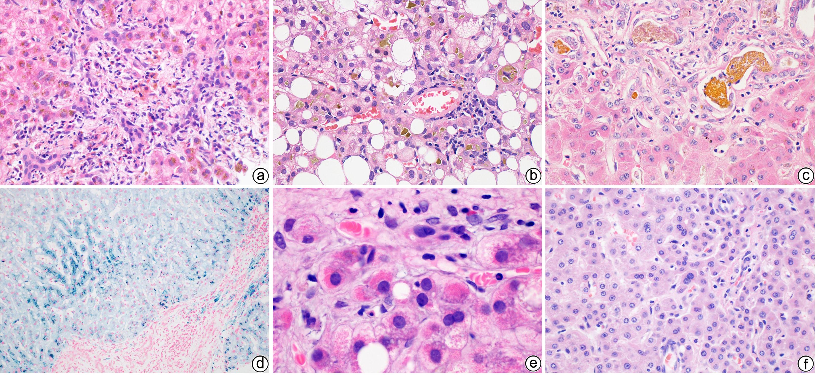
注: a,肝细胞大泡性脂肪变,绿色箭头示大泡性脂肪变中的大脂滴,红色箭头示小脂滴,但轮廓清晰仍属大泡性脂肪变,黑色箭头示脂质肉芽肿(HE染色,×400);b,肝细胞微泡性脂肪变(HE染色,×1 000);c,肝细胞气球样变伴Mallory小体形成(HE染色,×1 000);d,免疫组化CK8/18染色,箭头所示为染色阳性的绳索样Mallory小体(DAB显色,×1 000);e,巨线粒体(箭头所示)(HE染色,×1 000);f,肝实质内中性粒细胞浸润,部分包绕在气球样变的肝细胞周围,被称为“卫星现象”(HE染色,×400);g,ALD相关肝衰病例可见多量Mallory小体及中性粒细胞浸润(HE染色,×400);h,窦周纤维化(Masson染色,×400);i,中央静脉周围纤维化,箭头示胶原纤维在中央静脉外侧壁沉积,导致静脉壁明显增厚(HE染色,×400);j,小叶中心硬化性玻璃样坏死(HE染色,×200);k,小结节性酒精性肝硬化(网状纤维染色,×100);l,汇管区星芒状纤维化(Masson染色,×200)。
图 1 ALD的主要组织病理学特征
Figure 1. Major histopathological features of ALD
表 1 AHS与NASH的组织学特征比较
Table 1. Characteristic histologic features of ASH versus NASH
组织病理学 酒精性脂肪性 肝炎 非酒精性脂肪性肝炎 大泡性脂肪变 + ++ 微泡性脂肪变 +/- +/- 肝细胞气球样变 ++ + Mallory小体 ++ +/- 巨线粒体 +/-~+ +/- 中性粒细胞浸润 ++ + 肝细胞坏死/凋亡 ++ + 窦周纤维化 ++ + 中央静脉周围纤维化 +~++ + 硬化性玻璃样坏死 +/- - 胆汁淤积 +/-~+ - Lenta胆管炎 +/- - 糖原核 +/- + 汇管区细胆管反应 + +/- 汇管区炎症和纤维化 +~++ +/- 注:++,几乎总是存在;+,通常存在;+/-,偶然存在;-,通常缺乏。 -
[1] National Workshop on Fatty Liver and Alcoholic Liver Disease, Chinese Society of Hepatology, Chinese Medical Association; Fatty Liver Expert Committee, Chinese Medical Doctor Association. Guidelines of prevention and treatment for alcoholic liver disease: a 2018 update[J]. J Clin Hepatol, 2018, 34( 5): 939- 946. DOI: 10.3969/j.issn.1001-5256.2018.05.006.中华医学会肝病学分会脂肪肝和酒精性肝病学组, 中国医师协会脂肪性肝病专家委员会. 酒精性肝病防治指南(2018年更新版)[J]. 临床肝胆病杂志, 2018, 34( 5): 939- 946. DOI: 10.3969/j.issn.1001-5256.2018.05.006. [2] YANG S, XING HC, CHENG J. Comparison and interpretation of Chinese, American, and European guidelines on alcoholic liver diseases[J]. J Clin Hepatol, 2018, 34( 7): 1420- 1422. DOI: 10.3969/j.issn.1001-5256.2018.07.011.杨松, 邢卉春, 成军. 中美欧酒精性肝病相关指南的对比与解读[J]. 临床肝胆病杂志, 2018, 34( 7): 1420- 1422. DOI: 10.3969/j.issn.1001-5256.2018.07.011. [3] BEDOSSA P. Current histological classification of NAFLD: Strength and limitations[J]. Hepatol Int, 2013, 7( Suppl 2): 765- 770. DOI: 10.1007/s12072-013-9446-z. [4] BRUNT EM. Surgical assessment of significant steatosis in donor livers: The beginning of the end for frozen-section analysis?[J]. Liver Transpl, 2013, 19( 4): 360- 361. DOI: 10.1002/lt.23609. [5] DENK H, STUMPTNER C, ZATLOUKAL K. Mallory bodies revisited[J]. J Hepatol, 2000, 32( 4): 689- 702. DOI: 10.1016/s0168-8278(00)80233-0. [6] STRNAD P, ZATLOUKAL K, STUMPTNER C, et al. Mallory-Denk-bodies: Lessons from keratin-containing hepatic inclusion bodies[J]. Biochim Biophys Acta, 2008, 1782( 12): 764- 774. DOI: 10.1016/j.bbadis.2008.08.008. [7] ROBERTSON NJ, KENDALL CH. Liver giant mitochondria revisited[J]. J Clin Pathol, 1992, 45( 5): 412- 415. DOI: 10.1136/jcp.45.5.412. [8] BRUGUERA M, BERTRAN A, BOMBI JA, et al. Giant mitochondria in hepatocytes: A diagnostic hint for alcoholic liver disease[J]. Gastroenterology, 1977, 73( 6): 1383- 1387. [9] TELI MR, DAY CP, BURT AD, et al. Determinants of progression to cirrhosis or fibrosis in pure alcoholic fatty liver[J]. Lancet, 1995, 346( 8981): 987- 990. DOI: 10.1016/s0140-6736(95)91685-7. [10] JAESCHKE H. Neutrophil-mediated tissue injury in alcoholic hepatitis[J]. Alcohol, 2002, 27( 1): 23- 27. DOI: 10.1016/s0741-8329(02)00200-8. [11] RAMAIAH SK, JAESCHKE H. Role of neutrophils in the pathogenesis of acute inflammatory liver injury[J]. Toxicol Pathol, 2007, 35( 6): 757- 766. DOI: 10.1080/01926230701584163. [12] MA J, GUILLOT A, YANG ZH, et al. Distinct histopathological phenotypes of severe alcoholic hepatitis suggest different mechanisms driving liver injury and failure[J]. J Clin Invest, 2022, 132( 14): e157780. DOI: 10.1172/JCI157780. [13] BATALLER R, ARAB JP, SHAH VH. Alcohol-associated hepatitis[J]. N Engl J Med, 2022, 387( 26): 2436- 2448. DOI: 10.1056/NEJMra2207599. [14] ROTH NC, QIN J. Histopathology of alcohol-related liver diseases[J]. Clin Liver Dis, 2019, 23( 1): 11- 23. DOI: 10.1016/j.cld.2018.09.001. [15] MATHURIN P, BEUZIN F, LOUVET A, et al. Fibrosis progression occurs in a subgroup of heavy drinkers with typical histological features[J]. Aliment Pharmacol Ther, 2007, 25( 9): 1047- 1054. DOI: 10.1111/j.1365-2036.2007.03302.x. [16] WANLESS IR, SHIOTA K. The pathogenesis of nonalcoholic steatohepatitis and other fatty liver diseases: A four-step model including the role of lipid release and hepatic venular obstruction in the progression to cirrhosis[J]. Semin Liver Dis, 2004, 24( 1): 99- 106. DOI: 10.1055/s-2004-823104. [17] AIGELSREITER A, JANIG E, SOSTARIC J, et al. Clusterin expression in cholestasis, hepatocellular carcinoma and liver fibrosis[J]. Histopathology, 2009, 54( 5): 561- 570. DOI: 10.1111/j.1365-2559.2009.03258.x. [18] NAKANO M, WORNER TM, LIEBER CS. Perivenular fibrosis in alcoholic liver injury: Ultrastructure and histologic progression[J]. Gastroenterology, 1982, 83( 4): 777- 785. [19] van WAES L, LIEBER CS. Early perivenular sclerosis in alcoholic fatty liver: An index of progressive liver injury[J]. Gastroenterology, 1977, 73( 4 Pt 1): 646- 650. [20] EDMONDSON HA, PETERS RL, REYNOLDS TB, et al. Sclerosing hyaline necrosis of the liver in the chronic alcoholic. A recognizable clinical syndrome[J]. Ann Intern Med, 1963, 59: 646- 673. DOI: 10.7326/0003-4819-59-5-646. [21] FLEMING KA, MCGEE JO. Alcohol induced liver disease[J]. J Clin Pathol, 1984, 37( 7): 721- 733. DOI: 10.1136/jcp.37.7.721. [22] COLOMBAT M, CHARLOTTE F, RATZIU V, et al. Portal lymphocytic infiltrate in alcoholic liver disease[J]. Hum Pathol, 2002, 33( 12): 1170- 1174. DOI: 10.1053/hupa.2002.129414. [23] ROSKAMS T, DESMET V. Ductular reaction and its diagnostic significance[J]. Semin Diagn Pathol, 1998, 15( 4): 259- 269. [24] MORGAN MY, SHERLOCK S, SCHEUER PJ. Portal fibrosis in the livers of alcoholic patients[J]. Gut, 1978, 19( 11): 1015- 1021. DOI: 10.1136/gut.19.11.1015. [25] PHILLIPS GB, DAVIDSON CS. Liver disease of the chronic alcoholic simulating extrahepatic biliary obstruction[J]. Gastroenterology, 1957, 33( 2): 236- 244. [26] BRUNT EM. Nonalcoholic steatohepatitis: Definition and pathology[J]. Semin Liver Dis, 2001, 21( 1): 3- 16. DOI: 10.1055/s-2001-12925. [27] KATOONIZADEH A, LALEMAN W, VERSLYPE C, et al. Early features of acute-on-chronic alcoholic liver failure: A prospective cohort study[J]. Gut, 2010, 59( 11): 1561- 1569. DOI: 10.1136/gut.2009.189639. [28] BONAL M, MOURAD M, BANCEL B, et al. Cholangitis lenta: An underdiagnosed lesion associated with severe cholestasis following liver transplantation[J]. Clin Transplant, 2020, 34( 9): e14016. DOI: 10.1111/ctr.14016. [29] YAMADA S, TAKEZAWA J, TAKAGI H, et al. Histologic features and bile duct lesions in the alcoholic[J]. Jpn J Med, 1985, 24( 3): 223- 230. DOI: 10.2169/internalmedicine1962.24.223. [30] ZIMMER V, BITTENBRING J, FRIES P, et al. Severe mixed-type iron overload in alcoholic cirrhosis related to advanced spur cell anemia[J]. Ann Hepatol, 2014, 13( 3): 396- 398. [31] LI LX, GUO FF, LIU H, et al. Iron overload in alcoholic liver disease: Underlying mechanisms, detrimental effects, and potential therapeutic targets[J]. Cell Mol Life Sci, 2022, 79( 4): 201. DOI: 10.1007/s00018-022-04239-9. [32] COSTA MATOS L, BATISTA P, MONTEIRO N, et al. Iron stores assessment in alcoholic liver disease[J]. Scand J Gastroenterol, 2013, 48( 6): 712- 718. DOI: 10.3109/00365521.2013.781217. [33] NAHON P, SUTTON A, RUFAT P, et al. Liver iron, HFE gene mutations, and hepatocellular carcinoma occurrence in patients with cirrhosis[J]. Gastroenterology, 2008, 134( 1): 102- 110. DOI: 10.1053/j.gastro.2007.10.038. [34] YIP WW, BURT AD. Alcoholic liver disease[J]. Semin Diagn Pathol, 2006, 23( 3-4): 149- 160. DOI: 10.1053/j.semdp.2006.11.002. [35] SUZUKI Y, YOKOYAMA A, NAKANO M, et al. Cyanamide-induced liver dysfunction after abstinence in alcoholics: A long-term follow-up study on four cases[J]. Alcohol Clin Exp Res, 2000, 24( 4 Suppl): 100S- 105S. [36] TANJI K, BHAGAT G, VU TH, et al. Mitochondrial DNA dysfunction in oncocytic hepatocytes[J]. Liver Int, 2003, 23( 5): 397- 403. DOI: 10.1034/j.1478-3231.2003.00864.x. [37] HUANG DQ, MATHURIN P, CORTEZ-PINTO H, et al. Global epidemiology of alcohol-associated cirrhosis and HCC: Trends, projections and risk factors[J]. Nat Rev Gastroenterol Hepatol, 2023, 20( 1): 37- 49. DOI: 10.1038/s41575-022-00688-6. [38] HUANG DQ, TAN DJH, NG CH, et al. Hepatocellular carcinoma incidence in alcohol-associated cirrhosis: Systematic review and meta-analysis[J]. Clin Gastroenterol Hepatol, 2023, 21( 5): 1169- 1177. DOI: 10.1016/j.cgh.2022.06.032. [39] TERASAKI S, KANEKO S, KOBAYASHI K, et al. Histological features predicting malignant transformation of nonmalignant hepatocellular nodules: A prospective study[J]. Gastroenterology, 1998, 115( 5): 1216- 1222. DOI: 10.1016/s0016-5085(98)70093-9. [40] DELEMOS A, PATEL M, GAWRIEH S, et al. Distinctive features and outcomes of hepatocellular carcinoma in patients with alcohol-related liver disease: A US multicenter study[J]. Clin Transl Gastroenterol, 2020, 11( 3): e00139. DOI: 10.14309/ctg.0000000000000139. [41] LACKNER C, STAUBER RE, DAVIES S, et al. Development and prognostic relevance of a histologic grading and staging system for alcohol-related liver disease[J]. J Hepatol, 2021, 75( 4): 810- 819. DOI: 10.1016/j.jhep.2021.05.029. [42] PATEL V, SANYAL AJ. Drug-induced steatohepatitis[J]. Clin Liver Dis, 2013, 17( 4): 533- 546, vii. DOI: 10.1016/j.cld.2013.07.012. [43] RAMANATHAN R, IBDAH JA. Mitochondrial dysfunction and acute fatty liver of pregnancy[J]. Int J Mol Sci, 2022, 23( 7): 3595. DOI: 10.3390/ijms23073595. [44] KAYAÇETIN S, BAŞARANOĞLU M. Mallory-Denk bodies: Correlation with steatosis, severity, zonal distribution, and identification with ubiquitin[J]. Turk J Gastroenterol, 2015, 26( 6): 506- 510. DOI: 10.5152/tjg.2015.150199. [45] BASARANOGLU M, TURHAN N, SONSUZ A, et al. Mallory-Denk Bodies in chronic hepatitis[J]. World J Gastroenterol, 2011, 17( 17): 2172- 2177. DOI: 10.3748/wjg.v17.i17.2172. [46] GONZÁLEZ IA, FULLER LD, ZHANG XF, et al. Development of a scoring system to differentiate amiodarone-induced liver injury from alcoholic steatohepatitis[J]. Am J Clin Pathol, 2022, 157( 3): 434- 442. DOI: 10.1093/ajcp/aqab142. [47] OLIVEIRA THC, MARQUES PE, PROOST P, et al. Neutrophils: A cornerstone of liver ischemia and reperfusion injury[J]. Lab Invest, 2018, 98( 1): 51- 62. DOI: 10.1038/labinvest.2017.90. [48] VANSTAPEL MJ, DESMET VJ. Cytomegalovirus hepatitis: A histological and immunohistochemical study[J]. Appl Pathol, 1983, 1( 1): 41- 49. [49] TUNG BY, Jr CARITHERS RL. Cholestasis and alcoholic liver disease[J]. Clin Liver Dis, 1999, 3( 3): 585- 601. DOI: 10.1016/s1089-3261(05)70086-6. [50] STEWART RV, DINCSOY HP. The significance of giant mitochondria in liver biopsies as observed by light microscopy[J]. Am J Clin Pathol, 1982, 78( 3): 293- 298. DOI: 10.1093/ajcp/78.3.293. [51] SAKHUJA P. Pathology of alcoholic liver disease, can it be differentiated from nonalcoholic steatohepatitis?[J]. World J Gastroenterol, 2014, 20( 44): 16474- 16479. DOI: 10.3748/wjg.v20.i44.16474. [52] YEH MM, BRUNT EM. Pathological features of fatty liver disease[J]. Gastroenterology, 2014, 147( 4): 754- 764. DOI: 10.1053/j.gastro.2014.07.056. [53] DIEHL AM, GOODMAN Z, ISHAK KG. Alcohollike liver disease in nonalcoholics. A clinical and histologic comparison with alcohol-induced liver injury[J]. Gastroenterology, 1988, 95( 4): 1056- 1062. -


 PDF下载 ( 1853 KB)


 下载:
下载:


