PDF下载 ( 3361 KB)

肝硬化食管胃静脉曲张内镜下治疗后再出血的危险因素分析及列线图模型构建
DOI: 10.12449/JCH260817
Risk factors for rebleeding after endoscopic therapy for esophageal and gastric varices in liver cirrhosis patients and construction of a nomogram model
-
摘要:
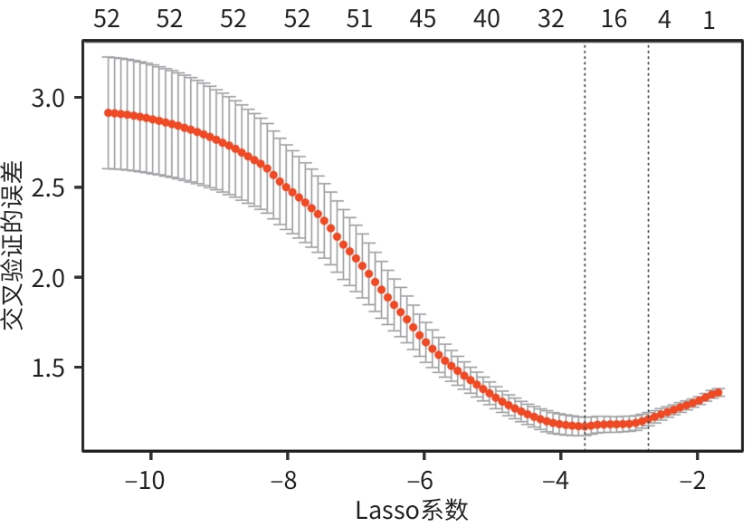
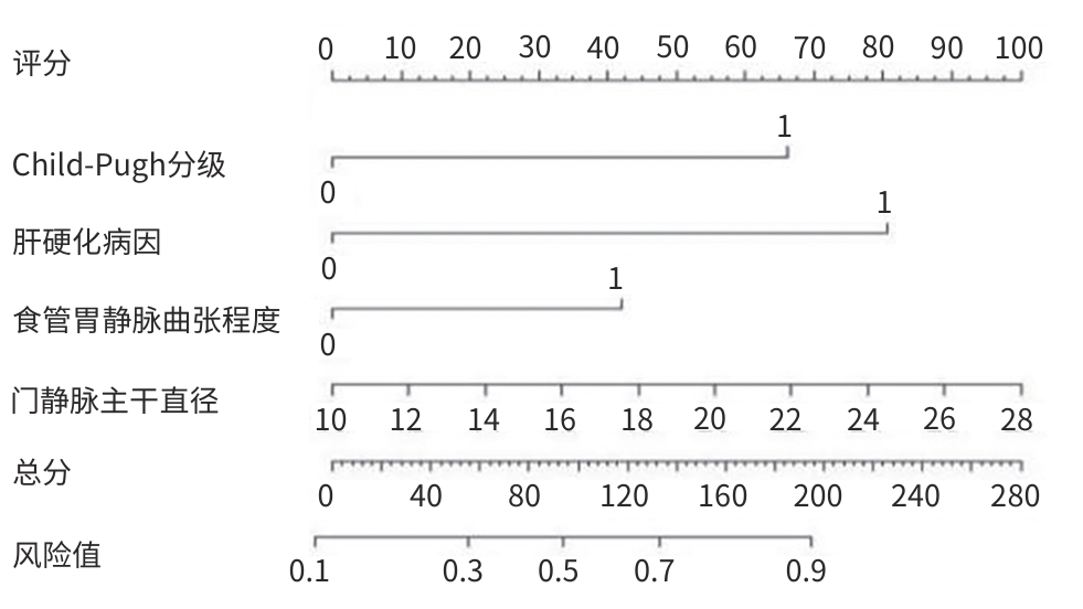
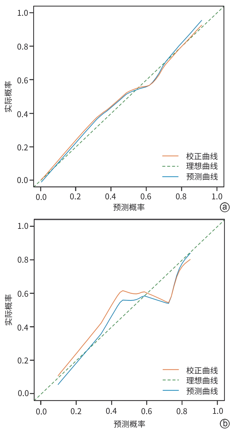
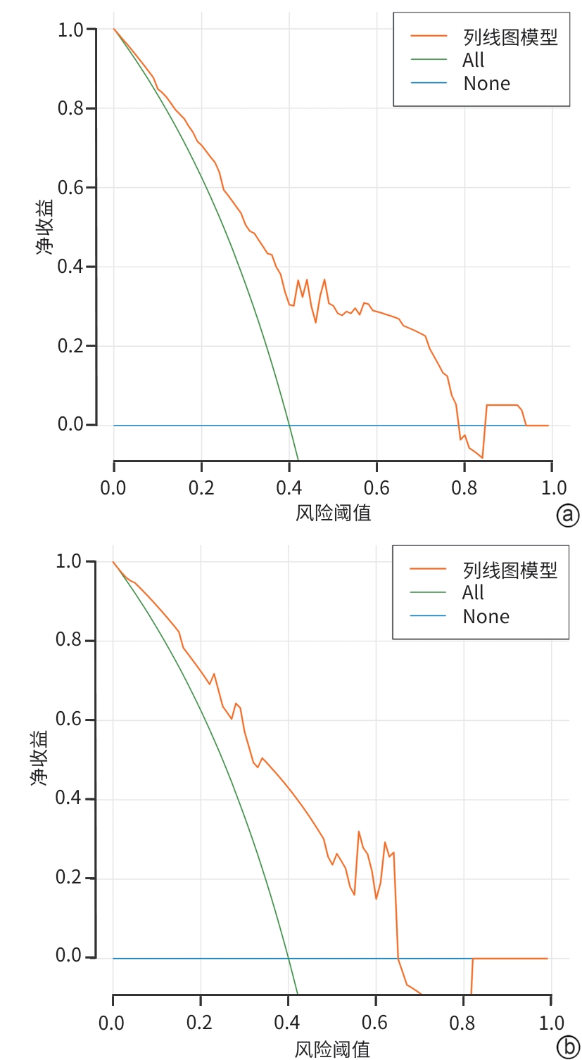
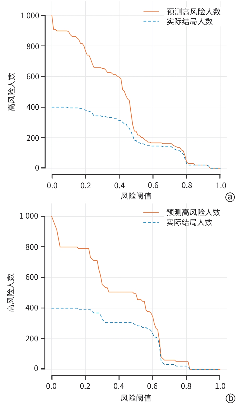
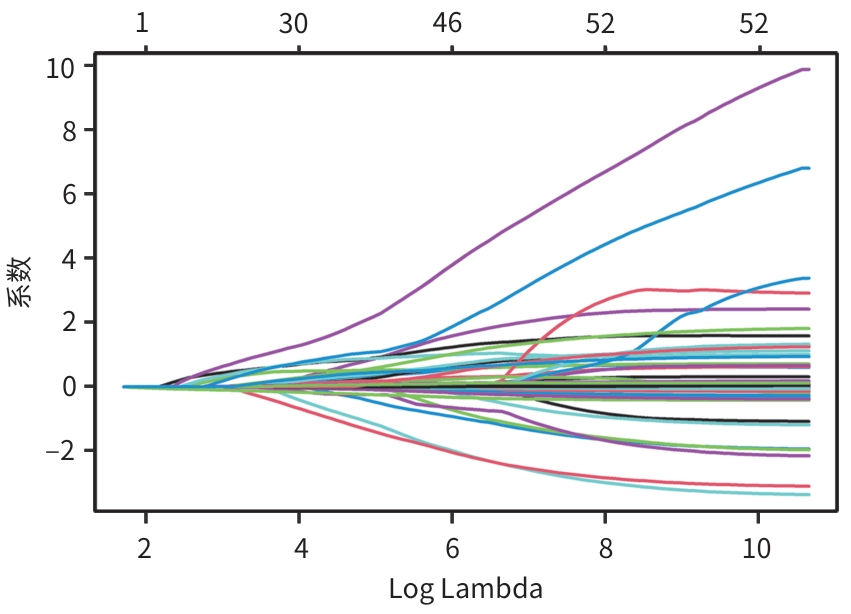
目的 分析肝硬化食管胃静脉曲张内镜治疗后再出血的危险因素并构建临床预测模型,为早期预测再出血、降低再出血发生率及改善患者临床转归提供参考。 方法 回顾性分析2021年1月1日—2025年5月31日在兰州市第二人民医院消化内科经内镜首次治疗的194例肝硬化食管胃静脉曲张破裂出血患者的临床资料,按照患者经内镜治疗后1年内是否发生消化道再出血分为再出血组和未出血组。计量资料两组间比较采用成组t检验或Mann-Whitney U检验。计数资料两组间比较采用χ2 检验或Fisher确切概率法。将入选患者按照7∶3比例随机分为训练集和验证集。在训练集中,通过Lasso回归分析筛选最优预测变量,然后将可能影响预后的因素纳入单因素和多因素Logistic回归分析,筛选肝硬化食管胃静脉曲张再出血的独立预测因素,并以筛选出的变量构建列线图模型。在训练集与验证集中,通过受试者操作特征曲线(ROC曲线)和校准曲线评估列线图模型的分辨力及校准度,决策曲线分析(DCA)和临床影响曲线(CIC)用以评估模型的临床实用性。 结果 194例肝硬化食管胃静脉曲张患者经内镜治疗后1年内,发生再出血者116例(59.79%),其中训练集和验证集再出血患者分别为76例和40例。在训练集中,经Lasso回归分析、单因素和多因素Logistic回归分析后,筛选出4个与肝硬化食管胃静脉曲张再出血相关的独立预测因子,分别为肝硬化病因[比值比(OR)=3.540,95%置信区间(95%CI):1.520~7.150,P<0.001]、蔡尔德-皮尤分级(OR=3.560,95%CI:1.380~9.500,P=0.019)、食管胃静脉曲张程度(OR=8.190,95%CI:3.568~17.850,P=0.026)和门静脉主干直径(OR=2.954,95%CI:1.349~15.030,P=0.044)。以上述独立预测因子构建列线图模型,ROC曲线分析显示,该列线图模型在训练集和验证集的曲线下面积分别为0.845(95%CI:0.778~0.912)和0.801(95%CI:0.798~0.868);训练集和验证集的一致性指数分别为0.832和0.820,提示区分度较好。Hosmer-Lemeshow检验结果显示P值分别为0.320和0.550,校准曲线显示,列线图模型预测概率与实际概率具有良好一致性,说明校准度较好;DCA和CIC分析结果表明,该模型具有良好的临床效用。 结论 基于肝硬化病因、蔡尔德-皮尤分级、食管胃静脉曲张程度和门静脉主干直径构建的列线图模型对肝硬化食管胃静脉曲张再出血风险预测具有一定的临床价值。 Abstract:Objective To investigate the risk factors for rebleeding after endoscopic therapy for esophageal and gastric varices in liver cirrhosis patients, to construct a clinical predictive model, and to provide a reference for predicting rebleeding in the early stage, reducing the incidence rate of rebleeding, and improving the clinical outcome of patients. Methods A retrospective analysis was performed for the clinical data of 194 liver cirrhosis patients with gastroesophageal variceal bleeding who received initial endoscopic therapy at Department of Gastroenterology, The Second People’s Hospital of Lanzhou, from January 1, 2021 to May 31, 2025. According to whether rebleeding occurred within 1 year after endoscopic therapy, the patients were divided into rebleeding group and non-rebleeding group. The independent-samples t test or the Mann-Whitney U test was used for comparison of continuous data between two groups, and the chi-square test or the Fisher’s exact test was used for comparison of categorical data between two groups. The patients enrolled were randomly divided into a training set and a validation set at a ratio of 7∶3. In the training set, the Lasso regression analysis was used to obtain optimal predictive variables, and the factors that might affect prognosis were included in the univariate and multivariate Logistic regression analyses to identify independent predictive factors for rebleeding after endoscopic therapy for esophageal and gastric varices in liver cirrhosis patients, which were used to construct a nomogram model. In both the training set and the validation set, the receiver operating characteristic (ROC) curve and the calibration curve were used to assess the discriminatory ability and calibration of the model, and decision curve analysis and the clinical impact curve were used to assess the clinical practicability of the model. Results Among the 194 liver cirrhosis patients with esophageal and gastric varices, 116 (59.79%) experienced rebleeding within 1 year after endoscopic therapy, with 76 patients in the training set and 40 patients in the validation set. In the training set, the Lasso regression analysis and the univariate and multivariate Logistic regression analyses showed that etiology of liver cirrhosis (odds ratio [OR]=3.540, 95% confidence interval [CI]: 1.520 — 7.150, P<0.001), Child-Pugh class (OR=3.560, 95%CI: 1.380 — 9.500, P=0.019), severity of esophageal and gastric varices (OR=8.190, 95%CI: 3.568 — 17.850, P=0.026), and main portal vein diameter (OR=2.954, 95%CI: 1.349 — 15.030, P=0.044) were independent predictive factors for rebleeding of esophageal and gastric varices in liver cirrhosis patients. A nomogram model was constructed based on the above independent predictive factors. The ROC curve analysis showed that this model had an area under the ROC curve of 0.845 (95%CI: 0.778 — 0.912) in the training set and 0.801 (95%CI: 0.798 — 0.868) in the validation set. The model had an index of concordance of 0.832 in the training set and 0.820 in the validation set, suggesting that the model had a good discriminatory ability. The Hosmer-Lemeshow test showed P values of 0.320 and 0.550 in the training set and validation set, respectively, the calibration curve indicated that the predicted probabilities of the nomogram model were in good concordance with the actual observed probabilities, suggesting that the model had good calibration. The decision curve analysis and the clinical impact curve showed that the model had good clinical utility. Conclusion The nomogram model based on etiology of liver cirrhosis, Child-Pugh class, severity of esophageal and gastric varices, and main portal vein diameter has a certain clinical value in predicting the risk of rebleeding from esophageal and gastric varices in liver cirrhosis. -
Key words:
- Liver Cirrhosis /
- Esophageal and Gastric Varices /
- Endoscopy /
- Hemorrhage /
- Risk Factors /
- Nomograms
-
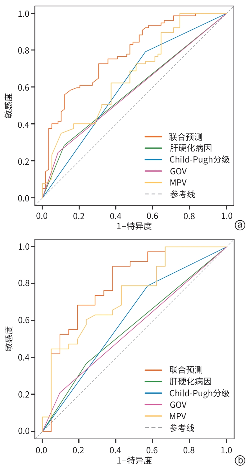
注: a,训练集;b,验证集。Child-Pugh分级,蔡尔德-皮尤分级;GOV,食管胃静脉曲张程度;MPV,门静脉主干直径。
图 4 受试者操作特征曲线
Figure 4. Receiver operator characteristic curve
表 1 训练集与验证集临床资料比较
Table 1. Clinical data between the training set and internal validation set
指标 训练集(n=136) 验证集(n=58) 统计值 P值 年龄(岁) 55.51±11.21 52.86±12.22 t=-1.468 0.144 性别[例(%)] χ2=7.563 0.006 女 69(50.74) 17(29.31) 男 67(49.26) 41(70.69) 体重指数(kg/m2) 22.48±3.48 22.33±4.28 t=-0.262 0.794 家族史[例(%)] χ2=0.087 0.768 无 115(84.56) 50(86.21) 有 21(15.44) 8(13.79) 吸烟史[例(%)] χ2=0.035 0.853 无 84(61.76) 35(60.34) 有 52(38.24) 23(39.66) 饮酒史[例(%)] χ2=0.003 0.956 无 92(67.65) 39(67.24) 有 44(32.35) 19(32.76) 肝硬化病因[例(%)] χ2=0.779 0.677 乙型肝炎 92(67.65) 41(70.69) 自身免疫性 26(19.12) 11(18.97) 酒精性 18(13.24) 6(10.34) 门静脉高压治疗史[例(%)] χ2=0.077 0.781 无 33(24.26) 13(22.41) 有 103(75.74) 45(77.59) 高血压[例(%)] χ2=0.016 0.899 无 102(75.00) 44(75.86) 有 34(25.00) 14(24.14) 糖尿病[例(%)] χ2=0.265 0.607 无 129(94.85) 56(96.55) 有 7(5.15) 2(3.45) Child-Pugh分级[例(%)] χ2=0.231 0.631 A级 111(81.62) 49(84.48) B级 25(18.38) 9(15.52) 肝性脑病[例(%)] χ2=0.233 0.630 无 129(94.85) 54(93.10) 有 7(5.15) 4(6.90) 腹水[例(%)] χ2=1.413 0.235 无 52(38.24) 17(29.31) 有 84(61.76) 41(70.69) 门静脉高压性胃病[例(%)] χ2=3.600 0.058 无 52(38.24) 14(24.14) 有 84(61.76) 44(75.86) 门静脉血栓[例(%)] χ2=0.392 0.531 无 113(83.09) 46(79.31) 有 23(16.91) 12(20.69) 腹膜炎[例(%)] χ2=0.230 0.631 无 131(96.32) 55(94.83) 有 5(3.68) 3(5.17)  下载: 导出CSV
表 1 (续)
Table 1. (continued)
指标 训练集(n=136) 验证集(n=58) 统计值 P值 电解质紊乱[例(%)] χ2=8.822 0.003 无 42(30.88) 31(53.45) 有 94(69.12) 27(46.55) 门静脉主干直径(mm) 16.23±4.24 16.05±4.78 t=-0.259 0.796 脾静脉直径(mm) 11.13±3.01 10.86±2.27 t=-0.631 0.541 门静脉血流速度(cm/s) 11.71±2.75 10.95±2.25 t=-1.870 0.063 食管胃静脉曲张程度[例(%)] χ2=0.492 0.782 轻度 81(59.56) 33(56.90) 中度 43(31.62) 21(36.21) 重度 12(8.82) 4(6.90) 红色征[例(%)] χ2=1.135 0.287 无 28(20.59) 16(27.59) 有 108(79.41) 42(72.41) 组织胶用量(mL) 1.00(0.50~1.50) 1.00(0.50~2.00) Z=-1.390 0.164 治疗方式[例(%)] χ2=0.672 0.412 EIS 53(38.97) 19(32.76) EIS+EVL 83(61.03) 39(67.24) 白细胞(×109/L) 3.00(2.47~4.78) 3.45(2.43~5.02) Z=-0.503 0.615 中性粒细胞(×109/L) 2.05(1.43~3.90) 2.10(1.52~3.17) Z=-0.560 0.575 中性粒细胞百分比(%) 0.69±0.12 0.67±0.10 t=-0.889 0.375 淋巴细胞(×109/L) 0.70(0.41~0.90) 0.70(0.50~1.00) Z=-0.708 0.479 单核细胞(×109/L) 0.26(0.17~0.38) 0.29(0.17~0.44) Z=-1.196 0.232 红细胞(×1012/L) 3.60±0.88 3.62±1.04 t=0.182 0.856 血小板(×1012/L) 57.00(42.25~91.00) 56.50(43.00~81.25) Z=-0.359 0.720 血红蛋白(g/L) 97.57±28.64 99.79±34.08 t=0.468 0.640 总胆红素(μmol/L) 27.90(20.43~41.53) 31.55(20.80~47.25) Z=-0.553 0.580 胆碱酯酶(U/L) 4 074.50(2 974.00~4 577.00) 3 897.50(3 016.25~4 879.50) Z=-0.370 0.711 AST(U/L) 31.00(22.00~43.00) 32.00(23.00~48.25) Z=-0.953 0.341 ALT(U/L) 23.00(18.00~33.00) 25.00(19.00~36.25) Z=-1.064 0.287 AST/ALT 1.31(1.00~1.66) 1.25(1.00~1.55) Z=-0.493 0.662 ALP(U/L) 97.00(72.25~140.75) 93.00(72.00~130.75) Z=-0.420 0.674 GGT(U/L) 27.00(16.00~50.75) 34.50(17.00~56.00) Z=-1.145 0.252 白蛋白(g/L) 36.84±6.12 35.80±6.02 t=-1.089 0.277 球蛋白(g/L) 28.09±7.24 28.19±5.66 t=0.099 0.921 前白蛋白(g/L) 116.56±41.71 114.38±45.23 t=-0.326 0.745 血清肌酐(μmol/L) 62.15(53.78~69.78) 59.30(53.70~67.95) Z=-0.809 0.419 尿酸(μmol/L) 268.49±115.47 270.22±106.72 t=0.098 0.922 总胆固醇(mmol/L) 2.92(2.36~3.56) 2.89(2.46~3.72) Z=-0.492 0.623 甘油三酯(mmol/L) 0.84(0.61~1.19) 0.98(0.64~1.37) Z=-1.407 0.160 凝血酶原时间(s) 15.00(13.73~16.80) 15.35(14.20~17.13) Z=-1.356 0.175 国际标准化比值 1.26(1.16~1.38) 1.29(1.17~1.41) Z=-0.914 0.361 活化部分凝血活酶时间(s) 29.45(27.10~34.18) 29.70(26.43~33.58) Z=-0.439 0.661 凝血酶时间(s) 18.15(17.03~19.45) 18.55(17.18~19.45) Z=-0.348 0.728 凝血酶原活动度(%) 62.15(53.78~69.78) 59.30(53.70~67.95) Z=-0.809 0.419 D-二聚体(mg/L) 0.80(0.33~1.92) 0.55(0.28~2.06) Z=-0.615 0.539 纤维蛋白原(g/L) 1.72(1.20~2.34) 1.87(1.43~2.37) Z=-1.007 0.314 注:Child-Pugh分级,蔡尔德-皮尤分级;EIS,内镜下硬化剂注射治疗;EVL,内镜下曲张静脉套扎术;AST,天冬氨酸氨基转移酶;ALT,丙氨酸氨基转移酶;AST/ALT,天冬氨酸氨基转移酶/丙氨酸氨基转移酶比值;ALP,碱性磷酸酶;GGT,γ-谷氨酰转移酶。
下载: 导出CSV
表 2 肝硬化食管胃静脉曲张再出血与未出血患者临床资料比较
Table 2. Clinical data between the rebleeding and no bleeding set
指标 再出血组(n=116) 未出血组(n=78) 统计值 P值 年龄(岁) 54.34±11.20 55.36±12.10 t=0.604 0.547 性别[例(%)] χ2=1.699 0.192 女 47(40.52) 39(50.00) 男 69(59.48) 39(50.00) 体重指数(kg/m2) 22.46±4.04 22.40±3.21 t=-0.119 0.905 家族史[例(%)] χ2=1.193 0.275 无 96(82.76) 69(88.46) 有 20(17.24) 9(11.54) 吸烟史[例(%)] χ2=0.065 0.799 无 72(62.07) 47(60.26) 有 44(37.93) 31(39.74) 饮酒史[例(%)] χ2=1.833 0.176 无 74(63.79) 57(73.08) 有 42(36.21) 21(26.92) 肝硬化病因[例(%)] χ2=11.344 0.003 乙型肝炎 90(77.59) 43(55.13) 自身免疫性 17(14.66) 20(25.64) 酒精性 9(7.76) 15(19.23) 门静脉高压治疗史[例(%)] χ2=0.265 0.607 无 29(25.00) 17(21.79) 有 87(75.00) 61(78.21) 高血压[例(%)] χ2=6.135 0.013 无 80(68.97) 66(84.62) 有 36(31.03) 12(15.38) 糖尿病[例(%)] χ2=0.071 0.791 无 111(95.69) 74(94.87) 有 5(4.31) 4(5.13) Child-Pugh分级[例(%)] χ2=6.600 0.010 A级 89(76.72) 71(91.03) B级 27(23.28) 7(8.97) 肝性脑病[例(%)] χ2=2.353 0.125 无 107(92.24) 76(97.44) 有 9(7.76) 2(2.56) 腹水[例(%)] χ2=2.586 0.108 无 36(31.03) 33(42.31) 有 80(68.97) 45(57.69) 门静脉高压性胃病[例(%)] χ2=2.852 0.091 无 34(29.31) 32(41.03) 有 82(70.69) 46(58.97) 门静脉血栓[例(%)] χ2=5.347 0.021 无 89(76.72) 70(89.74) 有 27(23.28) 8(10.26) 腹膜炎[例(%)] χ2=0.803 0.370 无 110(94.83) 76(97.44) 有 6(5.17) 2(2.56)
下载: 导出CSV
表 2 (续)
Table 2. (continued)
指标 再出血组(n=116) 未出血组(n=78) 统计值 P值 电解质紊乱[例(%)] χ2=1.975 0.160 无 39(33.62) 34(43.59) 有 77(66.38) 44(56.41) 门静脉主干直径(mm) 14.45±3.96 16.40±4.97 t=3.038 0.003 脾静脉直径(mm) 10.38±2.58 11.38±3.37 t=2.352 0.020 门静脉血流速度(cm/s) 10.89±2.32 11.78±2.67 t=2.475 0.014 食管胃静脉曲张程度[例(%)] χ2=13.580 0.001 轻度 56(48.28) 58(74.36) 中度 46(39.66) 18(23.08) 重度 14(12.07) 2(2.56) 红色征[例(%)] χ2=1.003 0.606 无 25(21.55) 19(24.36) 有 91(78.45) 59(75.64) 组织胶用量(mL) 1.50(0.50~1.70) 1.20(0.50~2.00) Z=-3.246 0.001 治疗方式[例(%)] χ2=2.344 0.126 EIS 38(32.76) 34(43.59) EIS+EVL 78(67.24) 44(56.41) 白细胞(×109/L) 3.00(2.45~4.76) 3.45(2.42~5.01) Z=-0.038 0.970 中性粒细胞(×109/L) 2.06(1.41~3.89) 2.11(1.53~3.18) Z=-0.194 0.846 中性粒细胞百分比(%) 0.69±0.12 0.67±0.11 t=-1.085 0.279 淋巴细胞(×109/L) 0.70(0.49~1.10) 0.65(0.44~0.90) Z=-0.253 0.801 单核细胞(×109/L) 0.26(0.17~0.44) 0.29(0.17~0.38) Z=-0.548 0.584 红细胞(×1012/L) 3.54±0.97 3.70±0.86 t=1.144 0.254 血小板(×1012/L) 55.50(41.25~86.25) 58.00(48.75~83.75) Z=-1.129 0.259 血红蛋白(g/L) 92.12±28.59 102.96±30.40 t=2.525 0.012 总胆红素(μmol/L) 28.60(20.05~41.68) 27.90(21.00~44.18) Z=-0.454 0.650 胆碱酯酶(U/L) 3 667.50(2 944.75~4 873.50) 4 074.50(3 122.00~4 316.00) Z=-0.112 0.911 AST(U/L) 31.00(22.00~44.00) 33.00(23.75~45.75) Z=-0.574 0.566 ALT(U/L) 23.00(19.00~51.30) 24.50(18.00~50.30) Z=-0.333 0.739 AST/ALT 1.25(1.00~1.55) 1.39(1.00~1.77) Z=-1.757 0.079 ALP(U/L) 91.50(70.25~129.50) 102.50(82.75~143.00) Z=-2.285 0.022 GGT(U/L) 27.00(17.00~48.00) 34.50(17.00~73.25) Z=-2.256 0.024 白蛋白(g/L) 36.13±6.86 37.13±4.71 t=1.119 0.265 球蛋白(g/L) 27.34±6.15 29.28±7.55 t=1.960 0.051 前白蛋白(g/L) 112.19±40.40 121.44±45.59 t=1.485 0.139 血清肌酐(μmol/L) 60.00(52.00~71.00) 57.50(49.00~72.25) Z=-2.950 0.003 尿酸(μmol/L) 263.04±119.32 277.88±102.06 t=0.899 0.370 总胆固醇(mmol/L) 2.89(2.32~3.73) 3.02(2.61~3.50) Z=-2.267 0.023 甘油三酯(mmol/L) 0.94(0.65~1.50) 0.70(0.54~1.09) Z=-3.230 0.001 凝血酶原时间(s) 15.25(13.93~17.00) 15.05(13.68~16.73) Z=-0.900 0.368 国际标准化比值 1.27(1.17~1.40) 1.26(1.16~1.35) Z=-0.914 0.360 活化部分凝血活酶时间(s) 31.26±5.78 29.35±4.87 t=-2.399 0.017 凝血酶时间(s) 18.45(17.25~21.19) 17.80(16.80~19.33) Z=-1.507 0.132 凝血酶原活动度(%) 59.80(52.00~68.05) 63.20(56.53~69.05) Z=-1.480 0.139 D-二聚体(mg/L) 0.50(0.28~1.40) 1.18(0.42~6.69) Z=-4.168 <0.001 纤维蛋白原(g/L) 1.64(1.16~1.98) 2.02(1.53~2.65) Z=-3.318 0.001 注:Child-Pugh分级,蔡尔德-皮尤分级;EIS,内镜下硬化剂注射治疗;EVL,内镜下曲张静脉套扎术;AST,天冬氨酸氨基转移酶;ALT,丙氨酸氨基转移酶;AST/ALT,天冬氨酸氨基转移酶/丙氨酸氨基转移酶比值;ALP,碱性磷酸酶;GGT,γ-谷氨酰转移酶。
下载: 导出CSV
表 3 肝硬化食管胃静脉曲张患者发生再出血单因素和多因素Logistic回归分析
Table 3. Logistic regression analysis of independent risk factors for rebleeding from esophageal and gastric varices in cirrhosis
变量 单因素 多因素 OR 95%CI P值 OR 95%CI P值 吸烟(否=0,是=1) 1.033 1.033~1.382 0.003 肝硬化病因(自身免疫性/酒精性=0,乙型肝炎=1) 5.025 1.213~30.819 0.026 3.540 1.520~7.150 <0.001 Child-Pugh分级(A级=0,B级=1) 2.530 1.497~12.863 0.026 3.560 1.380~9.500 0.019 血清肌酐(μmol/L) 1.073 1.013~1.137 0.016 血小板(×1012/L) 0.985 0.973~0.997 0.017 总胆固醇(mmol/L) 1.858 1.740~1.994 0.041 甘油三酯(mmol/L) 5.418 1.167~25.157 0.031 GGT(U/L) 0.978 0.965~0.992 0.002 食管胃静脉曲张程度(轻度/中度=0,重度=1) 7.812 2.563~19.231 0.025 8.190 3.568~17.850 0.026 门静脉主干直径(mm) 1.685 1.521~1.902 0.007 2.954 1.349~15.030 0.044 组织胶用量(mL) 0.907 0.814~1.012 0.097 注:Child-Pugh分级,蔡尔德-皮尤分级;GGT,γ-谷氨酰转移酶;OR,比值比;95%CI,95%置信区间。
下载: 导出CSV
表 4 各变量及联合预测模型评估肝硬化食管胃静脉曲张患者发生再出血的ROC曲线分析
Table 4. ROC curve analysis of variables and combined prediction model for evaluating rebleeding in patients with liver cirrhosis and esophagogastric varices
因素 训练集 验证集 AUC(95%CI) 敏感度
(%)特异度
(%)P值 AUC(95%CI) 敏感度
(%)特异度
(%)P值 肝硬化病因 0.718(0.629~0.807) 95.30 28.30 0.020 0.558(0.531~0.732) 42.50 76.50 0.031 Child-Pugh分级 0.574(0.478~0.670) 78.50 65.30 0.016 0.575(0.456~0.830) 57.10 75.00 0.020 门静脉主干直径 0.732(0.642~0.822) 28.80 46.50 0.039 0.530(0.413~0.630) 40.00 64.50 0.023 食管胃静脉曲张程度 0.621(0.525~0.717) 86.00 82.80 0.016 0.561(0.560~0.871) 60.00 64.70 0.020 联合预测 0.845(0.778~0.912) 90.60 79.10 0.002 0.801(0.798~0.868) 94.60 83.90 0.004 注:ROC曲线,受试者操作特征曲线;Child-Pugh分级,蔡尔德-皮尤分级;AUC,曲线下面积;95%CI,95%置信区间。
下载: 导出CSV
-
[1] Garcia-Tsao G, Abraldes J G, Berzigotti A, et al. Portal hypertensive bleeding in cirrhosis: Risk stratification, diagnosis, and management: 2016 practice guidance by the American Association for the Study of Liver Diseases[J]. Hepatology, 2017, 65( 1): 310- 335. DOI: 10.1002/hep.28906. [2] Jakab S S, Garcia-Tsao G. Evaluation and management of esophageal and gastric varices in patients with cirrhosis[J]. Clin Liver Dis, 2020, 24( 3): 335- 350. DOI: 10.1016/j.cld.2020.04.011. [3] de Franchis R, Bosch J, Garcia-Tsao G, et al. Baveno VII-Renewing consensus in portal hypertension[J]. J Hepatol, 2022, 76( 4): 959- 974. DOI: 10.1016/j.jhep.2021.12.022. [4] Zhan J Y, Chen J, Yu J Z, et al. Prognostic model for esophagogastric variceal rebleeding after endoscopic treatment in liver cirrhosis: A Chinese multicenter study[J]. World J Gastroenterol, 2025, 31( 2): 100234. DOI: 10.3748/wjg.v31.i2.100234. [5] Li J, Li J, Ji Q, et al. Nomogram based on spleen volume expansion rate predicts esophagogastric varices bleeding risk in patients with hepatitis B liver cirrhosis[J]. Front Surg, 2022, 9: 1019952. DOI: 10.3389/fsurg.2022.1019952. [6] Chinese Society of Spleen and Portal Hypertension Surgery, Chinese Society of Surgery, Chinese Medical Association. Expert consensus on the diagnosis and treatment of esophageal and gastric variceal rupture bleeding in cirrhotic portal hypertension(2025 edition)[J]. Chin J Dig Surg, 2025, 24( 3): 271- 280. DOI: 10.3760/cma.j.cn115610-20241228-00590.中华医学会外科学分会脾及门静脉高压外科学组. 肝硬化门静脉高压症食管、胃底静脉曲张破裂出血诊治专家共识(2025版)[J]. 中华消化外科杂志, 2025, 24( 3): 271- 280. DOI: 10.3760/cma.j.cn115610-20241228-00590. [7] Chinese Society of Hepatology, Chinese Society of Gastroenterology, Chinese Society of Digestive Endoscopology of Chinese Medical Association. Guidelines on the management of esophagogastric variceal bleeding in cirrhotic portal hypertension[J]. J Clin Hepatol, 2023, 39( 3): 527- 538.中华医学会肝病学分会, 中华医学会消化病学分会, 中华医学会消化内镜学分会. 肝硬化门静脉高压食管胃静脉曲张出血的防治指南[J]. 临床肝胆病杂志, 2023, 39( 3): 527- 538. [8] Zhang W H, Wang Y L, Chu J D, et al. Investigation report on endoscopic management of esophagogastric variceal bleeding by Chinese endoscopists[J]. Medicine, 2022, 101( 42): e31263. DOI: 10.1097/MD.0000000000031263. [9] Li Xinhui, Qi Xingshun, Wang Jing, et al. Construction and optimization of a quality control system for endoscopic management of esophageal and gastric varices in cirrhosis[J]. Clin J Med Off, 2026, 54( 1): 1- 5, 10. DOI: 10.16680/j.1671-3826.2026.01.01.李鑫会, 祁兴顺, 王静, 等. 肝硬化食管胃静脉曲张内镜管理质量控制体系构建与优化[J]. 临床军医杂志, 2026, 54( 1): 1- 5, 10. DOI: 10.16680/j.1671-3826.2026.01.01. [10] Yang J, Ge K, Chen L, et al. The efficacy comparison of carvedilol plus endoscopic variceal ligation and traditional, nonselective β-blockers plus endoscopic variceal ligation in cirrhosis patients for the prevention of variceal rebleeding: A meta-analysis[J]. Eur J Gastroenterol Hepatol, 2019, 31( 12): 1518- 1526. DOI: 10.1097/MEG.0000000000001442. [11] Duan X W, He X, Yan H Z, et al. Analysis of complications and risk factors other than bleeding before and after endoscopic treatment of esophagogastric variceal bleeding in patients with liver cirrhosis[J]. Can J Gastroenterol Hepatol, 2023, 2023: 7556408. DOI: 10.1155/2023/7556408. [12] Wu L, Fang Q Q, Huang X Q, et al. Risk factors associated with failure of endoscopic combined treatment to prevent varices rebleeding in patients with liver cirrhosis[J]. Expert Rev Gastroenterol Hepatol, 2023, 17( 3): 301- 308. DOI: 10.1080/17474124.2023.2181787. [13] Tao K, Shan X, He B B, et al. Sequential endoscopic treatment for esophageal and gastric variceal bleeding significantly reduces patient mortality and rebleeding rates[J]. Ther Adv Gastroenterol, 2024, 17: 17562848241299743. DOI: 10.1177/17562848241299743. [14] Ding S Y, Lu S Y, Cui L Y, et al. Association between duration of alcohol consumption and risk of hematemesis in patients with alcoholic cirrhosis complicated by esophagogastric variceal bleeding: A case-control study[J]. BMC Gastroenterol, 2025, 25( 1): 849. DOI: 10.1186/s12876-025-04444-3. [15] Taleb A, Ibrahim M. Gastrointestinal bleeding in liver cirrhosis[M]// Recent advances in liver cirrhosis related complications. Academic Press, 2025: 59- 69. [16] Lin C C, Huang Y S. Portal vein thrombosis and esophageal-gastric variceal bleeding in cirrhosis: Shared risk factors and causal relationship[J]. BMC Gastroenterol, 2025, 25( 1): 500. DOI: 10.1186/s12876-025-04094-5. [17] Yang Jianbo, Huang Xiaomei, He Yuanjing, et al. Outcomes of endoscopic treatment of esophagogastric variceal bleeding and construction and validation of a 1-year rebleeding risk prediction model[J]. J Sichuan Univ Med Sci, 2025, 56( 1): 284- 290. DOI: 10.12182/20250160106.杨建波, 黄小梅, 何远静, 等. 食管胃底静脉曲张破裂出血内镜治疗效果及1年内再出血风险预测模型的构建及验证[J]. 四川大学学报(医学版), 2025, 56( 1): 284- 290. DOI: 10.12182/20250160106. [18] Zhu Yingjing, Wu Jie, Tian Xinyue, et al. Endoscopic tissue glue injection and esophageal variceal ligation in treatment of patients with hepatitis B-induced liver cirrhosis and esophageal and gastric varices bleeding[J]. J Pract Hepatol, 2025, 28( 5): 731- 734. DOI: 10.3969/j.issn.1672-5069.2025.05.023.朱颖静, 吴洁, 田鑫越, 等. 内镜下组织胶注射治疗乙型肝炎肝硬化食管胃底静脉曲张破裂出血患者疗效分析[J]. 实用肝脏病杂志, 2025, 28( 5): 731- 734. DOI: 10.3969/j.issn.1672-5069.2025.05.023. -

 本文二维码
本文二维码
计量
- 文章访问数: 8
- HTML全文浏览量: 2
- PDF下载量: 0
- 被引次数: 0