PDF下载 ( 3007 KB)

急性胰腺炎新发糖尿病的危险因素分析及列线图模型构建
DOI: 10.12449/JCH260824
Risk factors for post-acute pancreatitis diabetes mellitus and construction of a nomogram prediction model
-
摘要:
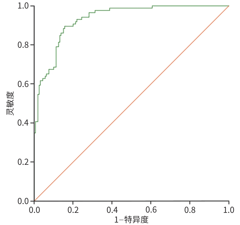
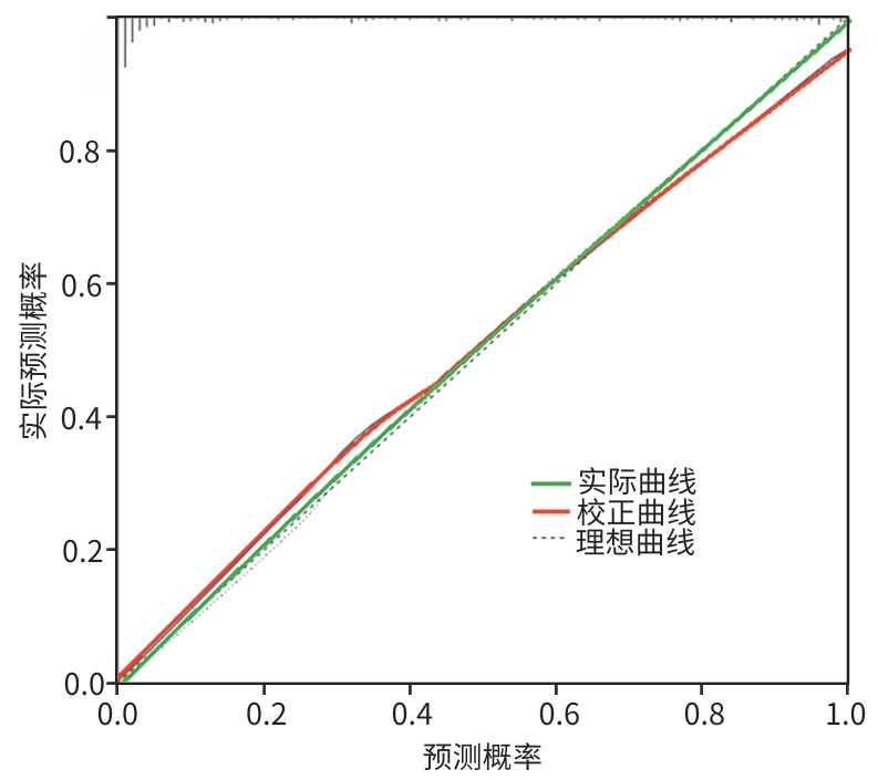
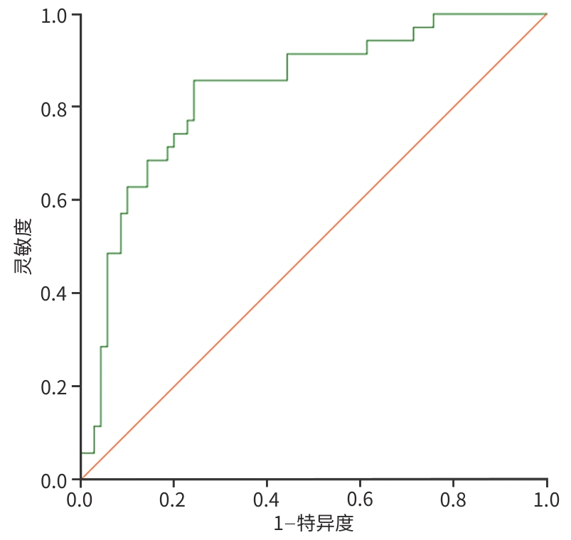
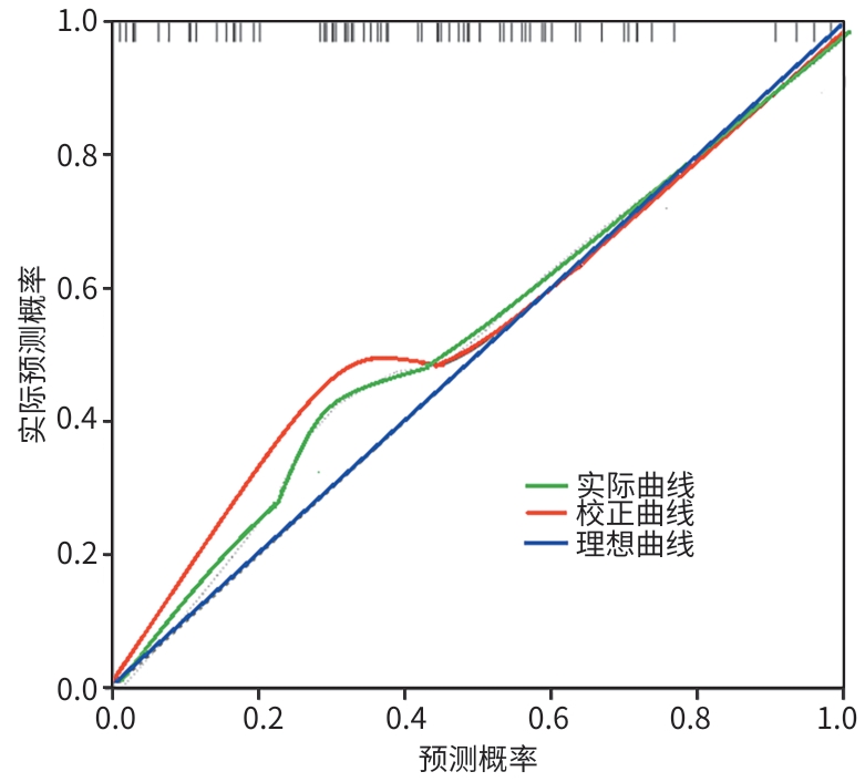
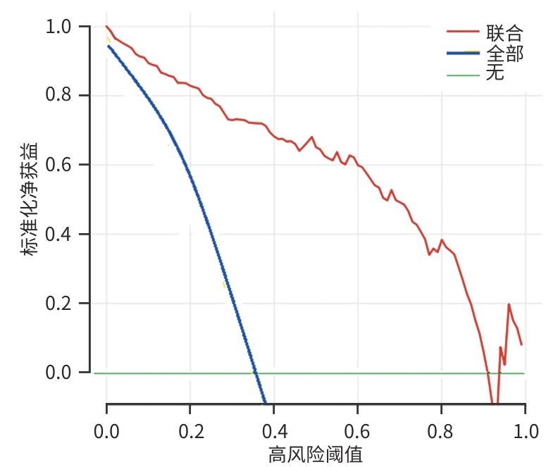
目的 分析急性胰腺炎(AP)患者新发糖尿病(PPDM-A)的危险因素,并构建列线图预测模型,为未来制订个体化治疗方案提供参考。 方法 前瞻性选取2021年6月—2025年1月徐州医科大学附属徐州市立医院收治的351例AP患者为研究对象,按照7∶3比例将患者随机分为建模组(n=246)与验证组(n=105)。根据AP患者是否发生PPDM-A,将建模组进一步分为PPDM-A组(n=86)和非PPDM-A组(n=160)。收集患者的临床资料,采用最小绝对收缩和选择算子(LASSO)回归分析筛选自变量,采用Logistic回归分析PPDM-A的影响因素,并利用R软件构建列线图模型。采用受试者操作特征曲线评估模型的区分度,使用Hosmer-Lemeshow(H-L)检验模型拟合度,校准曲线评估模型一致性;采用决策曲线分析(DCA)评估模型的临床应用价值。计量资料两组间比较采用成组t检验;计数资料两组间比较采用χ2检验。 结果 建模组246例患者中,有86例发生PPDM-A,发生率为34.96%。PPDM-A组与非PPDM-A组在年龄≥60岁(65.12% vs 40.62%)、男性(75.58% vs 55.63%)、体重指数(BMI)≥24 kg/m2(63.95% vs 37.50%)、酒精性AP(56.98% vs 36.87%)、中重度AP(54.65% vs 35.00%)、血钙[(1.46±0.35)mmol/L vs (1.89±0.37)mmol/L]、计算机体层成像严重程度指数(CTSI)评分≥4分(48.84% vs 28.75%)、随机血糖[(17.68±4.12)mmol/L vs (11.68±4.27)mmol/L]比较,差异均有统计学意义(P值均<0.05)。LASSO回归分析共筛选出8个自变量,将8个自变量进行Logistic回归分析,结果显示年龄、性别、BMI、酒精性AP、中重度AP、CTSI评分及随机血糖均是PPDM-A的危险因素(P值均<0.05),血钙为保护因素(P<0.05)。建模组的曲线下面积(AUC)为0.932(95%置信区间:0.903~0.962);H-L拟合优度检验结果为χ2=7.346(P=0.728),模型拟合的准确度较好;较准曲线显示预测概率与实际概率一致,说明一致性较好。验证组的AUC为0.835(95%置信区间:0.753~0.917),H-L拟合优度检验结果为χ2=7.014(P=0.711),模型拟合的准确度较好;较准曲线显示预测概率与实际概率一致,说明一致性较好。建模组DCA结果显示,当阈值概率在0.13~0.94时,该模型评估PPDM-A的临床价值较高。 结论 年龄、性别、BMI、酒精性AP、中重度AP、血钙、CTSI评分及随机血糖是PPDM-A的影响因素,基于上述影响因素构建的列线图模型对PPDM-A风险具有较好的预测能力,可为临床制订PPDM-A预防策略提供参考。 Abstract:Objective To investigate the risk factors for post-acute pancreatitis diabetes mellitus (PPDM-A) in patients with acute pancreatitis (AP), to construct a nomogram prediction model, and to provide a reference for the development of individualized treatment regimens. Methods A total of 351 patients with AP who were admitted to Xuzhou Municipal Hospital Affiliated to Xuzhou Medical University from June 2021 to January 2025 were prospectively enrolled, and they were randomly divided into modeling group with 246 patients and validation group with 105 patients at a ratio of 7∶3. According to the presence or absence of PPDM-A in the patients with AP, the modeling group was further divided into PPDM-A group with 86 patients and non-PPDM-A group with 160 patients. Clinical data were collected from all patients. The least absolute shrinkage and selection operator (LASSO) regression analysis was used to determine independent variables, and a Logistic regression analysis was used to investigate the influencing factors for PPDM-A. R software was used to construct a nomogram model. The receiver operating characteristic curve was used to assess the discriminatory ability of the model, and the Hosmer-Lemeshow test was used to test the model fitting degree, and the calibration curve was used to evaluate the model consistency; and decision curve analysis (DCA) was used to assess its clinical application value. The independent-samples t test was used for comparison of continuous data between two groups, and the chi-square test was used for comparison of categorical data between two groups. Results Among the 246 patients, 86 developed PPDM-A, resulting in an incidence rate of 34.96%. There were significant differences between the PPDM-A group and the non-PPDM-A group in the proportion of patients with an age of ≥60 years (65.12% vs 40.62%, P<0.05), male sex (75.58% vs 55.63%, P<0.05), a body mass index (BMI) of ≥24 kg/m2 (63.95% vs 37.50%, P<0.05), alcoholic AP (56.98% vs 36.87%, P<0.05), moderate-to-severe AP (54.65% vs 35.00%, P<0.05), or a computed tomography severity index (CTSI) score of ≥4 points (48.84% vs 28.75%, P<0.05), as well as significant differences in the levels of blood calcium (1.46±0.35 mmol/L vs 1.89±0.37 mmol/L, P<0.05) and random blood glucose (Glu) (17.68±4.12 mmol/L vs 11.68±4.27 mmol/L, P<0.05). The LASSO regression analysis obtained 8 independent variables. The Logistic regression analysis showed that age, sex, BMI, alcoholic AP, moderate-to-severe AP, CTSI score, and Glu were risk factors for PPDM-A (all P<0.05), while blood calcium was a protective factor (P<0.05). The model had an area under the ROC curve (AUC) of 0.932 (95% confidence interval [CI]: 0.903 — 0.962) in the modeling group, and the Hosmer-Lemeshow goodness-of-fit test yielded χ2=7.346 (P=0.728), the accuracy of model fitting was good; the calibration curve showed that the predicted probability was consistent with the actual probability, indicating that the consistency was good. The model had an AUC of 0.835 (95%CI: 0.753 — 0.917) in the validation group, and the Hosmer-Lemeshow goodness-of-fit test yielded χ2=7.014 (P=0.711), the accuracy of model fitting was good; the calibration curve showed that the predicted probability was consistent with the actual probability, indicating that the consistency was good. The DCA results of the modeling group showed that the model exhibited a high clinical value in evaluating PPDM-A when the threshold probability was 0.13 — 0.94. Conclusion Age, sex, BMI, alcoholic AP, moderate-to-severe AP, blood calcium, CTSI score, and Glu are influencing factors for PPDM-A. The nomogram model constructed based on the above influencing factors shows good performance in predicting the risk of PPDM-A and can thus provide a reference for developing prevention strategies for PPDM-A in clinical practice. -
Key words:
- Pancreatitis /
- Diabetes Mellitus /
- Risk Factors /
- Nomogram
-
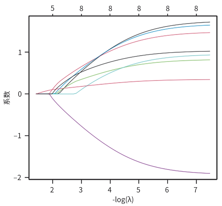
注: LASSO,最小绝对收缩和选择算子。
图 1 LASSO回归系数关系
Figure 1. LASSO regression coefficient relationships
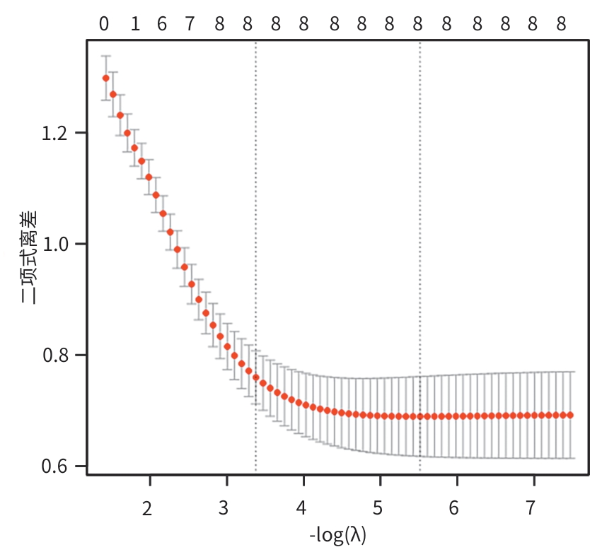
注: LASSO,最小绝对收缩和选择算子。
图 2 10折交叉验证LASSO回归降维图
Figure 2. LASSO regression dimensionality reduction with 10-fold cross-validation
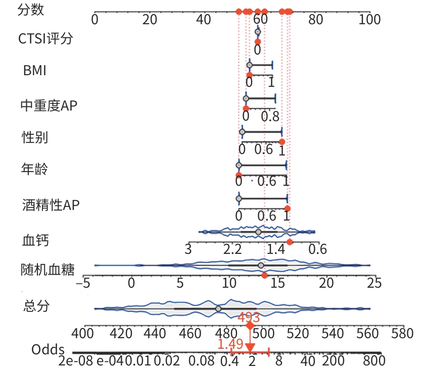
注: CTSI,计算机体层成像严重程度指数;BMI,体重指数;AP,急性胰腺炎;PPDM-A,急性胰腺炎新发糖尿病。
图 3 PPDM-A的列线图模型
Figure 3. The nomogram model of PPDM-A
表 1 建模组与验证组临床资料比较
Table 1. Comparison of clinical data between modeling group and validation group
因素 建模组(n=246) 验证组(n=105) 统计值 P值 年龄[例(%)] χ2=0.072 0.788 ≥60岁 121(49.19) 50(47.62) <60岁 125(50.81) 55(52.38) 性别[例(%)] χ2=0.046 0.830 男 154(62.60) 67(63.81) 女 92(37.40) 38(36.19) BMI[例(%)] χ2=0.256 0.613 ≥24 kg/m2 115(46.75) 46(43.81) <24 kg/m2 131(53.25) 59(56.19) 居住地[例(%)] χ2=0.138 0.711 农村 106(43.09) 43(40.95) 城镇 140(56.91) 62(59.05) 婚姻状况[例(%)] χ2=0.019 0.890 已婚 203(82.52) 86(81.90) 未婚/离异 43(17.48) 19(18.10) 文化程度[例(%)] χ2=0.101 0.751 高中以下 117(47.56) 48(45.71) 高中及以上 129(52.44) 57(54.29) 高血压[例(%)] 32(13.01) 13(12.38) χ2=0.026 0.872 吸烟史[例(%)] 91(36.99) 35(33.33) χ2=0.428 0.513 饮酒史[例(%)] 54(21.95) 24(22.86) χ2=0.035 0.852 高尿酸血症[例(%)] 45(18.29) 20(19.05) χ2=0.028 0.868 胆源性AP[例(%)] 88(35.77) 34(32.38) χ2=0.373 0.541 酒精性AP[例(%)] 108(43.90) 43(40.95) χ2=0.261 0.609 中重度AP[例(%)] 103(41.87) 42(40.00) χ2=0.106 0.745 TC(mmol/L) 4.66±1.24 4.67±1.25 t=0.069 0.945 TG(mmol/L) 3.23±0.35 3.20±0.31 t=0.760 0.448 LDL-C(mmol/L) 2.22±0.35 2.19±0.32 t=0.754 0.451 HDL-C(mmol/L) 1.59±0.33 1.62±0.34 t=0.773 0.440 总蛋白(g/L) 62.20±10.28 62.34±10.53 t=0.116 0.908 白蛋白(g/L) 35.68±5.90 35.86±5.86 t=0.262 0.793 球蛋白(g/L) 26.83±3.02 26.79±2.95 t=0.114 0.909 肌酐(μmol/L) 68.08±10.20 68.11±10.31 t=0.025 0.980 血尿酸(μmol/L) 254.62±15.00 254.75±14.98 t=0.074 0.941 血钙(mmol/L) 1.74±0.36 1.76±0.32 t=0.492 0.623 血钾(mmol/L) 3.90±0.46 3.86±0.43 t=0.760 0.448 胱抑素C(mg/L) 0.88±0.22 0.86±0.21 t=0.790 0.430 血脂肪酶(U/L) 387.99±37.70 387.86±37.13 t=0.030 0.976 血淀粉酶(U/L) 229.15±28.49 229.34±28.64 t=0.057 0.954 CTSI评分[例(%)] χ2=0.071 0.790 ≥4分 88(35.77) 36(34.29) <4分 158(64.23) 69(65.71) 随机血糖(mmol/L) 13.78±4.22 13.59±4.18 t=0.387 0.699 注:BMI,体重指数;TC,总胆固醇;TG,甘油三酯;LDL-C,低密度脂蛋白胆固醇;HDL-C,高密度脂蛋白胆固醇;CTSI,计算机体层成像严重程度指数;AP,急性胰腺炎。
 下载: 导出CSV
表 2 PPDM-A组和非PPDM-A组临床资料比较
Table 2. Comparison of clinical data between PPDM-A group and non-PPDM-A group
因素 PPDM-A组(n=86) 非PPDM-A组(n=160) 统计值 P值 年龄[例(%)] χ2=13.424 <0.001 ≥60岁 56(65.12) 65(40.62) <60岁 30(34.88) 95(59.38) 性别[例(%)] χ2=9.515 0.002 男 65(75.58) 89(55.63) 女 21(24.42) 71(44.37) BMI[例(%)] χ2=15.724 <0.001 ≥24 kg/m2 55(63.95) 60(37.50) <24 kg/m2 31(36.05) 100(62.50) 居住地[例(%)] χ2=0.065 0.799 农村 38(44.19) 68(42.50) 城镇 48(55.81) 92(57.50) 婚姻状况[例(%)] χ2=0.132 0.716 已婚 72(83.72) 131(81.88) 未婚/离异 14(16.28) 29(18.12) 文化程度[例(%)] χ2=0.086 0.769 高中以下 42(48.84) 75(46.87) 高中及以上 44(51.16) 85(53.13) 高血压[例(%)] 12(13.95) 20(12.50) χ2=0.104 0.747 吸烟史[例(%)] 34(39.53) 57(35.62) χ2=0.367 0.545 饮酒史[例(%)] 20(23.26) 34(21.25) χ2=0.131 0.717 高尿酸血症[例(%)] 18(20.93) 27(16.87) χ2=0.616 0.433 胆源性AP[例(%)] 32(37.21) 56(35.00) χ2=0.119 0.730 酒精性AP[例(%)] 49(56.98) 59(36.87) χ2=9.177 0.002 中重度AP[例(%)] 47(54.65) 56(35.00) χ2=8.875 0.003 TC(mmol/L) 4.71±1.28 4.63±1.22 t=0.482 0.630 TG(mmol/L) 3.26±0.38 3.22±0.34 t=0.844 0.399 LDL-C(mmol/L) 2.18±0.34 2.24±0.36 t=1.271 0.205 HDL-C(mmol/L) 1.62±0.34 1.58±0.33 t=0.887 0.376 总蛋白(g/L) 62.24±10.16 62.18±10.34 t=0.044 0.965 白蛋白(g/L) 34.98±6.10 36.05±5.79 t=1.356 0.176 球蛋白(g/L) 27.13±3.08 26.67±2.99 t=1.139 0.256 肌酐(μmol/L) 68.19±10.34 68.02±10.13 t=0.125 0.901 血尿酸(μmol/L) 253.64±14.32 255.14±15.37 t=0.747 0.456 血钙(mmol/L) 1.46±0.35 1.89±0.37 t=8.856 <0.001 血钾(mmol/L) 3.86±0.43 3.92±0.47 t=0.983 0.327 胱抑素C(mg/L) 0.91±0.23 0.87±0.21 t=1.377 0.170 血脂肪酶(U/L) 391.24±38.62 386.24±37.21 t=0.992 0.322 血淀粉酶(U/L) 230.67±29.65 228.34±27.86 t=0.612 0.541 CTSI评分[例(%)] χ2=9.823 0.002 ≥4分 42(48.84) 46(28.75) <4分 44(51.16) 114(71.25) 随机血糖(mmol/L) 17.68±4.12 11.68±4.27 t=10.638 <0.001 注:BMI,体重指数;TC,总胆固醇;TG,甘油三酯;LDL-C,低密度脂蛋白胆固醇;HDL-C,高密度脂蛋白胆固醇;CTSI,计算机体层成像严重程度指数;AP,急性胰腺炎;PPDM-A,急性胰腺炎新发糖尿病。
下载: 导出CSV
表 3 自变量赋值方式
Table 3. Methods of assigning values to independent variables
自变量 赋值方式 年龄 ≥60岁=1,<60岁=0 性别 男=1,女=0 BMI ≥24 kg/m2=1,<24 kg/m2=0 酒精性AP 有=1,无=0 中重度AP 有=1,无=0 血钙 连续变量 CTSI评分 ≥4分=1,<4分=0 随机血糖 连续变量 注:BMI,体重指数;CTSI,计算机体层成像严重程度指数;AP,急性胰腺炎。
下载: 导出CSV
表 4 PPDM-A的影响因素分析
Table 4. Analysis of influencing factors of PPDM-A
变量 B值 SE Wald χ2 P值 OR 95%CI 年龄 0.062 0.016 15.094 <0.001 1.064 1.031~1.097 性别 1.808 0.444 16.564 <0.001 6.099 2.553~14.568 BMI 1.086 0.469 5.364 0.011 2.963 1.182~7.431 酒精性AP 1.802 0.451 15.976 <0.001 6.061 2.505~14.665 中重度AP 0.964 0.436 4.901 0.027 2.623 1.117~6.161 血钙 -2.255 0.535 17.736 <0.001 0.105 0.037~0.300 CTSI评分 1.101 0.248 19.736 <0.001 3.006 1.850~4.885 随机血糖 0.361 0.064 31.730 <0.001 1.435 1.266~1.627 常量 -7.372 1.538 22.987 <0.001 0.001 — 注:BMI,体重指数;CTSI,计算机体层成像严重程度指数;AP,急性胰腺炎;PPDM-A,急性胰腺炎新发糖尿病;SE,标准误;OR,比值比;95%CI,95%置信区间。
下载: 导出CSV
-
[1] Xia Jun, Sun Liang, Wang Jun, et al. Value of C-reactive protein-albumin-lymphocyte index, C-reactive protein-to-lymphocyte ratio, and C-reactive protein-to-serum calcium ratio in evaluating the severity and prognosis of patients with acute pancreatitis[J]. J Clin Hepatol, 2025, 41( 12): 2597- 2606. DOI: 10.12449/JCH251222.夏军, 孙亮, 王军, 等. C反应蛋白-白蛋白-淋巴细胞指数、C反应蛋白与淋巴细胞比值和C反应蛋白与血清钙比值对急性胰腺炎患者病情程度及预后的评估价值[J]. 临床肝胆病杂志, 2025, 41( 12): 2597- 2606. DOI: 10.12449/JCH251222. [2] Yang Tao, Zhu Guangwen. The relationship between serum FABP4, miR-369-5p expressions and disease severity in patients with acute pancreatitis[J]. Labeled Immunoass Clin Med, 2021, 28( 2): 256- 261. DOI: 10.11748/bjmy.issn.1006-1703.2021.02.019.杨涛, 朱光文. 急性胰腺炎患者血清FABP4、miR-369-5p表达与疾病严重程度的关系[J]. 标记免疫分析与临床, 2021, 28( 2): 256- 261. DOI: 10.11748/bjmy.issn.1006-1703.2021.02.019. [3] Liu Hui, Li Xianqiu, Luo Gang, et al. Risk factors for new-onset diabetes after incipient acute pancreatitis[J]. J Clin Hepatol, 2020, 36( 12): 2771- 2776. DOI: 10.3969/j.issn.1001-5256.2020.12.026.刘慧, 李贤秋, 罗刚, 等. 初发急性胰腺炎后新发糖尿病的危险因素评估[J]. 临床肝胆病杂志, 2020, 36( 12): 2771- 2776. DOI: 10.3969/j.issn.1001-5256.2020.12.026. [4] Zhang J, Lv Y Q, Hou J Y, et al. Machine learning for post-acute pancreatitis diabetes mellitus prediction and personalized treatment recommendations[J]. Sci Rep, 2023, 13( 1): 4857. DOI: 10.1038/s41598-023-31947-4. [5] Hu R, Yang H, Zeng G F, et al. A radiomics model of contrast-enhanced computed tomography for predicting post-acute pancreatitis diabetes mellitus[J]. Quant Imaging Med Surg, 2024, 14( 3): 2267- 2279. DOI: 10.21037/qims-23-1232. [6] Pancreatic Disease Group, Digestive Disease Brunch, Chinese Medical Association; Editorial Board for Chinese Journal of Pancreatology; Editorial Board for Chinese Journal of Digestion. Guidelines for the diagnosis and treatment of acute pancreatitis in China(2019 Shenyang)[J]. Chin J Dig, 2019, 39( 11): 721- 730. DOI: 10.3760/cma.j.issn.0254-1432.2019.11.001.中华医学会消化病学分会胰腺疾病学组, 中华胰腺病杂志编辑委员会, 中华消化杂志编辑委员会. 中国急性胰腺炎诊治指南(2019年, 沈阳)[J]. 中华消化杂志, 2019, 39( 11): 721- 730. DOI: 10.3760/cma.j.issn.0254-1432.2019.11.001. [7] American Diabetes Association. 2. Classification and diagnosis of diabetes: Standards of medical care in diabetes-2019[J]. Diabetes Care, 2019, 42( Suppl 1): S13- S28. DOI: 10.2337/dc19-S002. [8] Noda Y, Goshima S, Fujimoto K, et al. Utility of the portal venous phase for diagnosing pancreatic necrosis in acute pancreatitis using the CT severity index[J]. Abdom Radiol, 2018, 43( 11): 3035- 3042. DOI: 10.1007/s00261-018-1579-z. [9] Xu Xin, Chen Zhangxing. Correlation of the serum levels of adiponectin, omentin, and visfatin with the severity of acute pancreatitis[J]. J Clin Hepatol, 2024, 40( 9): 1848- 1852. DOI: 10.12449/JCH240920.许新, 陈章兴. 血清脂联素、网膜素及内脂素水平与急性胰腺炎严重程度的相关性分析[J]. 临床肝胆病杂志, 2024, 40( 9): 1848- 1852. DOI: 10.12449/JCH240920. [10] Huang Bohan, Cao Feng, Ding Yixuan, et al. Analysis of influencing factors for splenomegaly secondary to acute pancreatitis and construc-tion of nomogram prediction model[J]. Chin J Dig Surg, 2024, 23( 5): 712- 719. DOI: 10.3760/cma.j.cn115610-20240221-00121.黄铂涵, 曹锋, 丁乙轩, 等. 急性胰腺炎继发脾大的影响因素分析及列线图预测模型构建[J]. 中华消化外科杂志, 2024, 23( 5): 712- 719. DOI: 10.3760/cma.j.cn115610-20240221-00121. [11] Tian Y, Huang Q, Ren Y T, et al. Visceral adipose tissue predicts severity and prognosis of acute pancreatitis in obese patients[J]. Hepatobiliary Pancreat Dis Int, 2024, 23( 5): 458- 462. DOI: 10.1016/j.hbpd.2023.08.014. [12] García-Compeán D, Jiménez-Rodríguez A R, Muñoz-Ayala J M, et al. Post-acute pancreatitis diabetes: A complication waiting for more recognition and understanding[J]. World J Gastroenterol, 2023, 29( 28): 4405- 4415. DOI: 10.3748/wjg.v29.i28.4405. [13] Jiang Mingfei, Hu Tangbin, Zuo Siyuan, et al. Risk factors for new-onset diabetes after incipient acute pancreatitis[J]. J Mudanjiang Med Univ, 2022, 43( 1): 44- 46, 50. DOI: 10.13799/j.cnki.mdjyxyxb.2022.01.010.江明菲, 胡唐彬, 左思源, 等. 初发急性胰腺炎后新发糖尿病的危险因素评估[J]. 牡丹江医学院学报, 2022, 43( 1): 44- 46, 50. DOI: 10.13799/j.cnki.mdjyxyxb.2022.01.010. [14] Ning C H, Ouyang H, Shen D C, et al. Prediction of survival in patients with infected pancreatic necrosis: A prospective cohort study[J]. Int J Surg, 2024, 110( 2): 777- 787. DOI: 10.1097/JS9.0000000000000844. [15] Li Zan, Liu Xiyang, He Zhuojia, et al. A survey study on the disease burden of diabetes in China from 1999 to 2019[J]. Med J Chin People’s Liberation Army, 2024, 49( 7): 776- 782. DOI: 10.11855/j.issn.0577-7402.0602.2024.0516.李赞, 刘喜洋, 贺卓佳, 等. 1999—2019年中国糖尿病疾病负担的调查研究[J]. 解放军医学杂志, 2024, 49( 7): 776- 782. DOI: 10.11855/j.issn.0577-7402.0602.2024.0516. [16] Yang Yulu, Li Mingxuan, Wang Zhongqiong. Risk factors and predictive models of diabetes mellitus after severe acute pancreatitis[J]. J Chengdu Med Coll, 2020, 15( 2): 232- 237. DOI: 10.3969/j.issn.1674-2257.2020.02.023.杨雨露, 李明轩, 王忠琼. 重症胰腺炎后并发糖尿病的相关危险因素及预测模型[J]. 成都医学院学报, 2020, 15( 2): 232- 237. DOI: 10.3969/j.issn.1674-2257.2020.02.023. [17] Luo Y X, Liu Z R, Luo J W, et al. BMI trajectories in late middle age, genetic risk, and incident diabetes in older adults: Evidence from a 26-year longitudinal study[J]. Am J Epidemiol, 2024, 193( 5): 685- 694. DOI: 10.1093/aje/kwad080. [18] Liang Yizhong, Zhang Weiqing, Guo Shaoju, et al. Risk factors for new onset type 2 diabetes after acute and chronic pancreatitis: A Meta-analysis[J]. Chin J Clin Res, 2021, 34( 3): 314- 318. DOI: 10.13429/j.cnki.cjcr.2021.03.006.梁艺钟, 张维晴, 郭绍举, 等. 急慢性胰腺炎后新发2型糖尿病的危险因素Meta分析[J]. 中国临床研究, 2021, 34( 3): 314- 318. DOI: 10.13429/j.cnki.cjcr.2021.03.006. [19] Li Bingqing, Zhang Qi, Mao Tao, et al. Meta-analysis of the prevalence and risk factors of new diabetes after chronic pancreatitis[J]. Chin J Pancreatol, 2023, 23( 5): 335- 341. DOI: 10.3760/cma.j.cn115667-20230225-00026.李冰清, 张琪, 毛涛, 等. 慢性胰腺炎后新发糖尿病的患病率及危险因素的荟萃分析[J]. 中华胰腺病杂志, 2023, 23( 5): 335- 341. DOI: 10.3760/cma.j.cn115667-20230225-00026. [20] Wang H L, Gao J H, Wen L L, et al. Ion channels in acinar cells in acute pancreatitis: Crosstalk of calcium, iron, and copper signals[J]. Front Immunol, 2024, 15: 1444272. DOI: 10.3389/fimmu.2024.1444272. [21] Uludağ S S, Güreş N, Şirolu S, et al. Investigating the correlation between severe acute pancreatitis and pancreatic necrosis with some serum parameters[J]. Ulus Travma Acil Cerrahi Derg, 2022, 28( 11): 1609- 1615. DOI: 10.14744/tjtes.2021.96782. [22] Zhong S T, Du Q L, Liu N, et al. Developing a CT-based radiomics nomogram for predicting post-acute pancreatitis diabetes mellitus incidence[J]. Br J Radiol, 2023, 96( 1152): 20230382. DOI: 10.1259/bjr.20230382. -

 本文二维码
本文二维码
计量
- 文章访问数: 6
- HTML全文浏览量: 1
- PDF下载量: 2
- 被引次数: 0