PDF下载 ( 14942 KB)

剪接因子3B亚基6在胰腺癌中的表达及其对胰腺癌细胞生物学行为的影响
DOI: 10.12449/JCH260825
Expression of splicing factor 3B subunit 6 in pancreatic cancer and its effect on the biological behaviors of pancreatic cancer cells
-
摘要:
目的 通过生物信息学、临床样本验证及体外功能实验,系统阐明剪接因子3B亚基6(SF3B6)在胰腺癌中的表达特征、临床预后价值及其对癌细胞恶性表型的调控作用,以评估其作为新型肿瘤标志物的价值。 方法 从癌症基因组图谱和加利福尼亚大学圣克鲁兹分校Xena公共数据库中下载转录组和临床信息数据,分析SF3B6在胰腺癌和正常胰腺组织中的表达差异,评估SF3B6表达水平与胰腺癌患者预后、临床指标的关联。培养3种胰腺导管腺癌细胞株和1种正常胰腺导管上皮细胞株,通过实时荧光定量聚合酶链式反应(RT-qPCR)实验初步检测SF3B6在胰腺癌中的表达水平。选取2024年1月—2025年12月在滨州医学院附属医院接受手术的43例胰腺癌患者为研究对象,通过蛋白质印迹实验和RT-qPCR实验进一步探究SF3B6在胰腺癌组织中的表达情况。根据RT-qPCR表达水平的中位数,将胰腺癌患者分为临床样本高、低表达组,评估SF3B6表达与临床特征和预后的关系。构建SF3B6稳定低表达的胰腺癌细胞株,并通过细胞计数试剂盒-8实验、Transwell迁移和侵袭实验,分析SF3B6对胰腺癌细胞株增殖、侵袭和迁移等生物学行为的影响。符合正态分布的计量资料两组间比较采用成组t检验,多组间比较采用单因素方差分析,进一步两两比较采用LSD-t检验或Games-Howell法;不符合正态分布的计量资料两组间比较采用Wilcoxon秩和检验。计数资料两组间比较采用χ2检验。采用Logistic回归分析SF3B6表达的影响因素;采用单因素及多因素Cox比例风险回归模型分析影响预后的危险因素。 结果 生物信息分析结果表明,SF3B6在胰腺癌组织中高表达(P<2.2×10⁻¹⁶),能显著影响胰腺癌患者的总生存期(P=0.011)和无进展生存期(P=0.002)。性别、年龄、临床分期和TNM分期均非SF3B6表达的影响因素(P值均>0.05),SF3B6为影响胰腺癌患者生存的独立危险因素(总生存期:风险比=2.26,95%置信区间:1.13~4.50,P=0.021;无进展生存期:风险比=2.37,95%置信区间:1.22~4.58,P=0.010)。细胞和组织实验证实,SF3B6在胰腺癌细胞株中均高表达(P值均<0.05),且表达水平随胰腺癌恶性程度升高而增高,在恶性程度最高的PANC-1细胞中相对表达量最高;SF3B6在胰腺癌实体组织中的表达水平显著高于癌旁组织(P<0.01)。SF3B6表达水平可影响胰腺癌患者的总生存期(P<0.05),但与年龄、性别、病理分级、肿瘤最大径和临床分期无关(P值均>0.05)。敲低SF3B6表达可显著抑制胰腺癌细胞的增殖、迁移和侵袭能力(P值均<0.01)。 结论 SF3B6在胰腺癌中高表达,并可促进胰腺癌细胞的增殖、迁移和侵袭,其可作为胰腺癌潜在的预后评价指标。 Abstract:Objective To systematically clarify the expression profile of splicing factor 3B subunit 6 (SF3B6) in pancreatic cancer, its clinical prognostic value, and its regulatory effect on the malignant phenotype of cancer cells through bioinformatics analysis, clinical sample validation, and in vitro functional experiments, and to assess its potential as a novel tumor biomarker. Methods Transcriptomic and clinical data were downloaded from The Cancer Genome Atlas and Xena public databases at The University of California Santa Cruz to investigate the difference in the expression of SF3B6 between pancreatic cancer tissue and normal pancreatic tissue, and the association of SF3B6 with the prognosis and clinical indices of patients with pancreatic cancer was analyzed. Three pancreatic ductal adenocarcinoma cell lines and one normal pancreatic ductal epithelial cell line were cultured, and RT-qPCR was used to measure the expression level of SF3B6 in pancreatic cancer. A total of 43 patients with pancreatic cancer who underwent surgery in Binzhou Medical University Hospital from January 2024 to December 2025 were enrolled as subjects, and Western blot and RT-qPCR were used to measure the expression of SF3B6 in pancreatic cancer tissue. Based on the median expression level of SF3B6 measured by RT-qPCR, the patients were divided into high and low expression groups. The association of SF3B6 expression with clinical features and prognosis was assessed. A pancreatic cancer cell line with stable low SF3B6 expression was constructed, and CCK-8 assay and Transwell migration and invasion assays were used to observe the impact of SF3B6 on the biological behaviors of the pancreatic cancer cell line, including proliferation, migration, and invasion. The independent-samples t test was used for comparison of normally distributed continuous data between two groups, and a one-way analysis of variance was used for comparison between multiple groups, while the least significant difference t-test or the Games-Howell test was used for further comparison between two groups; the Wilcoxon rank-sum test was used for comparison of non-normally distributed continuous data between two groups. The chi-square test was used for comparison of categorical data between two groups. A Logistic regression analysis was used to investigate the influencing factors for the expression of SF3B6, and the univariate and multivariate Cox proportional-hazards regression model analyses were used to investigate the risk factors for prognosis. Results The bioinformatics analysis showed that SF3B6 was highly expressed in pancreatic cancer tissue (P<2.2×10⁻¹⁶) and significantly affected the overall survival (P=0.011) and progression-free survival (P=0.002) of patients with pancreatic cancer. Sex, age, clinical stage, and TNM stage were not influencing factors for SF3B6 expression (all P>0.05), and SF3B6 could be used as an independent risk factor for the survival outcome of patients with pancreatic cancer (overall survival: hazard ratio=2.26, 95% confidence interval: 1.13 — 4.50, P=0.021; progression-free survival: hazard ratio=2.37,95% confidence interval: 1.22 — 4.58, P=0.010). Cellular and tissue experiments confirmed the high expression level of SF3B6 in pancreatic cancer cell lines (P<0.05), and the expression level of SF3B6 increased with the increase in the malignancy of pancreatic cancer, showing the highest relative expression level in PANC-1 cells with the highest degree of malignancy; the expression level of SF3B6 in the solid tissue of pancreatic cancer was significantly higher than that in paracancerous tissue (P<0.01). The expression level of SF3B6 might affect the overall survival of patients with pancreatic cancer (P<0.05), while it showed no significant association with age, sex, pathological grade, maximum tumor diameter, or clinical stage (all P>0.05). Knockdown of SF3B6 significantly inhibited the proliferation, migration, and invasion of pancreatic cancer cells (all P<0.01). Conclusion SF3B6 is highly expressed in pancreatic cancer, and the high expression of SF3B6 can promote the proliferation, migration, and invasion of pancreatic cancer cells. Therefore, SF3B6 can be used as a potential biomarker for prognostic evaluation. -
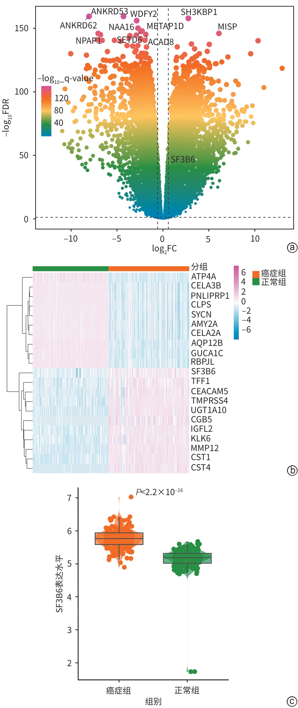
注: a,火山图;b,热图;c,箱式图。SF3B6,剪接因子3B亚基6。
图 1 SF3B6基因在胰腺癌和正常胰腺样本中的表达差异分析
Figure 1. Differentially expression analysis of SF3B6 between pancreatic cancer and normal samples
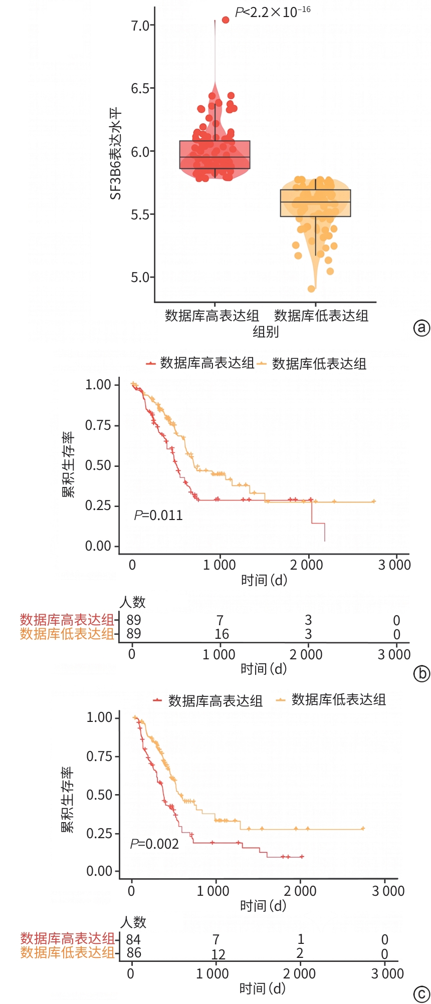
注: a,数据库高、低表达组SF3B6表达箱式图;b,SF3B6数据库高、低表达组胰腺癌患者总生存期比较;c,SF3B6数据库高、低表达组胰腺癌患者无进展生存期比较;SF3B6,剪接因子3B亚基6。
图 2 数据库中不同SF3B6表达组胰腺癌患者的生存分析
Figure 2. Survival analysis of pancreatic cancer patients with high and low SF3B6 expression from database
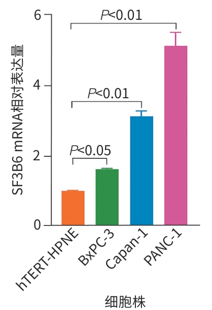
注: SF3B6,剪接因子3B亚基6;mRNA,信使RNA;hTERT-HPNE,人源正常胰腺导管上皮细胞株;BxPC-3,人源胰腺导管原位腺癌细胞株;Capan-1,高分化胰腺导管腺癌细胞株;PANC-1,低分化人源胰腺导管腺癌细胞株。
图 3 SF3B6 mRNA在不同细胞系中的表达情况
Figure 3. mRNA expression levels of SF3B6 across different cell lines
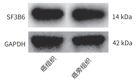
注: SF3B6,剪接因子3B亚基6;GAPDH,甘油醛-3-磷酸脱氢酶。
图 4 SF3B6在胰腺癌实体组织和癌旁组织中的表达
Figure 4. Expression of SF3B6 in pancreatic cancer tissues and adjacent normal tissues
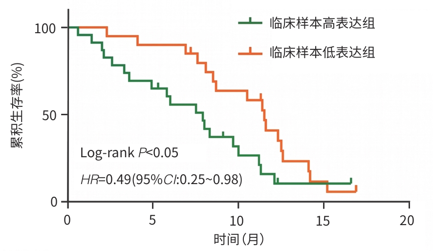
注: SF3B6,剪接因子3B亚基6;HR,风险比;CI,置信区间。
图 5 SF3B6表达水平对总生存期的影响
Figure 5. Effect of SF3B6 expression on overall survival
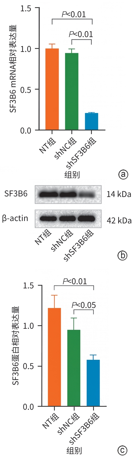
注: a,SF3B6 mRNA相对表达量;b,Western Blot实验结果;c,SF3B6蛋白相对表达量。SF3B6,剪接因子3B亚基6;mRNA,信使RNA;NT,未作任何处理的PANC-1细胞株;shNC,空白慢病毒转染的阴性对照PANC-1细胞株;shSF3B6,稳定低表达SF3B6的PANC-1细胞株;β-actin,β-肌动蛋白。
图 6 SF3B6在稳定低表达细胞株中的表达情况
Figure 6. Expression of SF3B6 in pancreatic cell lines with stable SF3B6 knockdown
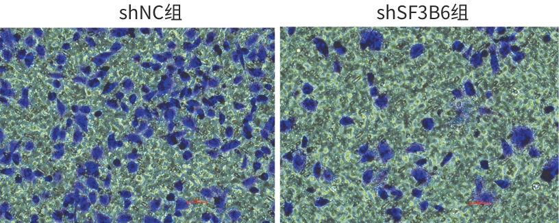
注: SF3B6,剪接因子3B亚基6;shNC,空白慢病毒转染的阴性对照PANC-1细胞株。
图 7 shSF3B6组和shNC组的Transwell小室迁移实验(×100)
Figure 7. Transwell migration assay of shSF3B6 and shNC groups(×100)
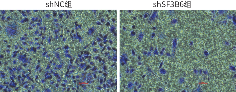
注: SF3B6,剪接因子3B亚基6;shNC,空白慢病毒转染的阴性对照PANC-1细胞株。
图 8 shSF3B6组和shNC组的Transwell小室侵袭实验(×100)
Figure 8. Transwell invasion assay of shSF3B6 and shNC groups(×100)
表 1 SF3B6表达水平的Logistic回归分析
Table 1. Logistic regression analysis of SF3B6 expression
指标 OR(95%CI) P值 性别(男 vs 女) 1.20(0.61~1.98) 0.763 年龄(>65岁 vs ≤65岁) 0.83(0.46~1.50) 0.548 临床分期(Ⅲ~Ⅳ期 vs Ⅰ~Ⅱ期) 0.16(0.01~0.96) 0.092 T分期(T3~T4 vs T1~T2) 1.08(0.50~2.37) 0.843 N分期(N1 vs N0) 0.98(0.50~7.90) 0.944 M分期(M1 vs M0) 0.33(0.02~2.73) 0.350 注:SF3B6,剪接因子3B亚基6;OR,比值比;CI,置信区间。
 下载: 导出CSV
表 2 OS为生存结局的单因素及多因素Cox分析
Table 2. Univariate and multivariate Cox proportional hazards regression analysis with OS as a endpoint
指标 单因素Cox分析 多因素Cox分析 HR 95%CI P值 HR 95%CI P值 性别 0.82 0.55~1.24 0.350 年龄 1.03 1.01~1.05 0.009 1.02 1.00~1.04 0.040 临床分期 1.32 0.90~1.93 0.150 T分期 1.57 1.01~2.43 0.044 1.05 0.63~1.73 0.861 N分期 2.11 1.26~3.55 0.005 1.96 1.14~3.37 0.015 M分期 1.05 0.25~4.39 0.947 SF3B6 2.71 1.42~5.16 0.002 2.26 1.13~4.50 0.021 注:OS,总生存期;SF3B6,剪接因子3B亚基6;HR,风险比;CI,置信区间。
下载: 导出CSV
表 3 PFS为生存结局的单因素及多因素Cox分析
Table 3. Univariate and multivariate Cox proportional hazards regression analysis with PFS as a endpoint
指标 单因素Cox分析 多因素Cox分析 HR 95%CI P值 HR 95%CI P值 性别 0.95 0.64~1.40 0.792 年龄 1.02 1.00~1.04 0.115 临床分期 1.42 1.03~2.00 0.036 1.03 0.62~1.71 0.911 T分期 1.75 1.15~2.67 0.010 1.32 0.77~2.25 0.317 N分期 1.74 1.11~2.73 0.016 1.53 0.97~2.41 0.069 M分期 0.84 0.26~2.70 0.768 SF3B6 3.09 1.63~5.84 0.001 2.37 1.22~4.58 0.010 注:PFS,无进展生存期;SF3B6,剪接因子3B亚基6;HR,风险比;CI,置信区间。
下载: 导出CSV
表 4 不同SF3B6表达组胰腺癌患者的一般临床资料比较
Table 4. Comparison of general clinical data of pancreatic cancer patients in different SF3B6 expression groups
指标 临床样本
高表达组
(n=23)临床样本
低表达组
(n=20)χ2值 P值 年龄[例(%)] 2.73 0.098 ≥60岁 15(65.22) 8(40.00) <60岁 8(34.78) 12(60.00) 性别[例(%)] 0.61 0.434 男 16(69.57) 16(80.00) 女 7(30.43) 4(20.00) 病理分级[例(%)] 0.98 0.323 高中分化 20(86.96) 14(70.00) 低分化 3(13.04) 6(30.00) 肿瘤最大径[例(%)] 0.40 0.526 ≤2 cm 17(73.91) 13(65.00) >2 cm 6(26.09) 7(35.00) 临床分期[例(%)] 3.31 0.069 Ⅰ+Ⅱ期 11(47.83) 15(75.00) Ⅲ+Ⅳ期 12(52.17) 5(25.00) 注:SF3B6,剪接因子3B亚基6。
下载: 导出CSV
-
[1] Farhangnia P, Khorramdelazad H, Nickho H, et al. Current and future immunotherapeutic approaches in pancreatic cancer treatment[J]. J Hematol Oncol, 2024, 17( 1): 40. DOI: 10.1186/s13045-024-01561-6. [2] Du Nan, Wei Miaoyan, Xu Jin. Advancements and frontiers in targeted therapy for pancreatic cancer[J]. Chin J Clin Pharmacol Ther, 2025, 30( 2): 183- 192. DOI: 10.12092/j.issn.1009-2501.2025.02.004.杜楠, 魏妙艳, 徐近. 胰腺癌的靶向治疗进展及前沿[J]. 中国临床药理学与治疗学, 2025, 30( 2): 183- 192. DOI: 10.12092/j.issn.1009-2501.2025.02.004. [3] Liu Yueze, Zhang Taiping, Zhao Yupei. Advances and thoughts in the diagnosis and treatment of pancreatic cancer[J]. J Clin Hepatol, 2025, 41( 4): 601- 604. DOI: 10.12449/JCH250401.刘悦泽, 张太平, 赵玉沛. 胰腺癌诊治的进展与思考[J]. 临床肝胆病杂志, 2025, 41( 4): 601- 604. DOI: 10.12449/JCH250401. [4] Angarola B L, Anczuków O. Splicing alterations in healthy aging and disease[J]. Wiley Interdiscip Rev RNA, 2021, 12( 4): e1643. DOI: 10.1002/wrna.1643. [5] Stanley R F, Abdel-Wahab O. Dysregulation and therapeutic targeting of RNA splicing in cancer[J]. Nat Cancer, 2022, 3( 5): 536- 546. DOI: 10.1038/s43018-022-00384-z. [6] Alors-Pérez E, Pedraza-Arevalo S, Blázquez-Encinas R, et al. Splicing alterations in pancreatic ductal adenocarcinoma: A new molecular landscape with translational potential[J]. J Exp Clin Cancer Res, 2023, 42( 1): 282. DOI: 10.1186/s13046-023-02858-z. [7] van der Feltz C, Hoskins A A. Structural and functional modularity of the U2 snRNP in pre-mRNA splicing[J]. Crit Rev Biochem Mol Biol, 2019, 54( 5): 443- 465. DOI: 10.1080/10409238.2019.1691497. [8] Sun C F. The SF3b complex: Splicing and beyond[J]. Cell Mol Life Sci, 2020, 77( 18): 3583- 3595. DOI: 10.1007/s00018-020-03493-z. [9] Stoop T F, Javed A A, Oba A, et al. Pancreatic cancer[J]. Lancet, 2025, 405( 10485): 1182- 1202. DOI: 10.1016/S0140-6736(25)00261-2. [10] Zhang Taiping, Ye Liyuan, Wang Yuanyang. Development and practice of precise treatment of pancreatic cancer[J]. Chin J Dig Surg, 2025, 24( 5): 567- 573. DOI: 10.3760/cma.j.cn115610-20250411-00147.张太平, 叶力源, 王苑阳. 胰腺癌精准治疗的发展与实践[J]. 中华消化外科杂志, 2025, 24( 5): 567- 573. DOI: 10.3760/cma.j.cn115610-20250411-00147. [11] Rahib L, Smith B D, Aizenberg R, et al. Projecting cancer incidence and deaths to 2030: The unexpected burden of thyroid, liver, and pancreas cancers in the United States[J]. Cancer Res, 2014, 74( 11): 2913- 2921. DOI: 10.1158/0008-5472.CAN-14-0155. [12] Baralle F E, Giudice J. Alternative splicing as a regulator of development and tissue identity[J]. Nat Rev Mol Cell Biol, 2017, 18( 7): 437- 451. DOI: 10.1038/nrm.2017.27. [13] Bonnal S C, López-Oreja I, Valcárcel J. Roles and mechanisms of alternative splicing in cancer: Implications for care[J]. Nat Rev Clin Oncol, 2020, 17( 8): 457- 474. DOI: 10.1038/s41571-020-0350-x. [14] Zhang Y J, Qian J J, Gu C Y, et al. Alternative splicing and cancer: A systematic review[J]. Sig Transduct Target Ther, 2021, 6: 78. DOI: 10.1038/s41392-021-00486-7. [15] Zhang Y D, Wu J Y, Qin T J, et al. Comparison of the revised 4th(2016) and 5th(2022) editions of the World Health Organization classification of myelodysplastic neoplasms[J]. Leukemia, 2022, 36( 12): 2875- 2882. DOI: 10.1038/s41375-022-01718-7. [16] Yang Chenmeng, Wang Chunmei. Splicing factor SF3b6 promotes proliferation and migration of gastric cancer cells through MAPK signaling pathway[J]. Chin J Cancer Biother, 2021, 28( 3): 247- 253. DOI: 10.3872/j.issn.1007-385x.2021.03.005.杨晨萌, 王春梅. 剪接因子SF3b6通过MAPK信号通路促进胃癌细胞的增殖和迁移[J]. 中国肿瘤生物治疗杂志, 2021, 28( 3): 247- 253. DOI: 10.3872/j.issn.1007-385x.2021.03.005. [17] Yuan C X, Gao Y Y, Huang Y, et al. SF3B6 promotes the oncogenic phenotypes of MDA-MB-231 cells by extensively interacting with and regulating transcripts expression and alternative splicing[J]. Sci Rep, 2025, 15( 1): 39840. DOI: 10.1038/s41598-025-23477-y. [18] Kato K, Udagawa R, Hayashi Y, et al. SF3B14 is involved in the centrosome regulation through splicing of TUBGCP6 pre-mRNA[J]. Biochem Biophys Res Commun, 2022, 588: 133- 139. DOI: 10.1016/j.bbrc.2021.12.059. -

 本文二维码
本文二维码
计量
- 文章访问数: 8
- HTML全文浏览量: 1
- PDF下载量: 0
- 被引次数: 0