益脾养肝方对大鼠肝癌前病变肝干细胞恶性转化的影响和作用机制
DOI: 10.3969/j.issn.1001-5256.2022.04.023
Effect of Yipi Yanggan prescription on malignant transformation of liver stem cells in rats with liver precancerous lesion and its mechanism of action
-
摘要:
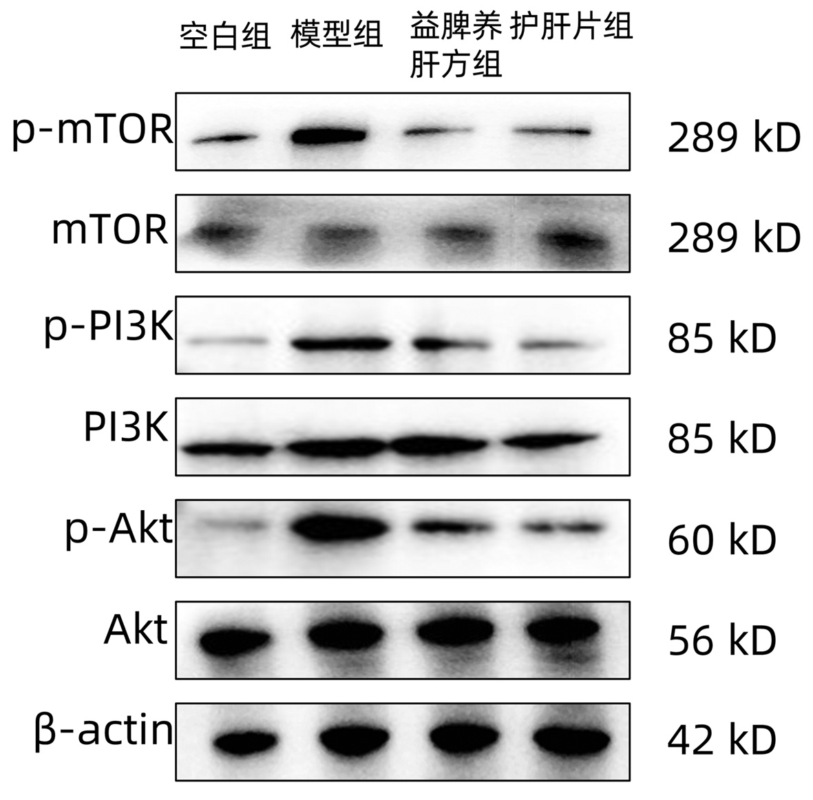
目的 探讨益脾养肝方对二乙基亚硝胺(DEN)诱导的肝癌前病变中肝干细胞恶性转化的影响及可能的分子机制。 方法 将35只雄性SD大鼠随机分为正常对照组(空白组)、DEN模型组(模型组)、DEN+益脾养肝方组(益脾养肝方组)和DEN+护肝片组(护肝片组),空白组5只,其余3组各10只。腹腔注射DEN诱导肝癌前病变模型,给药16周后处死。检测血清ALT、AST、Alb水平;取肝组织观察并记录其大小、外观等变化,计算肝重比(肝脏指数);HE染色、天狼星红染色观察大鼠肝组织病理形态学改变;免疫组化检测OV6和谷胱甘肽S转移酶(GST-Pi)的表达;实时定量PCR检测EpCAM、CD133和CD90 mRNA的表达;Western Blot检测PI3K、Akt、mTOR蛋白及其磷酸化水平的表达。计量资料多组间比较采用单因素方差分析,进一步两两比较采用LSD-t检验。 结果 与模型组相比,益脾养肝方组和护肝片组肝脏病理形态显著改善,肝脏指数、ALT和AST水平降低、Alb水平升高(P值均<0.05);同时GST-Pi、OV6、p-PI3K、p-Akt、p-mTOR蛋白和EpCAM、CD133、CD90 mRNA表达均明显降低(P值均<0.05)。与护肝片组相比,益脾养肝方组对肝脏的保护作用更显著,其肝脏指数、ALT和AST水平显著降低、Alb水平显著升高(P值均<0.05);且GST-Pi、OV6、p-PI3K、p-Akt、p-mTOR蛋白和EpCAM、CD133、CD90 mRNA表达水平均显著降低(P值均<0.05)。 结论 益脾养肝方可通过抑制肝干细胞恶性转化改善DEN诱导的大鼠肝癌前病变,其作用机制可能与PI3K/Akt/mTOR信号通路有关。 Abstract:Objective To investigate the effect of Yipi Yanggan prescription on the malignant transformation of liver stem cells in liver precancerous lesion induced by diethylnitrosamine (DEN) and its possible molecular mechanism. Methods A total of 35 male Sprague-Dawley rats were randomly divided into normal control group (blank group), DEN model group (model group), DEN+Yipi Yanggan prescription group (Yipi Yanggan prescription group), and DEN+Hugan tablet group (Hugan tablet group), with 5 rats in the blank group and 10 rats in the other three groups. Intraperitoneal injection of DEN was performed to establish a model of liver precancerous lesion, the rats were sacrificed after 16 weeks of administration. The serum levels of alanine aminotransferase (ALT), aspartate aminotransferase (AST), and albumin (Alb) were measured; liver tissue was collected to observe the changes in size and appearance and calculate liver weight ratio (liver index); HE staining and Sirius Red staining were used to observe the pathological and morphological changes of rat liver tissue; immunohistochemistry was used to measure the expression of OV6 and glutathione S-transferase-Pi (GST-Pi); RT-PCR was used to measure the mRNA expression of EpCAM, CD133, and CD90, and Western blot was used to measure the protein expression of PI3K, Akt, and mTOR and their phosphorylation level. A one-way analysis of variance was used for comparison between multiple groups, and the least significant difference t-test was used for further comparison between two groups. Results Compared with the model group, the Yipi Yanggan prescription group and the Hugan tablet group had significant improvements in liver pathology and morphology, significant reductions in liver index and the levels of ALT and AST, and a significant increase in the level of Alb (all P < 0.05), as well as significant reductions in the protein expression levels of GST-Pi, OV6, p-PI3K, p-Akt, and p-mTOR and the mRNA expression levels of EpCAM, CD133, and CD90 (all P < 0.05). Compared with the Hugan tablet group, the Yipi Yanggan prescription group showed a more significant protective effect on the liver, with significant reductions in liver index and the levels of ALT and AST, and a significant increase in the level of Alb (all P < 0.05), as well as significant reductions in the protein expression levels of GST-Pi, OV6, p-PI3K, p-Akt, and p-mTOR and the mRNA expression levels of EpCAM, CD133, and CD90 (all P < 0.05). Conclusion Yipi Yanggan prescription can improve liver precancerous lesion induced by DEN in rats by inhibiting the malignant transformation of liver stem cells, and its mechanism of action may be associated with the PI3K/Akt/mTOR signaling pathway. -
Key words:
- Liver Neoplasms /
- Yipi Yanggan Recipe /
- Pathologic Processes /
- Precancerous Conditions
-
表 1 引物序列表
Table 1. Primer sequences used in RT-PCR
基因 上游 下游 CD90 ATCCAGCATGAGTTCAGCCT ATCCTTGGTGGTGAAGTTGG CD133 AACCACAACGGAAGTCAGCT TGACATCCCCAGTTTGGTTT EpCAM CGCAGCTCAGAAAGACTGTG CTCGGGATCATACAGACCGT β-actin CAACCTTCTTGCAGCTCCTC TTCTGACCCATACCCACCAT 表 2 各组大鼠肝组织中胶原沉积半定量分析
Table 2. Semi-quantitative analysis of collagen deposition in liver tissues of rats in various groups
组别 动物数(只) 胶原沉积光密度值(107) 空白组 5 1.49±0.45 模型组 9 12.54±5.361) 护肝片组 9 4.59±1.042) 益脾养肝方组 10 2.32±0.512)3) F值 20.29 P值 <0.05 注:与空白组相比,1)P<0.05;与模型组相比,2)P<0.05;与护肝片组相比,3)P<0.05。 表 3 各组大鼠血清ALT、AST、Alb和肝脏指数比较
Table 3. Comparison of ALT, AST, Alb and liver index in various groups
组别 动物数(只) ALT(U/L) AST(U/L) Alb(g/L) 肝脏指数(%) 空白组 5 26.67±4.60 82.48±14.54 36.68±2.43 2.78±0.18 模型组 9 46.67±6.521) 228.89±16.881) 20.46±2.791) 5.79±0.621) 护肝片组 9 38.15±1.362) 196.91±17.972) 24.98±1.312) 4.65±0.392) 益脾养肝方组 10 29.97±3.832)3) 157.17±21.522)3) 27.81±1.472)3) 3.44±0.722)3) F值 16.28 49.15 38.90 22.32 P值 <0.05 <0.05 <0.05 <0.05 注:与空白组相比,1)P<0.01;与模型组相比,2)P<0.05;与护肝片组相比,3)P<0.05。 表 4 各组大鼠肝组织中OV6及GST-Pi含量半定量分析
Table 4. Semi-quantitative analysis of OV6 and GST-Pi in liver tissues of rats in various groups
组别 动物数(只) OV6光密度值(×107) GST-Pi光密度值(×108) 空白组 5 0.57±0.19 1.27±0.78 模型组 9 10.71±3.811) 6.71±1.321) 护肝片组 9 5.17±1.412) 4.11±1.272) 益脾养肝方组 10 2.21±0.922)3) 1.79±0.882)3) F值 38.21 33.81 P值 <0.05 <0.05 注:与空白组相比,1)P<0.05;与模型组相比,2)P<0.05;与护肝片组相比,3)P<0.05。 表 5 各组大鼠肝组织中EpCAM、CD133和CD90 mRNA相对表达量比较
Table 5. Comparison of mRNA expression levels of EpCAM, CD133 and CD90 in various groups
组别 动物数(只) EpCAM CD133 CD90 空白组 5 1.24±0.32 1.18±0.15 1.07±0.26 模型组 9 18.59±2.731) 20.66±3.841) 21.00±3.511) 护肝片组 9 10.37±2.362) 9.17±1.902) 12.04±1.612) 益脾养肝方组 10 6.13±1.602)3) 5.26±1.302)3) 9.11±2.142)3) F值 129.40 110.20 70.61 P值 <0.05 <0.05 <0.05 注:与空白组相比,1)P<0.05;与模型组相比,2)P<0.05;与护肝片组相比,3)P<0.05。 表 6 各组大鼠肝组织中PI3K/Akt/mTOR信号通路相关蛋白半定量分析
Table 6. Semi-quantitative analysis of PI3K/Akt/mTOR signaling pathway-related protein in liver tissues of rats in various groups
组别 动物数(只) p-mTOR mTOR p-PI3K PI3K p-Akt Akt 空白组 5 1 1 1 1 1 1 模型组 9 4.9±0.411) 1.08±0.16 10.05±0.981) 1.06±0.22 5.72±1.461) 1.31±0.07 护肝片组 10 2.64±0.282) 1.07±0.19 5.74±0.712) 1.12±0.32 2.54±0.212) 1.34±0.16 益脾养肝方组 9 2.08±0.142)3) 0.89±0.23 3.40±0.752)3) 0.93±0.32 1.86±0.192)3) 1.02±0.31 F值 40.35 0.27 58.61 0.22 15.38 1.19 P值 <0.05 >0.05 <0.05 >0.05 <0.05 >0.05 注:与空白组相比,1)P<0.05;与模型组相比,2)P<0.05;与护肝片组相比,3)P<0.05。 -
[1] LLOVET JM, KELLEY RK, VILLANUEVA A, et al. Hepatocellular carcinoma[J]. Nat Rev Dis Primers, 2021, 7(1): 6. DOI: 10.1038/s41572-020-00240-3. [2] JIAO JZ, LI JT, YAN SG, et al. Current research status of precancerous dysplastic nodules in hepatocellular carcinoma[J]. J Clin Hepatol, 2017, 33(5): 974-978. DOI: 10.3969/j.issn.1001-5256.2017.05.039.焦俊喆, 李京涛, 闫曙光, 等. 肝细胞癌癌前异型增生结节的研究现状[J]. 临床肝胆病杂志, 2017, 33(5): 974-978. DOI: 10.3969/j.issn.1001-5256.2017.05.039. [3] ADDANTE A, RONCERO C, ALMALÉ L, et al. Bone morphogenetic protein 9 as a key regulator of liver progenitor cells in DDC-induced cholestatic liver injury[J]. Liver Int, 2018, 38(9): 1664-1675. DOI: 10.1111/liv.13879. [4] WANG HY, YANG SL, LIANG HF, et al. HBx protein promotes oval cell proliferation by up-regulation of cyclin D1 via activation of the MEK/ERK and PI3K/Akt pathways[J]. Int J Mol Sci, 2014, 15(3): 3507-3518. DOI: 10.3390/ijms15033507. [5] J U D, LI JT, HAN M, et al. Research advances in the influence of hepatic stellate cells on precancerous lesions of liver cirrhosis and hepatocellular carcinoma via the TGFβ1-PI3K/AKT signaling pathway[J]. J Clin Hepatol, 2018, 34(1): 192-194. DOI: 10.3969/j.issn.1001-5256.2018.01.042.鞠迪, 李京涛, 韩曼, 等. 肝星状细胞通过TGFβ1-PI3K/AKT信号通路影响肝硬化-肝细胞癌癌前病变的研究进展[J]. 临床肝胆病杂志, 2018, 34(1): 192-194. DOI: 10.3969/j.issn.1001-5256.2018.01.042. [6] LIU YG, NAN R, LI JT, et al. Clinical study of YiPi Yanggan decoction combined with percutaneous radiofrequency ablation in treating hepatitis B-related small liver cancer[J]. J Guangzhou Univ Tradit Chin Med, 2021, 38(2): 223-229. DOI: 10.13359/j.cnki.gzxbtcm.2021.02.001.刘永刚, 南然, 李京涛, 等. 益脾养肝方联合经皮射频消融术治疗乙型肝炎相关小肝癌的临床研究[J]. 广州中医药大学学报, 2021, 38(2): 223-229. DOI: 10.13359/j.cnki.gzxbtcm.2021.02.001. [7] LIU YZ, LI JT, WEI HL, et al. Compound Yipi Yanggan decoction exerts inhibits liver precancerous lesion by mediating expression of PCNA and GGT proteins[J]. Shaanxi J Tradit Chin Med, 2017, 38(2): 264-266. DOI: 10.3969/j.issn.1000-7369.2017.02.058.刘亚珠, 李京涛, 魏海梁, 等. 益脾养肝方对大鼠肝癌癌前病变组织PCNA、GGT蛋白表达的影响[J]. 陕西中医, 2017, 38(2): 264-266. DOI: 10.3969/j.issn.1000-7369.2017.02.058. [8] GUO YJ, GUO Y, WEI HL, et al. Compound Yipi Yanggan Fang inhibits liver precancerous lesion and influences T cell subsets of spleen[J]. Chin J Integr Tradit West Med Liver Dis, 2017, 27(6): 360-361. DOI: 10.3969/j.issn.1005-0264.2017.06.014.郭英君, 郭英, 魏海梁, 等. 益脾养肝方对大鼠肝癌前病变病理形态和脾脏T细胞亚群的影响[J]. 中西医结合肝病杂志, 2017, 27(6): 360-361. DOI: 10.3969/j.issn.1005-0264.2017.06.014. [9] LI JT, YAN SG, WEI HL, et al. Compound YPYGF exerts inhibits liver precancerous lesion by mediating expressions of NF-κB and STAT3 proteins[J]. Liaoning J Tradit Chin Med, 2018, 45(1): 196-198. DOI: 10.13192/j.issn.1000-1719.2018.01.059.李京涛, 闫曙光, 魏海梁, 等. 益脾养肝方对大鼠肝癌癌前病变NF-κB、STAT3蛋白表达的影响[J]. 辽宁中医杂志, 2018, 45(1): 196-198. DOI: 10.13192/j.issn.1000-1719.2018.01.059. [10] LI JT, WEI HL, YAN SG, et al. Effect of Yipi Yanggan prescription to IL-6 and TNF-α contents in rats with hepatic precancerous lesion[J]. Mod J Integr Tradit Chin West Med, 2018, 27(3): 236-239. DOI: 10.3969/j.issn.1008-8849.2018.03.003.李京涛, 魏海梁, 闫曙光, 等. 益脾养肝方对肝癌前病变大鼠血清IL- 6、TNF-α的影响[J]. 现代中西医结合杂志, 2018, 27(3): 236-239. DOI: 10.3969/j.issn.1008-8849.2018.03.003. [11] WEI HL, CAO N, LI JT, et al. An experimental study on the effect of Yipi Yanggan Formula on precancerous lesions of hepatoma in rats based on miRNA-124[J]. J Shaanxi Coll Tradit Chin Med, 2020, 43(5): 60-65. DOI: 10.13424/j.cnki.jsctcm.2020.05.016.魏海梁, 曹妮, 李京涛, 等. 基于miRNA-124探讨益脾养肝方对大鼠肝癌前病变影响的实验研究[J]. 陕西中医药大学学报, 2020, 43(5): 60-65. DOI: 10.13424/j.cnki.jsctcm.2020.05.016. [12] BIAN Q, CAO N, LIU H, et al. Effect of Yipi Yanggan decoction on miR-221 expression in rats with hepatic precancerous lesions[J]. Shaanxi J Tradit Chin Med, 2020, 41(11): 1515-1519. DOI: 10.3969/j.issn.1000-7369.2020.11.001.边倩, 曹妮, 刘航, 等. 益脾养肝方对大鼠肝癌癌前病变miR-221表达的影响[J]. 陕西中医, 2020, 41(11): 1515-1519. DOI: 10.3969/j.issn.1000-7369.2020.11.001. [13] NING L, SUN JG. Research progress of traditional Chinese and Western medicine in the treatment of precancerous lesions of primary liver cancer[J]. World Sci Technol Modern Tradit Chin Med, 2021, 23(10): 3590-3598. DOI: 10.11842/wst.20210506008.宁麟, 孙建光. 原发性肝癌癌前病变中西医研究进展[J]. 世界科学技术-中医药现代化, 2021, 23(10): 3590-3598. DOI: 10.11842/wst.20210506008. [14] CUI GT, SHAO M, SHI HL. Shao Ming's experience in treating primary liver cancer[J]. J Changchun Univ Chin Med, 2020, 36(3): 439-441. DOI: 10.13463/j.cnki.cczyy.2020.03.010.崔桂婷, 邵铭, 史会连. 邵铭论治原发性肝癌[J]. 长春中医药大学学报, 2020, 36(3): 439-441. DOI: 10.13463/j.cnki.cczyy.2020.03.010. [15] LYU YH, WU SS, WANG ZC, et al. Clinical efficacy and hemodynamic effect of Rougan Huaxian Jiedu Granule in the treatment of advanced primary liver cancer[J]. Chin J Gerontol, 2022, 42(2): 277-280. DOI: 10.3969/j.issn.1005-9202.2022.02.007.吕艳杭, 吴姗姗, 王振常, 等. 柔肝化纤解毒颗粒治疗中晚期原发性肝癌的临床疗效及对血流动力学的影响[J]. 中国老年学杂志, 2022, 42(2): 277-280. DOI: 10.3969/j.issn.1005-9202.2022.02.007. [16] CHEN TY, CHENG Y, CHEN JJ. Research progress on prevention and treatment of precancerous lesions of liver cancer with traditional Chinese Medicine[J]. Guangxi Med J, 2019, 41(24): 3189-3192. DOI: 10.11675/j.issn.0253-4304.2019.24.22.陈天阳, 成扬, 陈建杰. 中医药防治肝癌前病变的研究进展[J]. 广西医学, 2019, 41(24): 3189-3192. DOI: 10.11675/j.issn.0253-4304.2019.24.22. [17] SOMA Y, SATOH K, SATO K. Purification and subunit-structural and immunological characterization of five glutathione S-transferases in human liver, and the acidic form as a hepatic tumor marker[J]. Biochim Biophys Acta, 1986, 869(3): 247-258. DOI: 10.1016/0167-4838(86)90064-6. [18] LI XF, CHEN C, XIANG DM, et al. Chronic inflammation-elicited liver progenitor cell conversion to liver cancer stem cell with clinical significance[J]. Hepatology, 2017, 66(6): 1934-1951. DOI: 10.1002/hep.29372. [19] BATLLE E, CLEVERS H. Cancer stem cells revisited[J]. Nat Med, 2017, 23(10): 1124-1134. DOI: 10.1038/nm.4409. [20] KAUR S, SIDDIQUI H, BHAT MH. Hepatic progenitor cells in action: Liver regeneration or fibrosis?[J]. Am J Pathol, 2015, 185(9): 2342-2350. DOI: 10.1016/j.ajpath.2015.06.004. [21] YANG W, WANG C, LIN Y, et al. OV6+tumor-initiating cells contribute to tumor progression and invasion in human hepatocellular carcinoma[J]. J Hepatol, 2012, 57(3): 613-620. DOI: 10.1016/j.jhep.2012.04.024. [22] LI L, LIU JD, GAO GD, et al. Puerarin 6″-O-xyloside suppressed HCC via regulating proliferation, stemness, and apoptosis with inhibited PI3K/AKT/mTOR[J]. Cancer Med, 2020, 9(17): 6399-6410. DOI: 10.1002/cam4.3285. [23] LIU Y, QI Y, BAI ZH, et al. A novel matrine derivate inhibits differentiated human hepatoma cells and hepatic cancer stem-like cells by suppressing PI3K/AKT signaling pathways[J]. Acta Pharmacol Sin, 2017, 38(1): 120-132. DOI: 10.1038/aps.2016.104. [24] WU C, HUANG L, MO LQ, et al. Anti-fibrotic effects of salvianolate on hepatic fibrosis in rats by regulating TGF-β1/Smad and PI3K/AKT/ mTOR signaling pathway[J]. Chin J Hosp Pharm, 2019, 39(7): 670-675. DOI: 10.13286/j.cnki.chinhosppharmacyj.2019.07.04. -


 PDF下载 ( 3795 KB)


 下载:
下载:


