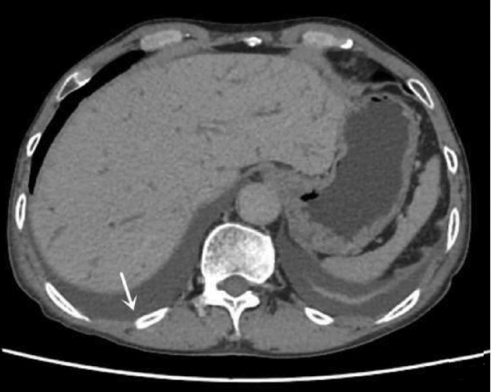
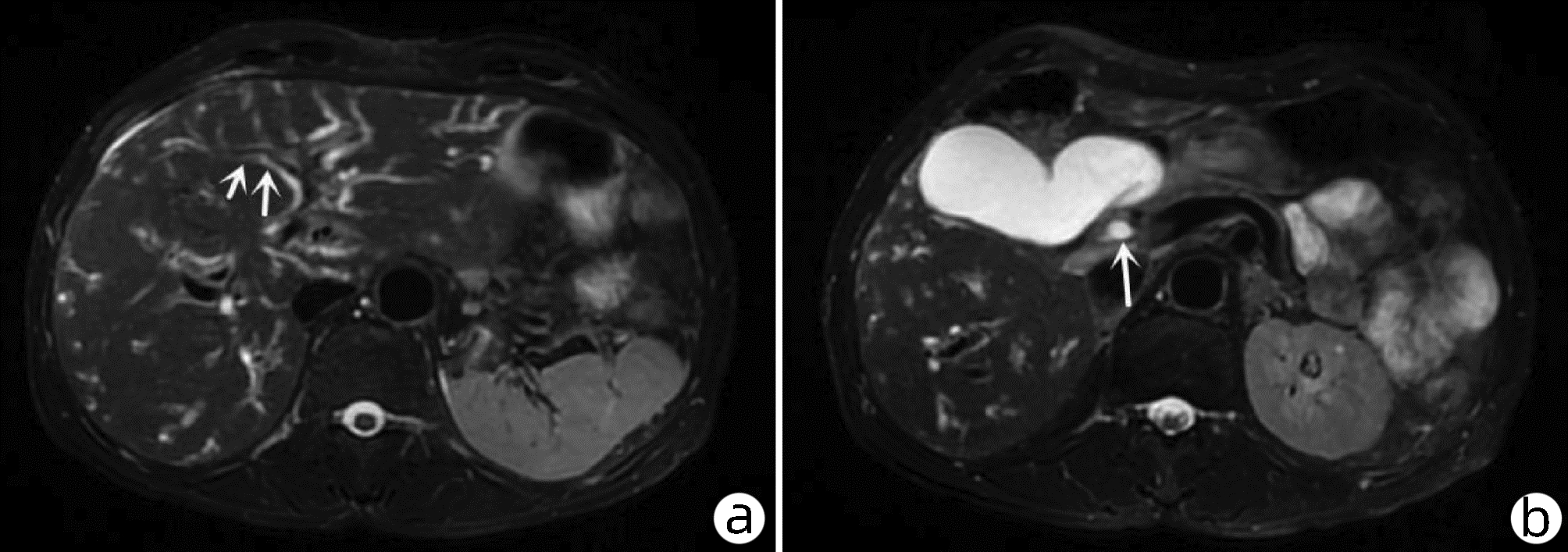
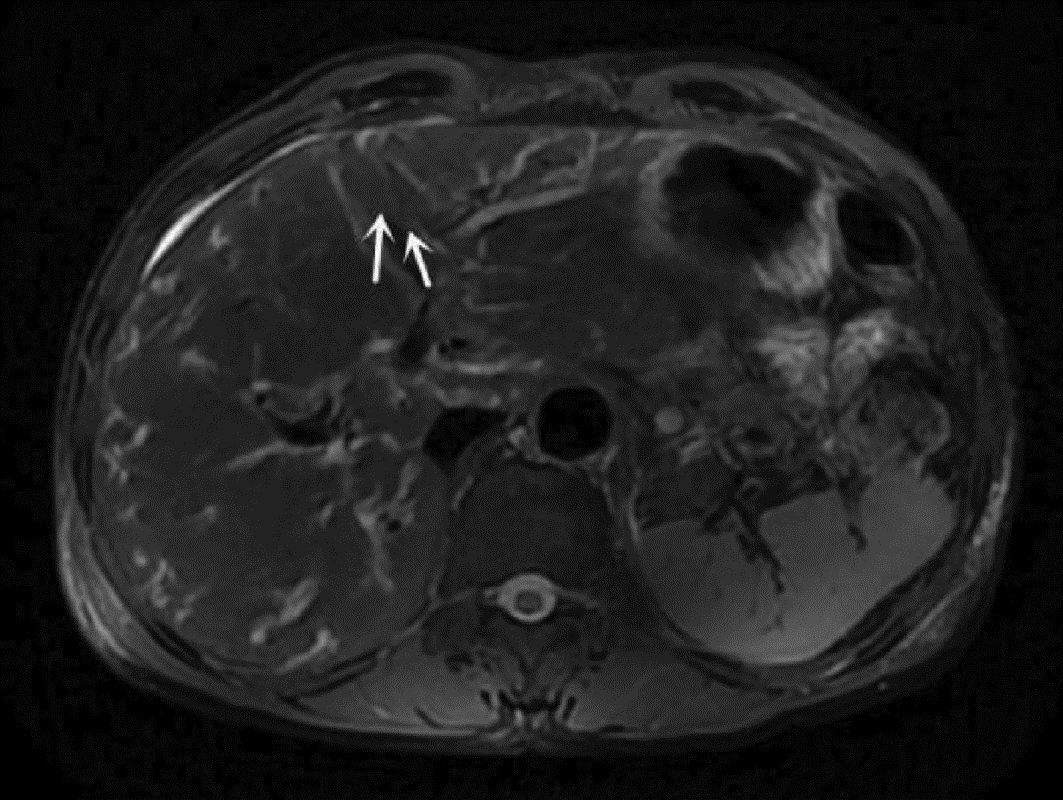
伴有胸腹腔积液及肝内外胆管扩张的肝吸虫病1例报告
DOI: 10.3969/j.issn.1001-5256.2022.06.033
伦理学声明:本例报告已获得患者知情同意。
利益冲突声明:所有作者均声明不存在利益冲突。
作者贡献声明:曾秋婷负责课题设计,资料分析,撰写论文;陈埏芳参与收集数据,修改论文;汤绍辉负责拟定写作思路,指导撰写文章并最后定稿。
Clonorchiasis with pleural and peritoneal effusion and dilatation of the intrahepatic and extrahepatic bile ducts: A case report
-
-
Key words:
- Fasciola Hepatica /
- Pleural Effusion /
- Ascites /
- Bile Duct Expansion
-
[1] YAO JK, DAI JR. The timeidemiology of clonorchiasis and the status of its treatment[J]. J Pathog Biol, 2020, 15(3): 364-370. DOI: 10.13350/j.cjpb.200326.姚甲凯, 戴建荣. 华支睾吸虫病的流行及治疗现状[J]. 中国病原生物学杂志, 2020, 15(3): 364-370. DOI: 10.13350/j.cjpb.200326. [2] JIANG SM, HUANG JK, LIANG QD, et al. Multiple low-density foci in the liver with pulmonary embolism diagnosed as clonorchiasis: A case report[J]. J Clin Hepatol, 2020, 36(10): 2285-2287. DOI: 10.3969/j.issn.1001-5256.2020.10.027.江善明, 黄继康, 梁其栋, 等. 肝内多发低密度灶合并肺栓塞诊断为肝吸虫病1例报告[J]. 临床肝胆病杂志, 2020, 36(10): 2285-2287. DOI: 10.3969/j.issn.1001-5256.2020.10.027. [3] WU GT, HE XW, ZHANG RJ, et al. Misdiagnosis analysis of recurrent severe upper abdominal pain presented as clonorchiasis[J]. Clin Misdiagn Misther, 2015, 28(12): 4-6. DOI: 10.3969/j.issn.1002-3429.2015.12.002.吴桂堂, 贺孝文, 张锐江, 等. 以反复剧烈上腹疼痛为表现的肝吸虫病误诊剖析[J]. 临床误诊误治, 2015, 28(12): 4-6. DOI: 10.3969/j.issn.1002-3429.2015.12.002. [4] XIE M, FENG QC. Analysis of the clinical features of 196 clonorchiasis patients and the causes of misdiagnosis[J]. China Trop Med, 2006, (11): 2002-2004. DOI: 10.3969/j.issn.1009-9727.2006.11.040.谢敏, 冯倩嫦. 华支睾吸虫病196例临床特点与误诊分析[J]. 中国热带医学, 2006, 6(11): 2002-2004. DOI: 10.3969/j.issn.1009-9727.2006.11.040. [5] WANG HX, CAI YJ, LI WY, et al. A case of eosinophilia secondary to infection with Clonorchis sinensis and Ascaris lumbricoides[J]. J Clin Hepatol, 2019, 35(4): 861-862. DOI: 10.3969/j.issn.1001-5256.2019.04.031.王海霞, 蔡艳俊, 李婉玉, 等. 华支睾吸虫、蛔虫合并感染继发嗜酸性粒细胞增多症1例报告[J]. 临床肝胆病杂志, 2019, 35(4): 861-862. DOI: 10.3969/j.issn.1001-5256.2019.04.031. [6] LIANG DR. CT manifestations of clonorchiasis sinensis[J]. Guangxi Med J, 2011, 33(5) : 638-640. DOI: 10.3969/j.issn.0253-4304.2011.05.051.梁德壬. 肝吸虫病的CT表现[J]. 广西医学, 2011, 33(5): 638-640. DOI: 10.3969/j.issn.0253-4304.2011.05.051. [7] HUANG JH, ZHENG FP, XU HX, et al. A case of atypical liver fluke disease with low-density space-occupying changes in the liver[J]. China Trop Med, 2019, 19(5): 501-502. DOI: 10.13604/j.cnki.46-1064/r.2019.05.25.黄嘉煌, 郑凤屏, 徐慧璇, 等. 以肝脏呈低密度占位性改变的1例非典型肝吸虫病例报道[J]. 中国热带医学, 2019, 19(5): 501-502. DOI: 10.13604/j.cnki.46-1064/r.2019.05.25. [8] NA BK, PAK JH, HONG SJ. Clonorchis sinensis and clonorchiasis[J]. Acta Trop, 2020, 203: 105309. DOI: 10.1016/j.actatropica.2019.105309. [9] ZOU YX. Parasitic infections involving the pleura[J]. Chin J Pract Pediatr, 2017, 32(3): 181-186. DOI: 10.19538/j.ek2017030607.邹映雪. 寄生虫性胸腔积液[J]. 中国实用儿科杂志, 2017, 32(3): 181-186. DOI: 10.19538/j.ek2017030607. [10] WEI CZ, HUANG LY, ZENG YH, et al. Liver fluke pleural effusion in 4 cases and literature review[J]. Clin Focus, 2009, 24(20): 1825-1827. https://www.cnki.com.cn/Article/CJFDTOTAL-LCFC200920048.htm韦彩周, 黄陆颖, 曾莹晖, 等. 肝吸虫相关胸腔积液4例并文献复习[J]. 临床荟萃, 2009, 24(20): 1825-1827. https://www.cnki.com.cn/Article/CJFDTOTAL-LCFC200920048.htm -


 PDF下载 ( 2003 KB)


 下载:
下载:


