
 PDF下载 ( 2160 KB)

内脏脂肪相关指数与急性胰腺炎严重程度的关系
DOI: 10.3969/j.issn.1001-5256.2022.10.021
Relationship between visceral fat related index and the severity of acute pancreatitis
-
摘要:
目的 探讨内脏脂肪相关指数与急性胰腺炎(AP)严重程度之间的关系。 方法 选取2014年9月—2021年10月于广西医科大学第一附属医院住院的308例诊断为AP的患者作为研究对象,按AP分级诊断标准将其分为轻症急性胰腺炎(MAP)(n=186)、中度重症急性胰腺炎(MSAP)(n=60)和重症急性胰腺炎(SAP)(n=62),通过收集腰围、身高、体质量、血脂、生化等指标对比它们在年龄、住院费用及天数、评分系统和人体测量学指标等方面的差异。符合正态分布且方差齐性的计量资料组间比较采用单因素方差分析,方差不齐的组间及组内两两比较均采用Kruskal-Wallis H检验检验;不符合正态分布的计量资料组间及组内两两比较均采用Kruskal-Wallis H检验。分类资料和计数资料组间比较采用Kruskal-Wallis H检验。采用Spearman秩相关分析方法对各指标与AP的严重程度进行相关性分析;对各指标构建受试者工作特征曲线,并计算曲线下面积(AUC),比较AUC大小;用单因素及多因素Logistic回归分析方法找出MSAP和SAP发生的独立危险因素。 结果 住院费用及天数、TG、HDL-C、NLR、WBC、Alb、Cr、BUN、SIRS、BISAP、MEWS、Glasgow、PANC-3在3组间的差异均有统计学意义(P值均<0.05)。进一步两两比较发现,与MAP组相比,MSAP和SAP组的CMI、LAP、WTI、CVAI明显升高,差异均有统计学意义(P值均<0.05)。在相关性分析中,CMI与AP严重程度之间有一定相关性(r=0.352,P<0.001)。通过对各AUC大小的比较发现,CMI对预测MSAP和SAP发生的效能最大(AUC=0.708,95%CI:0.651~0.765,P<0.001)。单因素Logistic回归分析显示CMI、LAP、WTI、CVAI、WC是MSAP和SAP发生的危险因素(P值均<0.05),校正混杂因素后,CMI、CVAI是MSAP和SAP发生的独立危险因素(OR值分别为3.740、2.380, 95%CI分别为1.983~7.056、1.110~5.104,P值均<0.05)。 结论 内脏脂肪与AP病情严重程度有关;在4个内脏脂肪相关指数中(CMI、LAP、WTI、CVAI),以心脏代谢指数(CMI)在预测AP病情严重程度方面的价值最大,优于其他指数,两者呈正相关;CMI是MSAP和SAP发生的独立危险因素,或可作为预测和评估AP病情严重程度的潜在参考指标。 Abstract:Objective To investigate the relationship between visceral fat related index and the severity of acute pancreatitis (AP). Methods A total of 308 patients hospitalized with AP at the First Affiliated Hospital of Guangxi Medical University from September 2014 to October 2021 were included. They were divided into mild acute pancreatitis (MAP) (n=186), moderate severe acute pancreatitis (MSAP) (n=60) and severe acute pancreatitis (SAP) (n=62) for comparison in age, hospitalization cost and days, scoring systems and body mass indexes. Comparison of normally distributed continuous data with homogeneity of variance between groups was made by one-way analysis of variance, and intergroup and intragroup pairwise comparison of data with heterogeneity of variance was made by the Kruskal-Wallis H test. The receiver operating characteristic curves (ROCs) for each index were constructed and area under the curve (AUC) was calculated to evaluate the performance of each index. Univariable and multivariable logistic regression analyses were used to identify the independent risk factors of MSAP and SAP. Results There were significant differences among the three groups in terms of hospitalization costs and durations, TG, HDL-C, NLR, WBC, Alb, Cr, BUN, scoring systems, CMI, LAP, WTI and CVAI. Further pairwise comparisons revealed that CMI, LAP, WTI and CVAI were significantly higher in the MAP group than in MSAP and SAP groups. We also found correlation between CMI and the severity of AP (r=0.352, P < 0.001). By comparing the AUCs, CMI was found to be the most accurate in predicting the occurrence of MSAP and SAP. Univariable logistic regression analysis showed that CMI, LAP, WTI, CVAI and WC were the risk factors of MSAP and SAP. After adjusting for confounding factors, CMI and CVAI were identified as the independent risk factors of MSAP and SAP. The risk of MSAP and SAP with CMI ≥ 0.801 was 3.740 times that with CMI < 0.801 (95%CI: 1.983~7.056, P < 0.001). Conclusions Visceral fat is related to the severity of AP. Among the four visceral fat related indexes (CMI, LAP, WTI and CVAI), cardiometabolic index is the most valuable in predicting the severity of AP and they are positively correlated. CMI, an independent risk factor for MSAP and SAP, can be used to predict and assess the severity of AP. -
Key words:
- Acute Pancreatitis /
- Cardiometabolic Index /
- Predictor
-
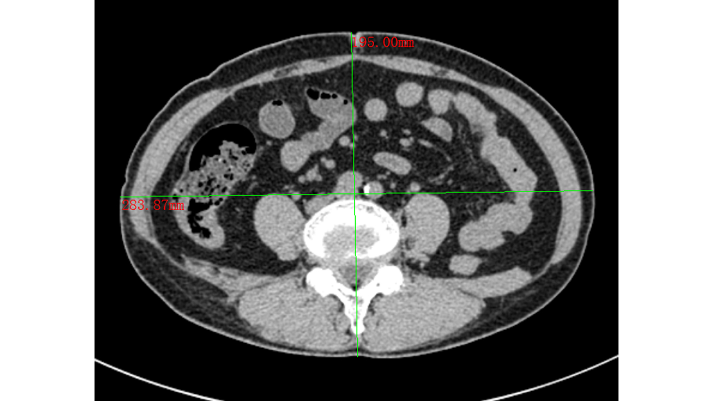
图 1 腰围测量方法示意图
Figure 1. Schematic diagram of the waist circumference measurement method
表 1 AP患者一般临床资料比较
Table 1. Comparison of the general clinical data of the patients with acute pancreatitis
指标 MAP(n=186) MSAP(n=60) SAP(n=62) 统计值 P值 性别[例(%)] H=0.458 0.795 男 149(80.11) 48(80.00) 52(83.87) 女 37(19.89) 12(20.00) 10(16.13) 年龄(岁) 45.23±13.86 43.68±12.68 47.45±14.32 F=1.175 0.310 病因[例(%)] H=1.240 0.538 胆源性 57(30.65) 9(15.00) 17(27.42) 脂源性 53(28.49) 25(41.67) 18(29.03) 酒精性 39(20.97) 13(21.67) 15(24.19) 混合性 12(6.45) 10(16.67) 7(11.29) 特发性 25(13.44) 3(5.00) 5(8.06) 住院费用(元) 12 057.06(7 768.13~20 279.21) 18 876.74(12 363.16~25 504.49)1) 47 561.88(29 534.51~82 790.38)1)2) H=94.670 <0.001 住院天数(d) 7.00(6.00~10.00) 10.00(8.00~13.00)1) 14.00(11.00~23.25)1)2) H=77.033 <0.001 注:与MAP组比较,1)P<0.05;与MSAP组比较,2)P<0.05。  下载: 导出CSV
表 2 不同严重程度AP患者之间实验室指标、评分系统和人体测量学指标的比较
Table 2. Comparison of laboratory indicators, scoring systems, and anthropometric indexes among patients with different severity of acute pancreatitis
项目 MAP(n=186) MSAP(n=60) SAP(n=62) 统计值 P值 实验室指标 TG(mmol/L) 1.90(0.91~5.05) 3.65(2.07~9.69)1) 4.15(1.81~7.30)1) H=24.012 <0.001 HDL-C(mmol/L) 1.06(0.81~1.34) 0.99(0.64~1.29) 0.72(0.45~1.11)1)2) H=24.568 <0.001 TC(mmol/L) 5.20(4.09~7.12) 6.24(4.37~9.45) 5.15(3.48~7.49) H=5.691 0.058 NLR 6.71(3.98~10.61) 8.80(6.43~11.98) 11.11(6.09~17.83)1) H=18.262 <0.001 WBC(×109/L) 11.74±4.33 12.58±4.50 14.53±5.491) F=12.509 0.002 Alb(g/L) 41.00±5.36 37.48±6.491) 35.02±7.131) F=40.855 <0.001 Cr(μmol/L) 69.50(55.75~82.00) 68.50(56.50~83.00) 79.50(59.75~179.25)1)2) H=11.585 0.003 BUN(mmol/L) 4.21(3.34~5.65) 4.15(3.25~5.34) 5.38(3.95~10.01)1)2) H=19.875 <0.001 评分系统(分) SIRS 1.00(0~2.00) 1.00(1.00~2.00) 2.00(1.00~3.00)1)2) H=28.707 <0.001 BISAP 0(0~1.00) 1.00(0~2.00)1) 2.00(1.00~3.00)1)2) H=69.555 <0.001 MEWS 1.00(1.00~2.00) 1.00(1.00~2.00) 3.00(2.00~5.25)1)2) H=64.019 <0.001 Glasgow 1.00(0~1.00) 1.00(1.00~2.00)1) 2.00(1.00~4.00)1)2) H=82.956 <0.001 PANC-3 1.00(0~1.00) 1.00(1.00~1.00)1) 1.00(1.00~1.00)1) H=24.835 <0.001 内脏脂肪相关指数 CMI 1.13(0.38~2.54) 2.29(1.12~4.71)1) 3.33(1.39~8.06)1) H=39.830 <0.001 LAP(cm·mmol/L) 46.21(15.22~133.73) 100.81(46.35~238.70)1) 96.96(38.80~197.96)1) H=19.207 <0.001 WTI(cm·mmol/L) 175.86(72.19~457.33) 319.49(183.33~803.15)1) 366.55(149.46~677.31)1) H=22.852 <0.001 CVAI 98.15±46.92 111.24±32.11 116.80±37.531) F=9.687 0.008 传统肥胖测量指数 BMI(kg/m2) 24.57±5.50 25.54±3.16 25.77±4.40 F=2.107 0.349 WC(cm) 84.14±16.11 87.48±7.20 87.70±8.61 F=4.409 0.110 注:与MAP组比较,1)P<0.05;与MSAP组比较,2)P<0.05。
下载: 导出CSV
表 3 人体测量学指标、实验室指标与非轻症急性胰腺炎的相关性分析
Table 3. Correlation analysis of anthropometric indexes, laboratory indicators with non-mild acute pancreatitis patients
指数 r值 P值 CMI 0.352 <0.001 LAP 0.249 <0.001 WTI 0.272 <0.001 CVAI 0.175 0.002 BMI 0.083 0.147 WC 0.120 0.036 TG 0.279 <0.001 HDL-C -0.224 <0.001 TC 0.058 0.312 NLR 0.232 <0.001 WBC 0.172 0.002 Alb -0.351 <0.001 Cr 0.135 0.018 BUN 0.140 0.014
下载: 导出CSV
表 4 各指标对非轻症急性胰腺炎预测能力的比较
Table 4. Comparison of predictive ability of each index for non-mild acute pancreatitis
指标 AUC 95%CI P值 CMI 0.708 0.651~0.765 <0.001 LAP 0.647 0.586~0.708 <0.001 WTI 0.661 0.601~0.720 <0.001 CVAI 0.603 0.541~0.666 0.002 BMI 0.549 0.485~0.613 0.147 WC 0.571 0.507~0.634 0.036 TG 0.665 0.605~0.724 <0.001 HDL-C 0.368 0.302~0.434 <0.001 TC 0.534 0.466~0.602 0.311 NLR 0.637 0, 575~0.699 <0.001 WBC 0.601 0.536~0.666 0.003 Alb 0.293 0.231~0.355 <0.001 Cr 0.579 0.512~0.647 0.018 BUN 0.583 0.517~0.649 0.014
下载: 导出CSV
表 5 非轻症急性胰腺炎危险因素的单因素Logistic回归分析
Table 5. Univariable logistic regression analysis of the risk factors in the non-mild acute pancreatitis
指标 OR 95%CI P值 CMI 1.086 1.032~1.143 0.002 LAP 1.002 1.000~1.003 0.008 WTI 1.001 1.000~1.001 0.004 CVAI 1.009 1.003~1.015 0.002 WC 1.023 1.002~1.044 0.034
下载: 导出CSV
表 6 非轻症急性胰腺炎危险因素的多因素Logistic回归分析
Table 6. Multivariable logistic regression analysis of the risk factors in the non-mild acute pancreatitis
指标 OR 95%CI P值 CMI(≥0.801 = 1, <0.801=0) 3.740 1.983~7.056 <0.001 CVAI(≥79.633=1, <79.633=0) 2.380 1.110~5.104 0.026
下载: 导出CSV
-
[1] LEE PJ, PAPACHRISTOU GI. New insights into acute pancreatitis[J]. Nat Rev Gastroenterol Hepatol, 2019, 16(8): 479-496. DOI: 10.1038/s41575-019-0158-2. [2] LANKISCH PG, APTE M, BANKS PA. Acute pancreatitis[J]. Lancet, 2015, 386(9988): 85-96. DOI: 10.1016/S0140-6736(14)60649-8. [3] ZHOU H, MEI X, HE X, et al. Severity stratification and prognostic prediction of patients with acute pancreatitis at early phase: A retrospective study[J]. Medicine (Baltimore), 2019, 98(16): e15275. DOI: 10.1097/MD.0000000000015275. [4] KONG W, HE Y, BAO H, et al. Diagnostic value of neutrophil-lymphocyte ratio for predicting the severity of acute pancreatitis: A meta-analysis[J]. Dis Markers, 2020, 2020: 9731854. DOI: 10.1155/2020/9731854. [5] LI M, XING XK, LU ZH, et al. Comparison of scoring systems in predicting severity and prognosis of hypertriglyceridemia-induced acute pancreatitis[J]. Dig Dis Sci, 2020, 65(4): 1206-1211. DOI: 10.1007/s10620-019-05827-9. [6] UNAMUNO X, GÓMEZ-AMBROSI J, RODRÍGUEZ A, et al. Adipokine dysregulation and adipose tissue inflammation in human obesity[J]. Eur J Clin Invest, 2018, 48(9): e12997. DOI: 10.1111/eci.12997. [7] CHEN SM, XIONG GS, WU SM. Is obesity an indicator of complications and mortality in acute pancreatitis? An updated meta-analysis[J]. J Dig Dis, 2012, 13(5): 244-251. DOI: 10.1111/j.1751-2980.2012.00587.x. [8] WAKABAYASHI I, DAIMON T. The "cardiometabolic index" as a new marker determined by adiposity and blood lipids for discrimination of diabetes mellitus[J]. Clin Chim Acta, 2015, 438: 274-278. DOI: 10.1016/j.cca.2014.08.042. [9] BANKS PA, BOLLEN TL, DERVENIS C, et al. Classification of acute pancreatitis--2012: revision of the Atlanta classification and definitions by international consensus[J]. Gut, 2013, 62(1): 102-111. DOI: 10.1136/gutjnl-2012-302779. [10] SINGH RG, CERVANTES A, KIM JU, et al. Intrapancreatic fat deposition and visceral fat volume are associated with the presence of diabetes after acute pancreatitis[J]. Am J Physiol Gastrointest Liver Physiol, 2019, 316(6): G806-G815. DOI: 10.1152/ajpgi.00385.2018. [11] PITT HA. Hepato-pancreato-biliary fat: the good, the bad and the ugly[J]. HPB (Oxford), 2007, 9(2): 92-97. DOI: 10.1080/13651820701286177. [12] FAIN JN, MADAN AK, HILER ML, et al. Comparison of the release of adipokines by adipose tissue, adipose tissue matrix, and adipocytes from visceral and subcutaneous abdominal adipose tissues of obese humans[J]. Endocrinology, 2004, 145(5): 2273-2282. DOI: 10.1210/en.2003-1336. [13] WANG C. Obesity, inflammation, and lung injury (OILI): the good[J]. Mediators Inflamm, 2014, 2014: 978463. [14] NATU A, STEVENS T, KANG L, et al. Visceral adiposity predicts severity of acute pancreatitis[J]. Pancreas, 2017, 46(6): 776-781. DOI: 10.1097/MPA.0000000000000845. [15] PATEL K, TRIVEDI RN, DURGAMPUDI C, et al. Lipolysis of visceral adipocyte triglyceride by pancreatic lipases converts mild acute pancreatitis to severe pancreatitis independent of necrosis and inflammation[J]. Am J Pathol, 2015, 185(3): 808-819. DOI: 10.1016/j.ajpath.2014.11.019. [16] KATUCHOVA J, BOBER J, HARBULAK P, et al. Obesity as a risk factor for severe acute pancreatitis patients[J]. Wien Klin Wochenschr, 2014, 126(7-8): 223-227. DOI: 10.1007/s00508-014-0507-7 [17] SADR-AZODI O, ORSINI N, ANDRÉN-SANDBERG Å, et al. Abdominal and total adiposity and the risk of acute pancreatitis: a population-based prospective cohort study[J]. Am J Gastroenterol, 2013, 108(1): 133-139. DOI: 10.1038/ajg.2012.381. [18] CHEN L, HUANG Y, YU H, et al. The association of parameters of body composition and laboratory markers with the severity of hypertriglyceridemia-induced pancreatitis[J]. Lipids Health Dis, 2021, 20(1): 9. DOI: 10.1186/s12944-021-01443-7 [19] YOON SB, CHOI MH, LEE IS, et al. Impact of body fat and muscle distribution on severity of acute pancreatitis[J]. Pancreatology, 2017, 17(2): 188-193. DOI: 10.1016/j.pan.2017.02.002 [20] FANG H, BERG E, CHENG X, et al. How to best assess abdominal obesity[J]. Curr Opin Clin Nutr Metab Care, 2018, 21(5): 360-365. DOI: 10.1097/MCO.0000000000000485 [21] O'LEARY DP, O'NEILL D, MCLAUGHLIN P, et al. Effects of abdominal fat distribution parameters on severity of acute pancreatitis[J]. World J Surg, 2012, 36(7): 1679-1685. DOI: 10.1007/s00268-011-1414-y. [22] BORKAN GA, GERZOF SG, ROBBINS AH, et al. Assessment of abdominal fat content by computed tomography[J]. Am J Clin Nutr, 1982, 36(1): 172-177. DOI: 10.1093/ajcn/36.1.172. [23] LEE SJ, JANSSEN I, HEYMSFIELD SB, et al. Relation between whole-body and regional measures of human skeletal muscle[J]. Am J Clin Nutr, 2004, 80(5): 1215-1221. DOI: 10.1093/ajcn/80.5.1215. [24] MADICO C, HERPE G, VESSELLE G, et al. Intra peritoneal abdominal fat area measured from computed tomography is an independent factor of severe acute pancreatitis[J]. Diagn Interv Imaging, 2019, 100(7-8): 421-426. DOI: 10.1016/j.diii.2019.03.008. [25] DUARTE-ROJO A, SOSA-LOZANO LA, SAU'L A, et al. Methods for measuring abdominal obesity in the prediction of severe acute pancreatitis, and their correlation with abdominal fat areas assessed by computed tomography[J]. Aliment Pharmacol Ther, 2010, 32(2): 244-253. DOI: 10.1111/j.1365-2036.2010.04321.x. [26] AMATO MC, GIORDANO C, GALIA M, et al. Visceral adiposity index: a reliable indicator of visceral fat function associated with cardiometabolic risk[J]. Diabetes Care, 2010, 33(4): 920-922. DOI: 10.2337/dc09-1825. [27] KAHN HS. The "lipid accumulation product" performs better than the body mass index for recognizing cardiovascular risk: a population-based comparison[J]. BMC Cardiovasc Disord, 2005, 5: 26. DOI: 10.1186/1471-2261-5-26. [28] LEMIEUX I, PASCOT A, COUILLARD C, et al. Hypertriglyceridemic waist: A marker of the atherogenic metabolic triad (hyperinsulinemia; hyperapolipoprotein B; small, dense LDL) in men?[J]. Circulation, 2000, 102(2): 179-184. DOI: 10.1161/01.cir.102.2.179. [29] XIA MF, CHEN Y, LIN HD, et al. A indicator of visceral adipose dysfunction to evaluate metabolic health in adult Chinese[J]. Sci Rep, 2016, 6: 38214. DOI: 10.1038/srep38214. [30] XIA W, YU H, HUANG Y, et al. The visceral adiposity index predicts the severity of hyperlipidaemic acute pancreatitis[J]. Intern Emerg Med, 2022, 17(2): 417-422. DOI: 10.1007/s11739-021-02819-4. [31] DING Y, ZHANG M, WANG L, et al. Association of the hypertriglyceridemic waist phenotype and severity of acute pancreatitis[J]. Lipids Health Dis, 2019, 18(1): 93. DOI: 10.1186/s12944-019-1019-2. [32] BEYDOGAN E, GULLE S, GEZER C, et al. Effect of abdominal fat distribution on severity of acute pancreatitis[J]. Clin Exp Hepatol, 2021, 7(3): 264-269. DOI: 10.5114/ceh.2021.109345 -


 本文二维码
本文二维码
计量
- 文章访问数: 389
- HTML全文浏览量: 195
- PDF下载量: 52
- 被引次数: 0

