PDF下载 ( 720 KB)

肝硬化伴分流的1型孤立性胃静脉曲张出血患者行内镜二级预防治疗的长期预后分析
DOI: 10.12449/JCH260718
Long-term prognosis of liver cirrhosis patients with spontaneous portosystemic shunt undergoing secondary endoscopic preventive therapy for type 1 isolated gastric variceal bleeding
-
摘要:
目的 通过观察肝硬化伴自发分流的1型孤立性胃静脉曲张出血患者,行内镜二级预防治疗后的长期生存结局及相关并发症的发生情况,分析影响长期预后的独立因素。 方法 纳入2015年1月5日—2019年11月29日首都医科大学附属北京地坛医院收治的70例肝硬化伴自发分流的1型孤立性胃静脉曲张出血患者为研究对象,收集患者年龄、性别、病因、血常规、肝功能、肾功能、凝血功能指标、蔡尔德-皮尤评分,以及是否合并胆汁淤积、肝癌、血栓、腹水、肝性脑病等基线资料。所有患者均行内镜二级预防再出血治疗,并随访5年。主要评价指标为异位栓塞率、全因死亡率,次要评价指标为肝病相关死亡和肝病相关并发症。计量资料2组间比较采用成组t检验或Mann-Whitney U检验;计数资料2组间比较采用χ2检验。采用Cox回归模型分析影响患者1年、3年及5年生存时间的预后因素,采用Logistic回归分析影响肝病相关并发症的独立危险因素。 结果 随访5年内,异位栓塞率为0。随访第1年全因死亡率为5.7%,第3年为24.3%,第5年为32.9%。肝病相关死亡第1年2例,第3年8例,第5年12例,5年肝病相关死亡率为17.14%(12/70)。随访第5年死亡患者中合并肝癌(17.39% vs 0.00%,χ2=8.669,P=0.003)和腹水(52.17% vs 38.30%,χ2=7.272,P=0.026)的发生率显著高于生存患者。生存患者的5年再出血(10.64% vs 100.00%,χ2=51.383,P<0.001)、腹水(8.51% vs 52.17%,χ2=21.574,P<0.001)及肝性脑病(0.00% vs 21.74%,χ2=12.029,P=0.002)的发生率均显著低于死亡患者。多因素Cox回归分析显示,在患者1年的随访中,术前合并肝癌[风险比(HR)=14.601, 95%CI: 2.049~104.098,P=0.007]、凝血酶原时间(HR=1.662, 95%CI: 1.090~2.535,P=0.018)是患者生存的独立危险因素,HGB水平(HR=0.863, 95%CI: 0.747~0.998,P=0.046)是延长患者生存的保护性因素;在3~5年的长期随访中,术前合并肝癌(3年:HR=40.174,95%CI:8.939~180.559,P<0.001;5年:HR=26.739,95%CI:6.993~102.243,P<0.001)、腹水(3年:HR=3.638,95%CI:1.751~7.575,P=0.001;5年:HR=2.555,95%CI:1.419~4.598,P=0.002)是患者死亡的独立危险因素。Logistic回归分析显示,随访3年:术前合并腹水可减少再出血的发生[比值比(OR)=0.255,95%CI:0.102~0.636,P=0.003],并发肝癌是发生再出血的独立危险因素(OR=16.231,95%CI:1.298~202.878,P=0.031);凝血酶原时间是患者发生肝性脑病的保护性因素(OR=0.790,95%CI:0.648~0.964,P=0.020)。随访5年:术后腹水加重是再出血的独立危险因素(OR=5.578,95%CI:1.474~21.103,P=0.011),术后腹水加重(OR=175.046,95%CI:14.306~2 141.909,P<0.001)是并发腹水的独立危险因素。 结论 术前合并肝癌、凝血酶原时间是影响患者1年生存的独立危险因素,高HGB水平是增加患者1年生存的保护因素。术前合并肝癌和腹水是影响伴有自发分流的1型孤立性胃静脉曲张出血患者长期预后的独立危险因素,可显著缩短生存时间。术前合并腹水可减少再出血的发生;术后并发肝癌及术后腹水加重是再出血的独立危险因素;术后腹水加重是未来并发腹水的独立危险因素。 Abstract:Objective To observe the long-term survival outcomes and complications of liver cirrhosis patients with spontaneous portosystemic shunt and type 1 isolated gastric varices (IGV-1) bleeding after secondary endoscopic preventive therapy, and to investigate the independent influencing factors for long-term prognosis. Methods A total of 70 liver cirrhosis patients with spontaneous portosystemic shunt and IGV-1 bleeding who were admitted to Beijing Ditan Hospital, Capital Medical University, from January 5, 2015 to November 29, 2019 were enrolled as subjects, and related baseline data were collected, including age, sex, etiology, routine blood test results, liver function parameters, renal function parameters, coagulation parameters, Child-Pugh score, and the presence or absence of comorbidities such as cholestasis, hepatocellular carcinoma, thrombosis, ascites, and hepatic encephalopathy. All patients underwent secondary endoscopic preventive therapy for rebleeding and were followed up for 5 years. The primary endpoints were ectopic embolism rate and all-cause mortality rate, and the secondary endpoints were liver-related mortality and liver-related complications. The independent-samples t test or the Mann-Whitney U test was used for comparison of continuous data between groups, and the chi-square test was used for comparison of categorical data between groups. The Cox regression analysis was used to investigate the prognostic factors for 1-, 3-, and 5-year survival time, and the Logistic regression analysis was used to identify the independent risk factors for liver disease-related complications. Results The incidence rate of ectopic embolism was 0% within 5 years of follow-up. The 1-, 3-, and 5-year all-cause mortality rates were 5.7%, 24.3%, and 32.9%, respectively. There were 2 cases of liver-related death in year 1, 8 cases in year 3, and 12 cases in year 5, resulting in a liver disease-related mortality rate of 17.14% (12/70). Compared with the survival group, the death group had significantly higher incidence rates of hepatocellular carcinoma (0.00% vs 17.39%, χ2=8.669, P=0.003) and ascites (38.30% vs 52.17%, χ2=7.272, P=0.026), and compared with the death group, the survival group had significantly lower 5-year incidence rates of rebleeding (10.64% vs 100.00%, χ2=51.383, P<0.001), ascites (8.51% vs 52.17%, χ2=21.574, P<0.001), and hepatic encephalopathy (0.00% vs 21.74%, χ2=12.029, P=0.002). The multivariate Cox regression analysis showed that during the 1-year follow-up, comorbidity with hepatocellular carcinoma before surgery (hazard ratio [HR]=14.601, 95% confidence interval [CI]: 2.049 — 104.098, P=0.007) and PT (HR=1.662, 95%CI: 1.090 — 2.535, P=0.018) were independent risk factors for survival, and HGB level (HR=0.863, 95%CI: 0.747 — 0.998, P=0.046) was a protective factor for prolonged survival; during the long-term follow-up for 3 or 5 years, preoperative comorbidities with hepatocellular carcinoma (3 years: HR=40.174, 95%CI: 8.939 — 180.559, P<0.001; 5 years: HR=26.739, 95%CI: 6.993 — 102.243, P<0.001) and ascites (3 years: HR=3.638, 95%CI: 1.751 — 7.575, P=0.001; 5 years: HR=2.555, 95%CI: 1.419 — 4.598, P=0.002) were independent risk factors for mortality. The Logistic regression analysis showed that during the follow-up for 3 years, comorbidity with ascites before surgery (odds ratio [OR]=0.255, 95%CI: 0.102 — 0.636, P=0.003) was associated with a lower risk of rebleeding, while comorbidity with hepatocellular carcinoma before surgery (OR=16.231, 95%CI: 1.298 — 202.878, P=0.031) was an independent risk factor for rebleeding; prothrombin time was a protective factor against hepatic encephalopathy (OR=0.790, 95%CI: 0.648 — 0.964, P=0.020); during the follow-up for 5 years, aggravation of ascites after surgery (OR=5.578, 95%CI: 1.474 — 21.103, P=0.011) was an independent risk factor for rebleeding, and aggravation of ascites after surgery (OR=175.046, 95%CI: 14.306 — 2 141.909, P<0.001) were independent risk factors for the development of ascites in the future. Conclusion Comorbidity with hepatocellular carcinoma before surgery and prothrombin time are independent risk factors for 1-year survival, whereas a high HGB level is a protective factor for increasing 1-year survival. Preoperative comorbidities with hepatocellular carcinoma and ascites are independent risk factors for the long-term prognosis of patients with spontaneous portosystemic shunt and IGV-1 bleeding and can significantly shorten survival time. Preoperative comorbidity with ascites can reduce the occurrence of rebleeding, while postoperative hepatocellular carcinoma and aggravation of ascites after surgery are independent risk factors for rebleeding; aggravation of ascites after surgery is an independent risk factor for the development of ascites in the future. -
Key words:
- Liver Cirrhosis /
- Esophageal and Gastric Varices /
- Endoscopy /
- Prognosis
-
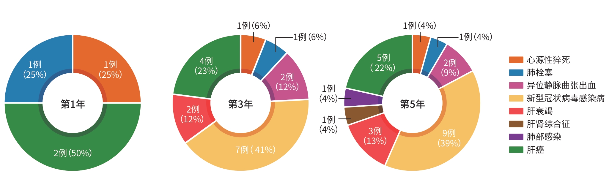
图 1 5年随访患者的死亡原因分析
Figure 1. Analysis of the causes of death in patients during the 5-year follow-up period
表 1 70例肝硬化伴有自发分流的1型孤立性胃静脉曲张出血患者的基线资料
Table 1. Baseline characteristics of 70 patients with type 1 isolated gastric variceal bleeding complicating liver cirrhosis with spontaneous shunts
项目 数值 项目 数值 性别[例(%)] 血栓形成[例(%)] 2(2.9) 男 36(51.4) 年龄(岁) 59.06±11.24 女 24(48.6) Alb(g/L) 32.554±6.538 肝癌[例(%)] 4(5.7) HGB(g/L) 83.5(70.0~101.0) 胆汁淤积[例(%)] 45(64.3) PLT(×109/L) 88.2(60.5~132.5) 腹水[例(%)] ALT(U/L) 23.100(12.375~30.325) 0级 40(57.2) AST(U/L) 23.050(15.875~38.150) 1级 22(31.4) TBil(μmol/L) 17.600(11.225~29.825) 2级 8(11.4) SCr(μmol/L) 64.500(56.225~77.350) 肝性脑病[例(%)] PT(s) 13.90(12.55~16.00) 0期 65(92.9) INR 1.270(1.145~1.558) 1期 4(5.7) Child-Pugh评分(分) 5.00(4.00~6.25) 2期 1(1.4) 注:Alb,白蛋白;HGB,血红蛋白;PLT,血小板;ALT,丙氨酸氨基转移酶;AST,天冬氨酸氨基转移酶;TBil,总胆红素;SCr,血清肌酐;PT,凝血酶原时间;INR,国际标准化比值。
 下载: 导出CSV
表 2 5年随访患者的全因死亡情况
Table 2. The all-cause mortality of patients during the 5-year follow-up period
随访时间 生存(例) 死亡(例) 全因死亡率(%) 第1年 66 4 5.7 第3年 53 17 24.3 第5年 47 23 32.9
下载: 导出CSV
表 3 随访第5年生存与死亡患者间的一般资料比较
Table 3. Comparison of baseline between surviving and deceased patients at the 5-year follow-up
项目 生存组(n=47) 死亡组(n=23) 统计值 P值 年龄(岁) 57.280±11.978 62.700±8.715 t=1.931 0.058 Alb(g/L) 32.857±6.843 31.935±6.770 t=-0.552 0.583 HGB(g/L) 86.0(71.0~101.0) 75.2(62.2~98.0) Z=-1.119 0.263 PLT(×109/L) 91.0(59.0~131.0) 86.0(63.3~135.0) Z=-0.094 0.925 ALT(U/L) 21.4(12.3~28.9) 24.2(12.9~35.5) Z=-0.456 0.648 AST(U/L) 26.1(17.7~35.7) 22.6(14.7~40.6) Z=-0.719 0.472 TBil(μmol/L) 16.5(10.6~29.9) 19.8(13.6~28.6) Z=-1.044 0.296 SCr(μmol/L) 65.2(56.4~77.8) 59.3(54.9~75.9) Z=-1.094 0.274 PT(s) 13.6(12.4~16.0) 14.4(13.4~17.5) Z=-1.589 0.112 INR 1.25(1.12~1.55) 1.34(1.24~1.59) Z=-1.157 0.247 Child-Pugh评分(分) 5(4~5) 5(4~7) Z=-1.283 0.200 性别[例(%)] χ2=0.356 0.551 男 23(48.94) 13(56.52) 女 24(51.06) 10(43.48) 肝癌[例(%)] 0(0.00) 4(17.39) χ2=8.669 0.003 胆汁淤积[例(%)] 14(29.79) 11(47.83) χ2=2.189 0.139 腹水[例(%)] 18(38.30) 12(52.17) χ2=7.272 0.026 肝性脑病[例(%)] 2(4.26) 3(13.04) χ2=3.383 0.147 术前血栓形成[例(%)] 1(2.13) 1(4.35) χ2=0.274 0.600 注:Alb,白蛋白;HGB,血红蛋白;PLT,血小板;ALT,丙氨酸氨基转移酶;AST,天冬氨酸氨基转移酶;TBil,总胆红素;SCr,血清肌酐;PT,凝血酶原时间;INR,国际标准化比值;Child-Pugh评分,蔡尔德-皮尤评分。
下载: 导出CSV
表 4 随访期间生存与死亡患者发生肝病相关并发症情况的比较
Table 4. Incidence of the hepatic complications and analysis of patients between two groups by all-cause mortality
并发症 总计 生存组 死亡组 χ2值 P值 随访1年(例) 70 66 4 早期再出血1)[例(%)] 8(11.43) 6(9.09) 2(50.00) 0.547 0.459 再出血[例(%)] 15(21.43) 13(19.70) 2(50.00) 0.010 0.921 腹水[例(%)] 8(11.43) 6(9.09) 2(50.00) 6.235 0.013 门静脉栓塞[例(%)] 5(7.14) 4(6.06) 1(25.00) 2.040 0.153 肝性脑病[例(%)] 3(4.26) 2(3.03) 1(25.00) 4.438 0.035 肝癌[例(%)] 4(5.71) 2(3.03) 2(50.0) 15.443 <0.001 随访3年(例) 70 53 17 再出血[例(%)] 19(27.14) 10(18.87) 9(52.94) 7.557 0.006 腹水[例(%)] 15(21.43) 6(11.32) 9(52.84) 13.243 <0.001 门静脉栓塞[例(%)] 3(4.29) 1(1.89) 2(11.76) 3.062 0.080 肝性脑病[例(%)] 4(5.71) 2(3.77) 2(11.76) 1.526 0.217 肝癌[例(%)] 8(11.43) 4(7.55) 4(23.53) 3.248 0.072 随访5年(例) 70 47 23 再出血[例(%)] 28(40.00) 5(10.64) 23(100.00) 51.383 <0.001 腹水[例(%)] 16(22.86) 4(8.51) 12(52.17) 21.574 <0.001 门静脉栓塞[例(%)] 7(10.00) 3(6.38) 4(17.39) 2.172 0.141 肝性脑病[例(%)] 5(7.14) 0(0.00) 5(21.74) 12.029 0.002 肝癌[例(%)] 12(17.14) 7(14.89) 5(21.74) 1.065 0.587 注:1)指治疗后3月内发生的再出血。
下载: 导出CSV
表 5 生存时间的多因素Cox回归分析
Table 5. Survival time of Cox multivariate regression
影响因素 β值 SE Wald值 HR(95%CI) P值 随访1年 术前合并肝癌(无=0,有=1) 2.681 1.002 7.159 14.601(2.049~104.098) 0.007 PT(s) 0.508 0.215 5.564 1.662(1.090~2.535) 0.018 HGB(g/L) -0.147 0.074 3.967 0.863(0.747~0.998) 0.046 随访3年 术前合并肝癌(无=0,有=1) 3.693 0.767 23.200 40.174(8.939~180.559) <0.001 术前合并腹水(无=0,有=1) 1.291 0.373 11.982 3.638(1.751~7.557) 0.001 随访5年 术前合并肝癌(无=0,有=1) 3.268 0.684 23.061 26.739(6.993~102.243) <0.001 术前合并腹水(无=0,有=1) 0.938 0.300 9.786 2.555(1.419~4.598) 0.002 注:PT,凝血酶原时间; HGB,血红蛋白;HR,风险比;CI,置信区间。
下载: 导出CSV
表 6 肝病相关并发症的Logistic回归分析
Table 6. Logistic regression anlysis of hepatic complications
影响因素 β值 SE Wald值 OR(95%CI) P值 随访3年再出血 术前合并腹水(无=0,有=1) -1.367 0.467 8.576 0.255(0.102~0.636) 0.003 术后并发肝癌(无=0,有=1) 2.787 1.289 4.677 16.231(1.298~202.878) 0.031 随访3年肝性脑病 PT(s) -0.236 0.101 5.406 0.790(0.648~0.964) 0.020 随访5年再出血 术后腹水加重(无=0,有=1) 1.719 0.674 6.409 5.578(1.474~21.103) 0.011 随访5年腹水 术后腹水加重(无=0,有=1) 5.165 1.278 16.339 175.046(14.306~2 141.909) <0.001 注:PT,凝血酶原时间;OR,比值比;CI,置信区间。
下载: 导出CSV
-
[1] Sarin S K, Lahoti D, Saxena S P, et al. Prevalence, classification and natural history of gastric varices: A long-term follow-up study in 568 portal hypertension patients[J]. Hepatology, 1992, 16( 6): 1343- 1349. DOI: 10.1002/hep.1840160607. [2] Garcia-Tsao G, Abraldes J G, Berzigotti A, et al. Portal hypertensive bleeding in cirrhosis: Risk stratification, diagnosis, and management: 2016 practice guidance by the American Association for the study of liver diseases[J]. Hepatology, 2017, 65( 1): 310- 335. DOI: 10.1002/hep.28906. [3] Lee E W, Shahrouki P, Alanis L, et al. Management options for gastric variceal hemorrhage[J]. JAMA Surg, 2019, 154( 6): 540. DOI: 10.1001/jamasurg.2019.0407. [4] Chinese Society of Hepatology, Chinese Society of Gastroenterology, Chinese Society of Digestive Endoscopology of Chinese Medical Association. Guidelines on the management of esophagogastric variceal bleeding in cirrhotic portal hypertension[J]. Chin J Intern Med, 2023, 62( 1): 7- 22. DOI: 10.3760/cma.j.cn501113-20220824-00436.中华医学会肝病学分会, 中华医学会消化病学分会, 中华医学会消化内镜学分会. 肝硬化门静脉高压食管胃静脉曲张出血的防治指南[J]. 中华内科杂志, 2023, 62( 1): 7- 22. DOI: 10.3760/cma.j.cn501113-20220824-00436. [5] de Franchis R, Bosch J, Garcia-Tsao G, et al. Baveno VII–Renewing consensus in portal hypertension[J]. J Hepatol, 2022, 76( 4): 959- 974. DOI: 10.1016/j.jhep.2021.12.022. [6] Gralnek I M, Camus Duboc M, Garcia-Pagan J C, et al. Endoscopic diagnosis and management of esophagogastric variceal hemorrhage: European Society of Gastrointestinal Endoscopy(ESGE) Guideline[J]. Endoscopy, 2022, 54( 11): 1094- 1120. DOI: 10.1055/a-1939-4887. [7] Ma Jiali, Ai Zhenglin, Hu Julong, et al. The preliminary therapeutic effect of endoscopic ultrasound-guided tissue adhesive injection with the assistance of metal clips in treatment of cirrhotic patients with gastric varices and gastric-renal shunt[J]. J Clin Hepatol, 2024, 40( 4): 734- 738. DOI: 10.12449/JCH240415.马佳丽, 艾正琳, 胡居龙, 等. 金属夹辅助内镜超声引导组织胶注射治疗合并胃肾分流的肝硬化胃静脉曲张患者的效果初探[J]. 临床肝胆病杂志, 2024, 40( 4): 734- 738. DOI: 10.12449/JCH240415. [8] Ai Zhenglin, Min Min, Hu Julong, et al. Efficacy evaluation of three endoscopic therapies of isolated gastric varices with modified tissue adhesive[J]. J Sichuan Univ Med Sci, 2022, 53( 3): 386- 390. DOI: 10.12182/20220560102.艾正琳, 闵敏, 胡居龙, 等. 三种内镜下改良组织胶方案治疗孤立性胃静脉曲张的疗效评价[J]. 四川大学学报(医学版), 2022, 53( 3): 386- 390. DOI: 10.12182/20220560102. [9] Ai Z L, Li P. Loop combined endoscopic clip and cyanoacrylate injection to treat severe gastric varices with spleno-renal shunt[J]. VideoGIE, 2020, 5( 12): 652- 654. DOI: 10.1016/j.vgie.2020.07.020. [10] Chinese Society of Hepatology, Chinese Medical Association. Guidelines on the management of ascites in cirrhosis(2023 version)[J]. Chin J Hepatol, 2023, 31( 8): 813- 826. DOI: 10.3760/cma.j.cn501113-20230719-00011.中华医学会肝病学分会. 肝硬化腹水诊疗指南(2023年版)[J]. 中华肝脏病杂志, 2023, 31( 8): 813- 826. DOI: 10.3760/cma.j.cn501113-20230719-00011. [11] Chinese Society of Hepatology, Chinese Medical Association. Chinese guidelines on the management of hepatic encephalopathy in cirrhosis(2024)[J]. Chin J Hepatol, 2024, 32( 9): 799- 812. DOI: 10.3760/cma.j.cn501113-20240630-00309.中华医学会肝病学分会. 肝硬化肝性脑病诊疗指南(2024年版)[J]. 中华肝脏病杂志, 2024, 32( 9): 799- 812. DOI: 10.3760/cma.j.cn501113-20240630-00309. [12] Xia Y F, Tie J, Wang G C, et al. Small transjugular intrahepatic portosystemic shunt plus variceal embolization for gastric varices: A multicenter cohort study[J]. Clin Gastroenterol Hepatol, 2025, 23( 6): 965- 977.e20. DOI: 10.1016/j.cgh.2024.04.037. [13] Zhu J Y, Xia Y F, Wang G C, et al. TIPS versus endoscopic cyanoacrylate injection for the treatment of gastric fundal variceal rebleeding: A propensity score-matched retrospective cohort study[J]. Surg Endosc, 2025, 39( 6): 3662- 3670. DOI: 10.1007/s00464-025-11761-x. [14] Li M, Wei Z H, Su J T, et al. Changing spectrum and mortality disparities of etiology of liver cirrhosis in Beijing, China[J]. J Med Virol, 2024, 96( 1): e29405. DOI: 10.1002/jmv.29405. [15] Zheng Wenmin, Tian Yuanyuan. Age-period-cohort analysis and prediction of incidence and mortality of cirrhosis in China, 1990-2019[J]. Dis Surveillance, 2024, 39( 11): 1489- 1494. DOI: 10.3784/jbjc.202401290061.郑文敏, 田元元. 1990—2019年中国肝硬化发病和死亡年龄-时期-队列分析及预测[J]. 疾病监测, 2024, 39( 11): 1489- 1494. DOI: 10.3784/jbjc.202401290061. [16] Huang D Q, Terrault N A, Tacke F, et al. Global epidemiology of cirrhosis: Aetiology, trends and predictions[J]. Nat Rev Gastroenterol Hepatol, 2023, 20( 6): 388- 398. DOI: 10.1038/s41575-023-00759-2. [17] Fadlallah H, El Masri D, Bahmad H F, et al. Update on the complications and management of liver cirrhosis[J]. Med Sci, 2025, 13( 1): 13. DOI: 10.3390/medsci13010013. [18] Hernaez R, Liu Y, Kramer J R, et al. Model for end-stage liver disease-sodium underestimates 90-day mortality risk in patients with acute-on-chronic liver failure[J]. J Hepatol, 2020, 73( 6): 1425- 1433. DOI: 10.1016/j.jhep.2020.06.005. [19] Ginès P, Krag A, Abraldes J G, et al. Liver cirrhosis[J]. Lancet, 2021, 398( 10308): 1359- 1376. DOI: 10.1016/S0140-6736(21)01374-X. [20] Patidar K R, Belcher J M, Regner K R, et al. Incidence and outcomes of acute kidney injury including hepatorenal syndrome in hospitalized patients with cirrhosis in the US[J]. J Hepatol, 2023, 79( 6): 1408- 1417. DOI: 10.1016/j.jhep.2023.07.010. [21] Liu N, Wu S, Guo J. Spillover health losses of COVID-19 in China: Evidence from non-COVID-19-related mortality[J]. Public Heal, 2024, 235: 8- 14. DOI: 10.1016/j.puhe.2024.06.026. [22] Gupta T, Sharma H. COVID-19 and the liver: Are footprints still there?[J]. World J Gastroenterol, 2023, 29( 4): 656- 669. DOI: 10.3748/wjg.v29.i4.656. [23] Su F. COVID-19 and cirrhosis: A combination we must strive to prevent[J]. Gastroenterology, 2021, 161( 5): 1371- 1373. DOI: 10.1053/j.gastro.2021.08.037. [24] Rodrigues S G, Mendoza Y P, Bosch J. Beta-blockers in cirrhosis: Evidence-based indications and limitations[J]. JHEP Rep, 2020, 2( 1): 100063. DOI: 10.1016/j.jhepr.2019.12.001. [25] Møller S, Wiese S, Barløse M, et al. How non-alcoholic fatty liver disease and cirrhosis affect the heart[J]. Hepatol Int, 2023, 17( 6): 1333- 1349. DOI: 10.1007/s12072-023-10590-1. [26] Xian J C, Tang Y Z, Shao H, et al. Effect of portal vein thrombosis on the prognosis of patients with cirrhosis without a liver transplant: A systematic review and meta-analysis[J]. Medicine, 2021, 100( 16): e25439. DOI: 10.1097/MD.0000000000025439. [27] Verma N, Singh S, Roy A, et al. Cirrhosis and fungal infections-a cocktail for catastrophe: A systematic review and meta-analysis with machine learning[J]. Mycoses, 2022, 65( 9): 844- 858. DOI: 10.1111/myc.13482. [28] Verma N, Singh S, Syal A, et al. Invasive aspergillosis is a critical determinant of mortality in cirrhosis: A systematic review with meta-analysis[J]. Med Mycol, 2021, 59( 11): 1092- 1100. DOI: 10.1093/mmy/myab044. [29] Ardevol A, Alvarado-Tapias E, Garcia-Guix M, et al. Early rebleeding increases mortality of variecal bleeders on secondary prophylaxis with β-blockers and ligation[J]. Dig Liver Dis, 2020, 52( 9): 1017- 1025. DOI: 10.1016/j.dld.2020.06.005. [30] O’Shea R S, Davitkov P, Ko C W, et al. AGA clinical practice guideline on the management of coagulation disorders in patients with cirrhosis[J]. Gastroenterology, 2021, 161( 5): 1615- 1627.e1. DOI: 10.1053/j.gastro.2021.08.015. [31] Guerrero A, del Campo L, Piscaglia F, et al. Anticoagulation improves survival in patients with cirrhosis and portal vein thrombosis: The IMPORTAL competing-risk meta-analysis[J]. J Hepatol, 2023, 79( 1): 69- 78. DOI: 10.1016/j.jhep.2023.02.023. [32] Gupta A, Rana R, Agarwal S, et al. Assessing the risk of further decompensation and survival in patients with cirrhosis with variceal bleeding as their first decompensation event[J]. Am J Gastroenterol, 2023, 118( 5): 833- 839. DOI: 10.14309/ajg.0000000000002018. [33] Tapper E B, Parikh N D. Diagnosis and management of cirrhosis and its complications: A review[J]. JAMA, 2023, 329( 18): 1589- 1602. DOI: 10.1001/jama.2023.5997. [34] Chinese Society of Spleen and Portal Hypertension Surgery, Chinese Society of Surgery, Chinese Medical Association. Expert consensus on the diagnosis and treatment of esophageal and gastric variceal rupture bleeding in cirrhotic portal hypertension(2025 edition)[J]. Chin J Dig Surg, 2025, 24( 3): 271- 280. DOI: 10.3760/cma.j.cn115610-20241228-00590.中华医学会外科学分会脾及门静脉高压外科学组. 肝硬化门静脉高压症食管、胃底静脉曲张破裂出血诊治专家共识(2025版)[J]. 中华消化外科杂志, 2025, 24( 3): 271- 280. DOI: 10.3760/cma.j.cn115610-20241228-00590. [35] Yokoyama S, Honda T, Ishizu Y, et al. Predicting early rebleeding and mortality after endoscopic hemostasis of esophagogastric varices: Diagnostic performance of aspartate aminotransferase-to-platelet ratio index and model for end-stage liver disease-Na score[J]. J Hepato Biliary Pancreat Sci, 2024, 31( 11): 830- 839. DOI: 10.1002/jhbp.12057. -

 本文二维码
本文二维码
计量
- 文章访问数: 4
- HTML全文浏览量: 0
- PDF下载量: 0
- 被引次数: 0