PDF下载 ( 2655 KB)

细胞因子联合终末期肝病模型评分对终末期肝病并发肝性脑病的诊断价值
DOI: 10.12449/JCH260721
Diagnostic value of cytokines combined with Model for End-Stage Liver Disease score in predicting hepatic encephalopathy in end-stage liver disease
-
摘要:
目的 通过联合细胞因子及终末期肝病模型(MELD)评分等关键指标,构建并验证终末期肝病(ESLD)患者并发肝性脑病(HE)的预测模型,为临床早期识别高危人群、优化干预策略提供循证依据。 方法 回顾性选取2022年1月—2024年12月昆明市第三人民医院收治的2 167例ESLD患者为研究对象,采用分层随机抽样法将ESLD患者按7∶3比例分为训练集(n=1 517)与验证集(n=650)。对训练集采用单因素分析及最小绝对收缩和选择算子(LASSO)回归筛选潜在影响变量,随后构建二元Logistic回归模型以识别ESLD患者并发HE的独立影响因素,基于该模型建立列线图预测模型,进行霍斯默-莱梅肖(H-L)拟合优度检验。通过对验证集采用受试者操作特征曲线(ROC曲线)、校准曲线、决策曲线分析(DCA)、临床影响曲线(CIC)对训练集预测模型的拟合度、准确性、一致性和临床实用性等进行多维度综合评价。非正态分布的的计量资料两组间比较采用Mann-Whitney U检验。计数资料2组间比较采用χ2检验或Fisher确切概率法。 结果 单因素分析结果显示,研究组与对照组在年龄、性别、血氨(NH3)、淋巴细胞、血红蛋白、血小板计数、凝血酶原时间、纤维蛋白原、国际标准化比值(INR)、总胆红素、天冬氨酸氨基转移酶、总蛋白、白蛋白、前白蛋白、碱性磷酸酶、胆碱酯酶、总胆汁酸、甘油三酯、总胆固醇、高密度脂蛋白、低密度脂蛋白、血糖、CD3+ T细胞计数、CD4+ T细胞计数、CD8+ T细胞计数、超敏C反应蛋白、癌胚抗原、三碘甲状腺原氨酸、甲状腺素(T4)、游离三碘甲状腺原氨酸、游离甲状腺素、白细胞介素(IL)-1β、IL-5、IL-6、IL-8、IL-12p70、IL-17、干扰素-γ以及MELD评分方面,差异均有统计学意义(P值<0.05)。LASSO回归分析筛选出年龄、NH3、白蛋白、前白蛋白、胆碱酯酶、总胆固醇、T4、IL-17、MELD评分等9个变量;进一步二元Logistic回归分析显示,NH3、IL-17和MELD评分为ESLD患者发生HE的独立危险因素,年龄、T4为独立保护因素(P值<0.05)。基于上述5个变量构建列线图预测模型,训练集的H-L拟合优度检验显示模型拟合良好(P=0.207),McFadden伪R2为0.184,提示模型具有可接受的解释力;内部验证集的H-L拟合优度检验进一步验证了模型的校准稳定性(P=0.067),提示模型在独立数据中仍具有较好的拟合效果。验证集ROC曲线下面积为0.784、敏感度为0.710、特异度为0.752,校准曲线的平均绝对误差为0.067,DCA和CIC分析显示列线图预测模型具有正向净获益和良好的临床实用性。 结论 基于年龄、NH3、T4、IL-17和MELD评分构建的ESLD患者发生HE的列线图预测模型具有较好的拟合度、准确性、一致性,同时具有良好的临床实用性。 Abstract:Objective To construct and validate a predictive model for hepatic encephalopathy (HE) in patients with end-stage liver disease (ESLD) by combining key indicators such as cytokines and Model for End-Stage Liver Disease (MELD) score, and to provide evidence-based support for the early identification of high-risk populations and the optimization of intervention strategies in clinical practice. Methods A retrospective analysis was performed for 2 167 patients with ESLD who were admitted to The Third People’s Hospital of Kunming from January 2022 to December 2024, the patients were divided into a training set with 1 517 patients and a validation set with 650 patients at a ratio of 7∶3 using the stratified random sampling method. A univariate analysis and a least absolute shrinkage and selection operator (LASSO) regression analysis were performed for the training set to identify potential influencing variables, and then a binary Logistic regression model was constructed to investigate the independent influencing factors for HE in ESLD patients. A nomogram prediction model was established based on this regression model, and the Hosmer-Lemeshow goodness-of-fit test was performed. The receiver operating characteristic (ROC) curve, calibration curves, decision curve analysis, and clinical impact curve were used to comprehensively evaluate the degree of fit, accuracy, consistency, and clinical practicability of the predictive model derived from the training set. The Mann-Whitney U test was used for comparison of non-normally distributed quantitative data between two groups. The chi-square test or Fisher exact test was used for comparison of categorical data between two groups. Results The univariate analysis showed that there were significant differences between the study group and the control group in age, sex, blood ammonia, lymphocytes, hemoglobin, platelet count, prothrombin time, fibrinogen, international normalized ratio, total bilirubin, aspartate aminotransferase, total protein, albumin, prealbumin, alkaline phosphatase, cholinesterase, total bile acid, triglyceride, total cholesterol, high-density lipoprotein, low-density lipoprotein, blood glucose, CD3+ T cells, CD4+ T cells, CD8+ T cells, high-sensitivity C-reactive protein, carcinoembryonic antigen, triiodothyronine, thyroxine, free triiodothyronine, free thyroxine, interleukin-1β, interleukin-5, interleukin-6, interleukin-8, interleukin-12p70, interleukin-17, interferon-γ, and MELD score (all P<0.05). The LASSO regression analysis identified nine variables of age, blood ammonia, albumin, prealbumin, cholinesterase, total cholesterol, thyroxine, interleukin-17, and MELD score, and the binary Logistic regression analysis showed that blood ammonia, interleukin-17, and MELD score were independent risk factors for HE in ESLD patients, while age and thyroxine were independent protective factors (all P<0.05). A nomogram model was constructed based on these five variables. The Hosmer-Lemeshow goodness-of-fit test in the training set showed a good degree of fit (P=0.207), with a McFadden’s pseudo-R2 of 0.184, suggesting that the model had acceptable explanatory power; the Hosmer-Lemeshow goodness-of-fit test in the internal validation set further confirmed the calibration stability of the model (P=0.067), suggesting that the model had a good degree of fit in independent data. The model had an area under the ROC curve of 0.784 in the validation set, with a sensitivity of 0.710 and a specificity of 0.752. The mean absolute error of the calibration curve was 0.067, and decision curve analysis and clinical impact curve showed that the nomogram model had positive net benefit and good clinical practicability. Conclusion The nomogram model constructed based on age, blood ammonia, thyroxine, interleukin-17, and MELD score for predicting HE in patients with ESLD has a good degree of fit, high accuracy and consistency, and excellent clinical practicability. -
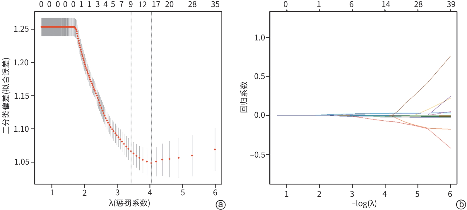
注: a,LASSO交叉验证偏差曲线;b,LASSO回归系数变化曲线。
图 1 终末期肝病患者发生肝性脑病的LASSO回归分析
Figure 1. Least absolute shrinkage and selection operator regression analysis of hepatic encephalopathy in patients with end-stage liver disease
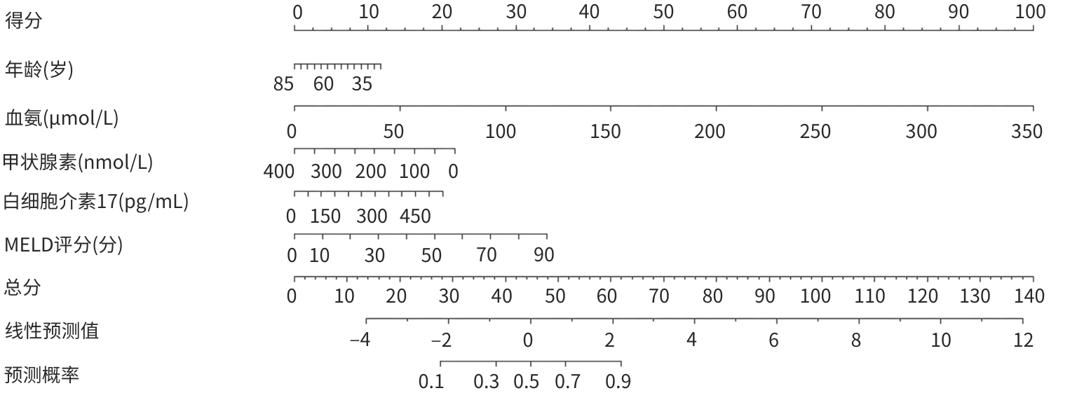
注: MELD,终末期肝病模型。
图 2 终末期肝病患者发生肝性脑病的列线图预测模型
Figure 2. Nomogram prediction model for hepatic encephalopathy in patients with end-stage liver disease
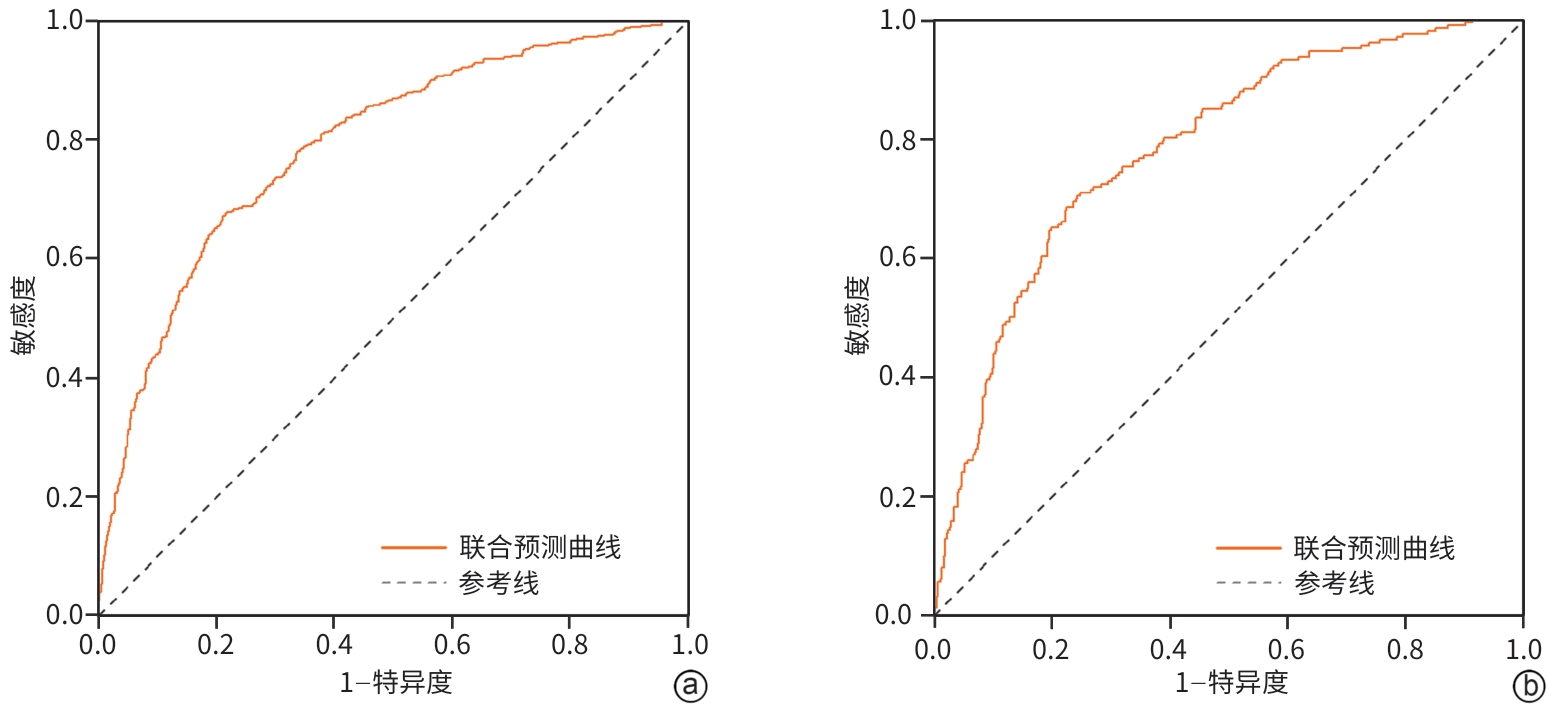
注: a,训练集;b验证集。
图 3 终末期肝病患者发生肝性脑病的受试者操作特征曲线
Figure 3. ROC curve of hepatic encephalopathy in patients with end-stage liver disease
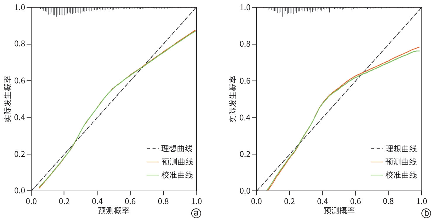
注: a,训练集;b验证集。
图 4 终末期肝病患者发生肝性脑病的校准曲线
Figure 4. Calibration curves of hepatic encephalopathy in patients with end-stage liver disease
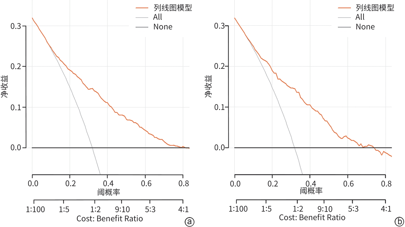
注: a,训练集;b验证集。
图 5 终末期肝病患者发生肝性脑病的临床决策曲线
Figure 5. Decision curve analysis curves of hepatic encephalopathy in patients with end-stage liver disease
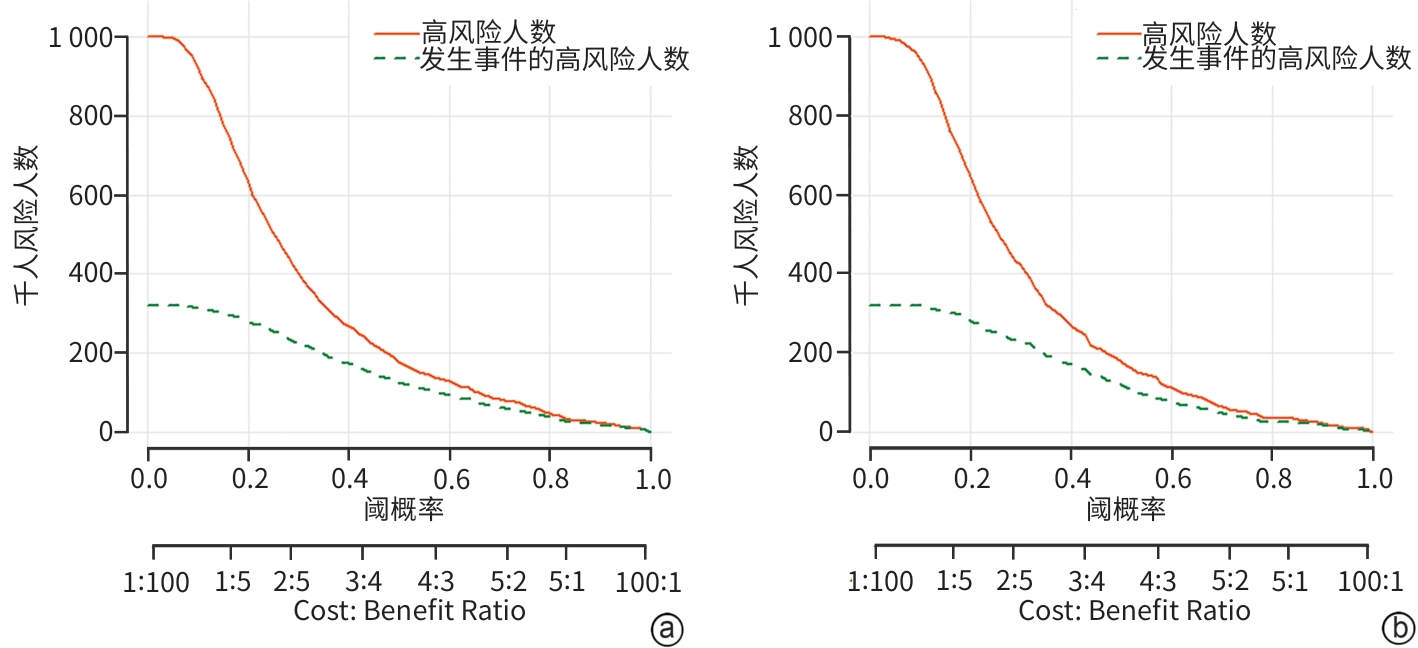
注: a,训练集;b验证集。
图 6 终末期肝病患者发生肝性脑病的临床影响曲线
Figure 6. Clinical impact curve of hepatic encephalopathy in patients with end-stage liver disease
表 1 训练集中研究组与对照组的临床资料比较
Table 1. Comparison of clinical data between the study group and the control group in the training set
变量 对照组(n=1 033) 研究组(n=484) 统计值 P值 年龄(岁) 55.00(49.00~61.00) 53.00(48.00~59.00) Z=-3.574 <0.001 性别[例(%)] χ2=5.271 0.022 男 782(75.70) 392(80.99) 女 251(24.30) 92(19.09) 吸烟史[例(%)] χ2=0.817 0.366 有 523(50.63) 233(48.14) 无 510(49.37) 251(51.86) 饮酒史[例(%)] χ2=0.018 0.893 有 470(45.50) 222(45.87) 无 563(54.50) 262(54.13) NH3(μmol/L) 32.20(22.30~43.50) 49.75(38.20~67.73) Z=-16.478 <0.001 WBC(×109/L) 4.30(3.14~5.83) 4.09(2.99~5.83) Z=-1.182 0.237 Lym(×109/L) 0.97(0.65~1.37) 0.85(0.57~1.26) Z=-3.803 <0.001 NE(×109/L) 2.64(1.85~3.83) 2.55(1.73~4.07) Z=-0.546 0.585 Hb(g/L) 132.00(109.00~151.00) 121.50(97.00~158.50) Z=-5.656 <0.001 PLT(×109/L) 97.00(67.00~146.00) 80.00(55.00~119.00) Z=-5.650 <0.001 PT(s) 15.60(14.30~17.30) 16.90(15.50~19.00) Z=-11.213 <0.001 FIB(g/L) 2.46(1.91~3.15) 2.02(1.55~2.71) Z=-7.966 <0.001 INR 1.26(1.14~1.45) 1.43(1.27~1.65) Z=-12.275 <0.001 TBil(μmol/L) 22.90(15.20~38.90) 34.85(21.40~63.25) Z=-9.064 <0.001 AST(U/L) 44.00(30.00~77.00) 49.00(35.00~95.00) Z=-4.109 <0.001 ALT(U/L) 32.00(21.00~52.00) 34.00(22.00~59.00) Z=-1.913 0.056 TP(g/L) 65.80(59.75~71.55) 62.70(56.65~68.40) Z=-6.413 <0.001 Alb(g/L) 34.20(28.60~39.20) 29.60(25.83~34.68) Z=-9.922 <0.001 PA(mg/L) 112.70(71.50~173.00) 77.50(49.15~117.40) Z=-9.863 <0.001 GGT(U/L) 69.00(34.00~147.00) 63.50(33.00~161.00) Z=-0.421 0.674 ALP(U/L) 125.00(92.00~182.50) 143.00(102.00~219.75) Z=-4.309 <0.001 ChE(U/L) 4 238.00(2 735.00~6 123.00) 2 978.00(2 025.50~4 355.00) Z=-9.829 <0.001 TBA(μmol/L) 17.70(5.95~44.60) 36.60(15.63~86.25) Z=-8.686 <0.001 TG(mmol/L) 0.90(0.65~1.28) 0.75(0.53~1.16) Z=-5.257 <0.001 TC(mmol/L) 3.70(2.97~4.42) 3.21(2.48~3.91) Z=-7.581 <0.001 HDL(mmol/L) 0.95(0.67~1.20) 0.86(0.48~1.19) Z=-3.809 <0.001 LDL(mmol/L) 2.11(1.60~2.79) 1.78(1.28~2.33) Z=-7.222 <0.001 Cr(μmol/L) 63.00(53.00~76.00) 62.50(49.00~78.75) Z=-0.035 0.972 UA(μmol/L) 321.00(262.00~394.50) 308.00(247.00~397.75) Z=-1.659 0.097 GLU(mmol/L) 5.40(4.89~6.46) 5.55(4.95~7.20) Z=-2.594 0.009 CD3+T细胞(个/μL) 659.92(438.39~986.99) 591.57(395.91~837.72) Z=-4.433 <0.001 CD4+T细胞(个/μL) 394.09(257.61~598.76) 325.93(207.77~531.55) Z=-2.588 <0.001 CD8+T细胞(个/μL) 216.75(131.91~351.68) 198.59(118.99~318.88) Z=-0.422 0.010 CD19+B细胞(个/μL) 136.50(80.66~214.98) 141.11(76.77~228.95) Z=-3.724 0.673 hs-CRP(mg/L) 3.56(1.03~14.84) 5.16(1.41~19.09) Z=-2.926 0.003 AFP(ng/mL) 6.22(2.93~126.23) 7.77(3.08~110.32) Z=-0.889 0.374 CEA(ng/mL) 2.93(1.84~4.38) 3.27(2.24~4.85) Z=-4.261 <0.001 TSH(μIU/mL) 2.25(1.41~3.57) 2.18(1.35~3.26) Z=-1.570 0.116 T3(nmol/L) 1.45(1.15~1.80) 1.25(0.99~1.65) Z=-6.060 <0.001  下载: 导出CSV
表 2 终末期肝病患者发生肝性脑病的二元Logistic回归分析
Table 2. Binary Logistic regression analysis of hepatic encephalopathy in patients with end-stage liver disease
变量 β值 SE OR 95%CI Wald P值 年龄 -0.023 0.006 0.978 0.965~0.990 12.329 <0.001 NH3 0.034 0.003 1.035 1.029~1.041 121.924 <0.001 Alb -0.024 0.014 0.976 0.951~1.003 3.096 0.078 PA -0.002 0.002 0.998 0.995~1.001 1.112 0.292 ChE 0.000 0.000 1.000 1.000~1.000 0.035 0.851 TC -0.108 0.060 0.898 0.798~1.009 3.272 0.070 T4 -0.005 0.002 0.995 0.991~0.999 7.744 0.005 IL-17 0.005 0.002 1.005 1.001~1.008 8.141 0.004 MELD评分 0.030 0.010 1.030 1.010~1.051 8.655 0.003 注:NH3,血氨;Alb,白蛋白;PA,前白蛋白;ChE,胆碱酯酶;TC,总胆固醇;T4,甲状腺素;IL-17,白细胞介素17;MELD,终末期肝病模型;OR,比值比;CI,置信区间。
下载: 导出CSV
Table . (continued)
变量 对照组(n=1 033) 研究组(n=484) 统计值 P值 T4(nmol/L) 94.59(75.34~117.70) 78.92(60.48~101.64) Z=-8.050 <0.001 FT3(pmol/L) 3.85(3.10~4.57) 3.64(2.86~4.47) Z=-3.166 0.002 FT4(pmol/L) 15.36(13.11~17.53) 14.83(12.23~17.02) Z=-3.503 <0.001 IL-1β(pg/mL) 2.00(1.04~5.97) 2.24(1.27~7.23) Z=-2.147 0.032 IL-2(pg/mL) 1.63(0.94~2.83) 1.69(0.99~2.98) Z=-1.582 0.114 IL-4(pg/mL) 1.44(0.86~2.25) 1.45(0.86~2.17) Z=-0.159 0.874 IL-5(pg/mL) 1.25(0.72~2.37) 1.55(0.93~2.75) Z=-3.995 <0.001 IL-6(pg/mL) 11.10(4.87~29.88) 14.92(6.38~38.85) Z=-3.527 <0.001 IL-8(pg/mL) 21.96(9.09~54.86) 27.31(8.13~77.32) Z=-2.097 0.036 IL-10(pg/mL) 4.67(2.82~7.49) 4.76(2.49~8.18) Z=-0.292 0.770 IL-12p70(pg/mL) 1.93(1.33~2.96) 1.99(1.45~3.28) Z=-2.182 0.029 IL-17(pg/mL) 7.96(3.03~15.29) 10.36(3.43~23.22) Z=-3.939 <0.001 IFN-γ(pg/mL) 2.10(0.99~5.23) 3.13(1.59~6.92) Z=-5.242 <0.001 IFN-α(pg/mL) 1.74(1.07~2.93) 1.86(1.18~3.13) Z=-1.879 0.060 TNF-α(pg/mL) 2.00(1.02~4.28) 1.95(1.04~3.91) Z=-0.254 0.799 MELD评分(分) 7.00(4.00~11.00) 10.00(6.00~15.00) Z=-9.617 <0.001 注:NH3,血氨;WBC,白细胞计数;Lym,淋巴细胞;NE,中性粒细胞;Hb,血红蛋白;PLT,血小板计数;PT,凝血酶原时间;FIB,纤维蛋白原;TBil,总胆红素;AST,天冬氨酸氨基转移酶;ALT,丙氨酸氨基转移酶;TP,总蛋白;Alb,白蛋白;PA,前白蛋白;GGT,γ-谷氨酰转移酶;ALP,碱性磷酸酶;ChE,胆碱酯酶;TBA,总胆汁酸;TG,甘油三酯;TC,总胆固醇;HDL,高密度脂蛋白;LDL,低密度脂蛋白;Cr,肌酐;UA,尿酸;GLU,血糖;hs-CRP,超敏C反应蛋白;AFP,甲胎蛋白;CEA,癌胚抗原;TSH,促甲状腺素;T3,三碘甲状腺原氨酸;T4,甲状腺素;FT3,游离三碘甲状腺原氨酸;FT4,游离甲状腺素;IL-1β,白细胞介素1β;IL-2,白细胞介素2;IL-4,白细胞介素4;IL-5,白细胞介素5;IL-6,白细胞介素6;IL-8,白细胞介素8;IL-10,白细胞介素10;IL-12p70,白细胞介素12p70;IL-17,白细胞介素17;IFN-γ,干扰素γ;IFN-α,干扰素α;TNF-α,肿瘤坏死因子α;MELD,终末期肝病模型。
下载: 导出CSV
-
[1] Chen T, Chen G, Wang G Q, et al. Expert consensus on the diagnosis and treatment of end-stage liver disease complicated by infections[J]. Hepatol Int, 2024, 18( 3): 817- 832. DOI: 10.1007/s12072-023-10637-3. [2] Vilstrup H, Amodio P, Bajaj J, et al. Hepatic encephalopathy in chronic liver disease: 2014 Practice Guideline by the American Association for the Study of Liver Diseases and the European Association for the Study of the Liver[J]. Hepatology, 2014, 60( 2): 715- 735. DOI: 10.1002/hep.27210. [3] Romero-gómez M, Boza F, García-valdecasas M S, et al. Subclinical hepatic encephalopathy predicts the development of overt hepatic encephalopathy[J]. Am J Gastroenterol, 2001, 96( 9): 2718- 2723. DOI: 10.1111/j.1572-0241.2001.04130.x. [4] Ridola L, Cardinale V, Riggio O. The burden of minimal hepatic encephalopathy: From diagnosis to therapeutic strategies[J]. Ann Gastroenterol, 2018, 31( 2): 151- 164. DOI: 10.20524/aog.2018.0232. [5] Ridola L, Nardelli S, Gioia S, et al. Quality of life in patients with minimal hepatic encephalopathy[J]. World J Gastroenterol, 2018, 24( 48): 5446- 5453. DOI: 10.3748/wjg.v24.i48.5446. [6] Society of Infectious Diseases, Chinese Medical Association. Expert consensus on diagnosis and treatment of end-stage liver disease complicated with infections(2021 version)[J]. J Clin Hepatol, 2022, 38( 2): 304- 310. DOI: 10.3969/j.issn.1001-5256.2022.02.010.中华医学会感染病学分会. 终末期肝病合并感染诊治专家共识(2021年版)[J]. 临床肝胆病杂志, 2022, 38( 2): 304- 310. DOI: 10.3969/j.issn.1001-5256.2022.02.010. [7] Chen Tao, Ning Qin. Highlights of diagnosis and treatment for end stage of liver disease with infection[J]. Chin J Clin Infect Dis, 2017, 10( 5): 389- 393. DOI: 10.3760/cma.j.issn.1674-2397.2017.05.014.陈韬, 宁琴. 终末期肝病合并感染诊治的热点与难点[J]. 中华临床感染病杂志, 2017, 10( 5): 389- 393. DOI: 10.3760/cma.j.issn.1674-2397.2017.05.014. [8] Liver Failure and Artificial Liver Group, Chinese Society of Infectious Diseases, Chinese Medical Association; Severe Liver Disease and Artificial Liver Group, Chinese Society of Hepatology, Chinese Medical Association. Guideline for diagnosis and treatment of liver failure(2018)[J]. J Clin Hepatol, 2019, 35( 1): 38- 44. DOI: 10.3969/j.issn.1001-5256.2019.01.007.中华医学会感染病学分会肝衰竭与人工肝学组, 中华医学会肝病学分会重型肝病与人工肝学组. 肝衰竭诊治指南(2018年版)[J]. 临床肝胆病杂志, 2019, 35( 1): 38- 44. DOI: 10.3969/j.issn.1001-5256.2019.01.007. [9] Chinese Society of Hepatology, Chinese Medical Association. Chinese guidelines on the management of liver cirrhosis[J]. J Clin Hepatol, 2019, 35( 11): 2408- 2425. DOI: 10.3969/j.issn.1001-5256.2019.11.006.中华医学会肝病学分会. 肝硬化诊治指南[J]. 临床肝胆病杂志, 2019, 35( 11): 2408- 2425. DOI: 10.3969/j.issn.1001-5256.2019.11.006. [10] Chinese Society of Hepatology, Chinese Medical Association. Guidelines for diagnosis and treatment of primary liver cancer(2022 edition)[J]. Chin J Dig Surg, 2022, 21( 2): 143- 168. DOI: 10.3760/cma.j.cn115610-20220124-00053.中华人民共和国国家卫生健康委员会医政医管局. 原发性肝癌诊疗指南(2022年版)[J]. 中华消化外科杂志, 2022, 21( 2): 143- 168. DOI: 10.3760/cma.j.cn115610-20220124-00053. [11] Chinese Society of Hepatology, Chinese Medical Association. Guidelines on the management of hepatic encephalopathy in cirrhosis[J]. J Clin Hepatol, 2018, 34( 10): 2076- 2089. DOI: 10.3969/j.issn.1001-5256.2018.10.007.中华医学会肝病学分会. 肝硬化肝性脑病诊疗指南[J]. 临床肝胆病杂志, 2018, 34( 10): 2076- 2089. DOI: 10.3969/j.issn.1001-5256.2018.10.007. [12] Liu Huimin, Huang Yuan. Recommendations for EASL clinical practice guidelines on the management of hepatic encephalopathy(2022)[J]. J Clin Hepatol, 2022, 38( 9): 1997- 1998. DOI: 10.3969/j.issn.1001-5256.2022.09.010.刘慧敏, 黄缘.《2022年欧洲肝病学会肝性脑病管理临床实践指南》推荐意见[J]. 临床肝胆病杂志, 2022, 38( 9): 1997- 1998. DOI: 10.3969/j.issn.1001-5256.2022.09.010. [13] Li N. Research progress in hepatic encephalopathy in recent 10 years: A Web of Science-based literature analysis[J]. Neural Regen Res, 2012, 7( 34): 2727- 2733. DOI: 10.3969/j.issn.1673-5374.2012.34.011. [14] Wijdicks E F M. Hepatic encephalopathy[J]. N Engl J Med, 2016, 375( 17): 1660- 1670. DOI: 10.1056/nejmra1600561. [15] Hu Xiangdang, Ba Xiaotong, Zhu Ping. Risk factors and prognosis analysis of secondary hepatic encephalopathy in decompensated liver cirrhosis[J]. J Clin Exp Med, 2024, 23( 1): 9- 12. DOI: 10. 3969/j.issn.1671-4695.2024.01.003.胡向党, 巴晓彤, 朱萍. 肝硬化失代偿期继发肝性脑病的危险因素及预后分析[J]. 临床和实验医学杂志, 2024, 23( 1): 9- 12. DOI: 10. 3969/ j. issn. 1671- 4695. 2024. 01. 003. [16] Betrapally N S, Gillevet P M, Bajaj J S. Gut microbiome and liver disease[J]. Transl Res, 2017, 179: 49- 59. DOI: 10.1016/j.trsl.2016.07.005. [17] Gao Xiaoqin, Zhou Dan, Wang Shan, et al. Role of microbiota-gut-liver-brain axis on liver cirrhosis-associated neuropsychiatric diseases[J/CD]. Chin J Liver Dis(Electronic Version), 2025, 17( 3): 28- 33. DOI: 10.3969/j.issn.1674-7380.2025.03.004.高晓琴, 周丹, 王珊, 等. 肠道微生物群-肠-肝-脑轴在肝硬化相关神经精神疾病中的作用[J/CD]. 中国肝脏病杂志(电子版), 2025, 17( 3): 28- 33. DOI: 10.3969/j.issn.1674-7380.2025.03.004. [18] Yang Zhihui, Chen Meijuan, Zhang Mengxiao, et al. Research progress of multi-dimensional liver function evaluation in predicting liver cirrhosis compensation[J]. Clin J Med Offic, 2026, 54( 2): 111- 114, 120.杨志慧, 陈美娟, 张濛销, 等. 多维度肝功能评估预测肝硬化再代偿研究进展[J]. 临床军医杂志, 2026, 54( 2): 111- 114, 120. [19] Odeh M, Sabo E, Srugo I, et al. Relationship between tumor necrosis factor-alpha and ammonia in patients with hepatic encephalopathy due to chronic liver failure[J]. Ann Med, 2005, 37( 8): 603- 612. DOI: 10.1080/07853890500317414. [20] Yang Xuhong, Yang Yong, Wang Minglei, et al. Value of blood ammonia and cholinesterase in the early diagnosis of liver cirrhosis with minimal hepatic encephalopathy[J]. J Clin Hepatol, 2023, 39( 2): 339- 344. DOI: 10.3969/j.issn.1001-5256.2023.02.013.杨旭宏, 杨勇, 王明磊, 等. 血氨及胆碱酯酶对肝硬化伴轻微肝性脑病的早期诊断价值[J]. 临床肝胆病杂志, 2023, 39( 2): 339- 344. DOI: 10.3969/j.issn.1001-5256.2023.02.013. [21] Le Yingyu, Zhang Rongzhen, Xiao Weisong, et al. Research progress on pathogenesis of hepatic encephalopathy[J]. Chin J Integr Tradit West Med Liver Dis, 2022, 32( 5): 468- 472. DOI: 10.3969/j.issn.1005-0264.2022.05.023.乐滢玉, 张荣臻, 肖伟松, 等. 肝性脑病发病机制的研究进展[J]. 中西医结合肝病杂志, 2022, 32( 5): 468- 472. DOI: 10.3969/j.issn.1005-0264.2022.05.023. [22] Odeh M. Pathogenesis of hepatic encephalopathy: The tumour necrosis factor-alpha theory[J]. Eur J Clin Invest, 2007, 37( 4): 291- 304. DOI: 10.1111/j.1365-2362.2007.01778.x. [23] Tranah T H, Vijay G K M, Ryan J M, et al. Systemic inflammation and ammonia in hepatic encephalopathy[J]. Metab Brain Dis, 2013, 28( 1): 1- 5. DOI: 10.1007/s11011-012-9370-2. [24] Arenas Y M, Montoliu C, Llansola M, et al. A transient blood IL-17 increase triggers neuroinflammation in cerebellum and motor incoordination in hyperammonemic rats[J]. J Neuroinflammation, 2024, 21( 1): 314. DOI: 10.1186/s12974-024-03310-5. [25] Sun X M, Lv Y, Huang L, et al. Pro-inflammatory cytokines serve as communicating molecules between the liver and brain for hepatic encephalopathy pathogenesis and Lycium barbarum polysaccharides protection[J]. J Ethnopharmacol, 2020, 248: 112357. DOI: 10.1016/j.jep.2019.112357. [26] Li J Q, Liang X, You S L, et al. Development and validation of a new prognostic score for hepatitis B virus-related acute-on-chronic liver failure[J]. J Hepatol, 2021, 75( 5): 1104- 1115. DOI: 10.1016/j.jhep.2021.05.026. [27] Zhang Yuan, Wu Chengyun, Chen Bo, et al. Comparison of therapeutic effects of different artificial liver models on chronic and acute liver failure associated with hepatitis B virus[J]. Chin J Integr Tradit West Nephrol, 2021, 22( 5): 451- 453. DOI: 10.3969/j.issn.1009-587X.2021.05.024.张育安, 吴成云, 陈波, 等. 不同人工肝模式治疗乙型肝炎病毒相关慢加急性肝衰竭的疗效比较[J]. 中国中西医结合肾病杂志, 2021, 22( 5): 451- 453. DOI: 10.3969/j.issn.1009-587X.2021.05.024. [28] Liu Changjiang, Wang Li, Zhang Liyuan, et al. Analysis the model for end-stage liver disease of hepatic encephalopathy in patients with cirrhosis[J]. Chin J Postgrad Med, 2014, 37( 22): 45- 47. DOI: 10.3760/cma.j.issn.1673-4904.2014.22.014.刘昌江, 王莉, 张丽媛, 等. 肝硬化患者肝性脑病的发生率与终末期肝病模型评分分析[J]. 中国医师进修杂志, 2014, 37( 22): 45- 47. DOI: 10.3760/cma.j.issn.1673-4904.2014.22.014. [29] Guo Xiuli, Zhao Hui, Liang Pixia, et al. Clinical significance of the level of serum thyroxin in type-C HE patients[J]. J Clin Hepatol, 2011, 27( 10): 1077- 1080. DOI: 10.3969/j.issn.1001-5256.2011.10.018.郭秀丽, 赵晖, 梁丕霞, 等. 血清甲状腺素水平对C型肝性脑病评估的临床意义[J]. 临床肝胆病杂志, 2011, 27( 10): 1077- 1080. DOI: 10.3969/j.issn.1001-5256.2011.10.018. [30] Bajaj J S, Tandon P, O’leary J G, et al. Admission serum metabolites and thyroxine predict advanced hepatic encephalopathy in a multicenter inpatient cirrhosis cohort[J]. Clin Gastroenterol Hepatol, 2023, 21( 4): 1031- 1040. e 3. DOI: 10.1016/j.cgh.2022.03.046. -

 本文二维码
本文二维码
计量
- 文章访问数: 5
- HTML全文浏览量: 1
- PDF下载量: 0
- 被引次数: 0