PDF下载 ( 4358 KB)

肿瘤相关急性胰腺炎的临床特征及机器学习预测模型构建
DOI: 10.12449/JCH260724
Clinical features of tumor-induced acute pancreatitis and construction of a machine learning prediction model
-
摘要:
目的 分析肿瘤相关急性胰腺炎(TIAP)患者的临床特征,构建并验证TIAP预测模型,以辅助临床早期识别。 方法 回顾性收集2020年1月—2026年1月新疆医科大学第一附属医院收治的3 051例急性胰腺炎(AP)患者资料,其中TIAP患者72例(TIAP组),为减少类别不平衡,从非TIAP患者中(2 979例)按性别分层随机抽取216例作为常规组,并将TIAP组和常规组共288例患者按7∶3比例随机分为训练集(n=201)和测试集(n=87)。比较两组患者的一般资料及实验室指标差异。不符合正态分布的计量资料两组间比较采用Mann-Whitney U检验;计数资料两组间比较采用χ2检验。采用递归特征消除和最小绝对收缩与选择算子回归筛选特征变量,分别构建逻辑回归、随机森林、支持向量机、极端梯度提升及轻量梯度提升机模型。通过受试者操作特征曲线、精确率-召回率曲线评估模型性能,采用校准曲线评价模型拟合优度,应用决策曲线分析评价模型的临床实用性,并采用沙普利加性解释对模型进行可解释性分析。 结果 训练集和测试集中的TIAP患者分别有52例(25.9%)和20例(23.0%)。与常规组相比,TIAP组患者存在胰管扩张比例(χ2=79.474)更高,年龄(Z=-5.838)更大,白细胞计数(Z=5.630)、淀粉酶(Z=2.606)、血红蛋白(Z=5.038)、中性粒细胞百分比(Z=5.269)水平更低,差异均有统计学意义(P值均<0.05),直接胆红素(Z=0.936)水平有降低趋势,但未表现出统计学差异(P>0.05)。基于上述7个变量构建的5种机器学习模型均具有一定预测效能,其中逻辑回归模型在测试集中表现最佳,曲线下面积为0.893(95%置信区间:0.821~0.953),精确率-召回率曲线的平均精确率为0.654,校准度良好,决策曲线分析显示其具有较好的临床获益。 结论 基于胰管扩张、年龄、白细胞计数、淀粉酶、血红蛋白、中性粒细胞百分比和直接胆红素构建的TIAP预测模型有较良好的预测性能,可为TIAP的早期诊断提供重要策略指导。 Abstract:Objective To investigate the clinical features of tumor-induced acute pancreatitis (TIAP), to construct and validate a predictive model for TIAP, and to provide help for early identification in clinical practice. Methods A retrospective analysis was performed for the clinical data of 3 051 patients with acute pancreatitis (AP) who were admitted to The First Affiliated Hospital of Xinjiang Medical University from January 2020 to January 2026, among whom there were 72 patients with TIAP. To reduce class imbalance, 216 patients with non-tumor-related AP (2 979 patients) were randomly selected as conventional group using sex-stratified sampling, resulting in a cohort of 288 patients, and this cohort was randomly divided into a training set with 201 patients and a test set with 87 patients at a ratio of 7∶3. The two groups were compared in terms of general information and laboratory markers. The independent-samples t test was used for comparison of normally distributed continuous data between two groups, and the Mann-Whitney U test was used for comparison of non-normally distributed continuous data between two groups; the chi-square test was used for comparison of categorical data between two groups. Recursive feature elimination and least absolute shrinkage and selection operator regression were used for screening of characteristic variables, and five machine learning models were constructed, i.e., logistic regression model, random forest model, support vector machine model, extreme gradient boosting model, and light gradient boosting machine model. The receiver operating characteristic curve and the precision-recall curve were used to assess model performance; the calibration curve was used to assess goodness of fit; decision curve analysis was used to evaluate clinical applicability and practicality; Shapley additive explanations were used to assess model interpretability. Results In the training set of 201 patients, there were 52 patients (25.9%) in the TIAP group, and in the test set of 87 patients, there were 20 patients (23.0%) in the TIAP group. Compared with the conventional group, the TIAP group had a significantly higher proportion of patients with pancreatic duct dilatation (χ2=79.474, P<0.05), a significantly higher age (Z=-5.838, P<0.05), and significantly lower levels of white blood cell count (Z=5.630, P<0.05), amylase (Z=2.606, P<0.05), hemoglobin (Z=5.038, P<0.05), and neutrophil percentage (Z=5.269, P<0.05), as well as a lower level of direct bilirubin (Z=0.936, P>0.05). The five machine learning models constructed based on these seven variables had a certain predictive ability, among which the logistic regression model had the best performance in the test set, with an area under the curve of 0.893 (95% confidence interval: 0.821 — 0.953), an average precision of 0.654 in the precision-recall curve, good calibration, and good clinical benefits based on the decision curve analysis. Conclusion The predictive model for TIAP based on pancreatic duct dilatation, age, white blood cell count, amylase, hemoglobin, neutrophil percentage, and direct bilirubin shows good predictive performance and can provide important guidance for the early diagnosis of TIAP. -
Key words:
- Pancreatitis /
- Digestive System Tumors /
- Machine Learning /
- Models, Statistical
-
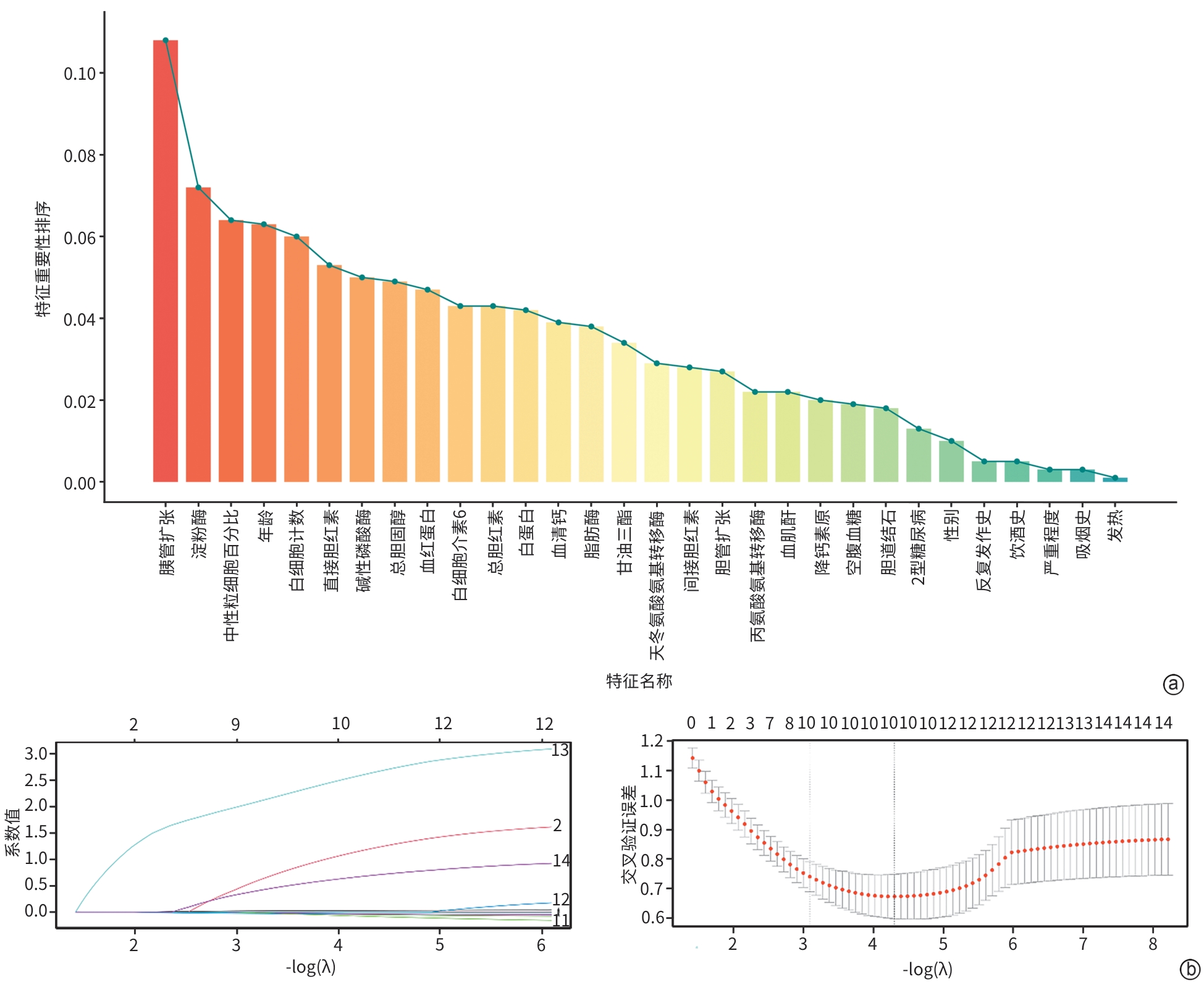
注: a,递归特征消除,特征按照重要性进行前后排序,从第9个特征开始,后续特征的重要性逐渐减小;b,最小绝对收缩与选择算子算法的系数路径图与交叉验证图。
图 1 特征筛选结果
Figure 1. Feature selection results
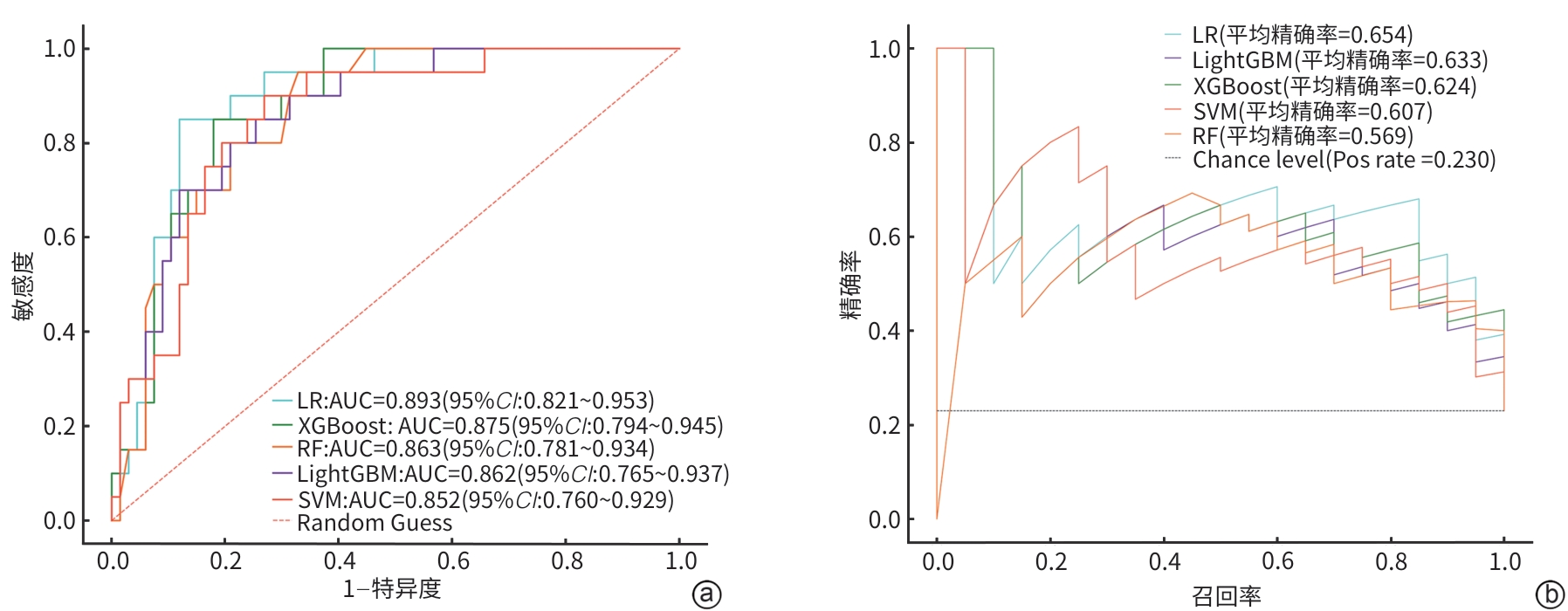
注: a,ROC曲线;b,精确率-召回率曲线。ROC曲线,受试者操作特征曲线;AUC,曲线下面积;RF,随机森林;LR,逻辑回归;XGBoost,极限梯度提升;LightGBM,轻量级梯度提升机;SVM,支持向量机;CI,置信区间。
图 2 5种机器学习模型在测试集上的ROC曲线和精确率-召回率曲线
Figure 2. ROC curves and precision-recall curves for five machine learning models on the test set
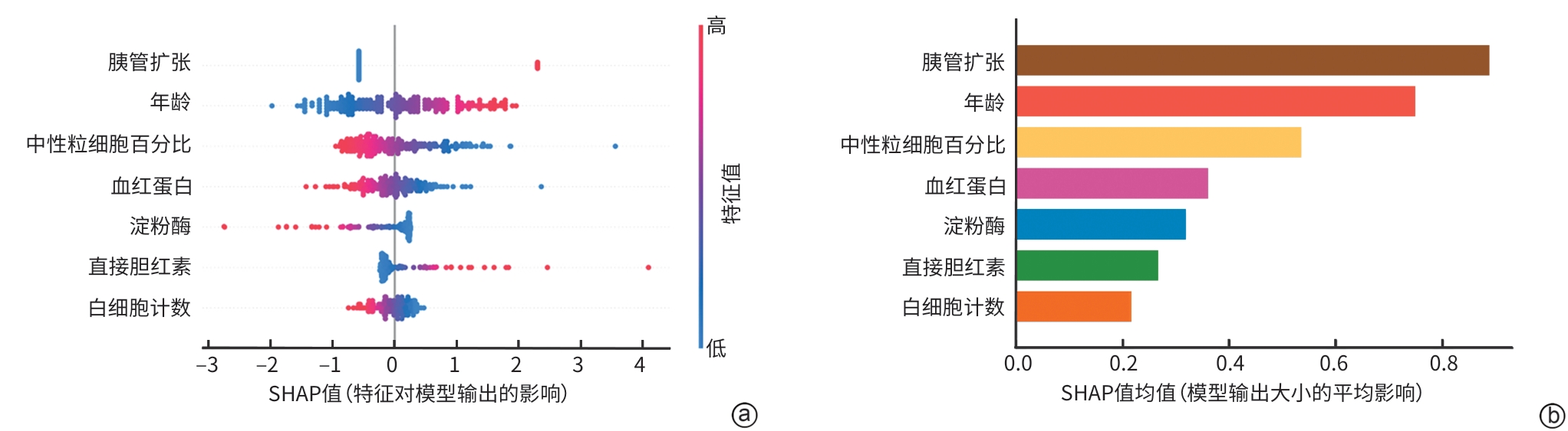
注: a,SHAP 特征重要性汇总图,展示每个样本的SHAP值和特征值分布;b,平均SHAP值柱状图,展示整体特征重要性排序。SHAP,沙普利加性解释。
图 3 SHAP值的模型解释图
Figure 3. Model interpretation diagram of shapley additive explanations values
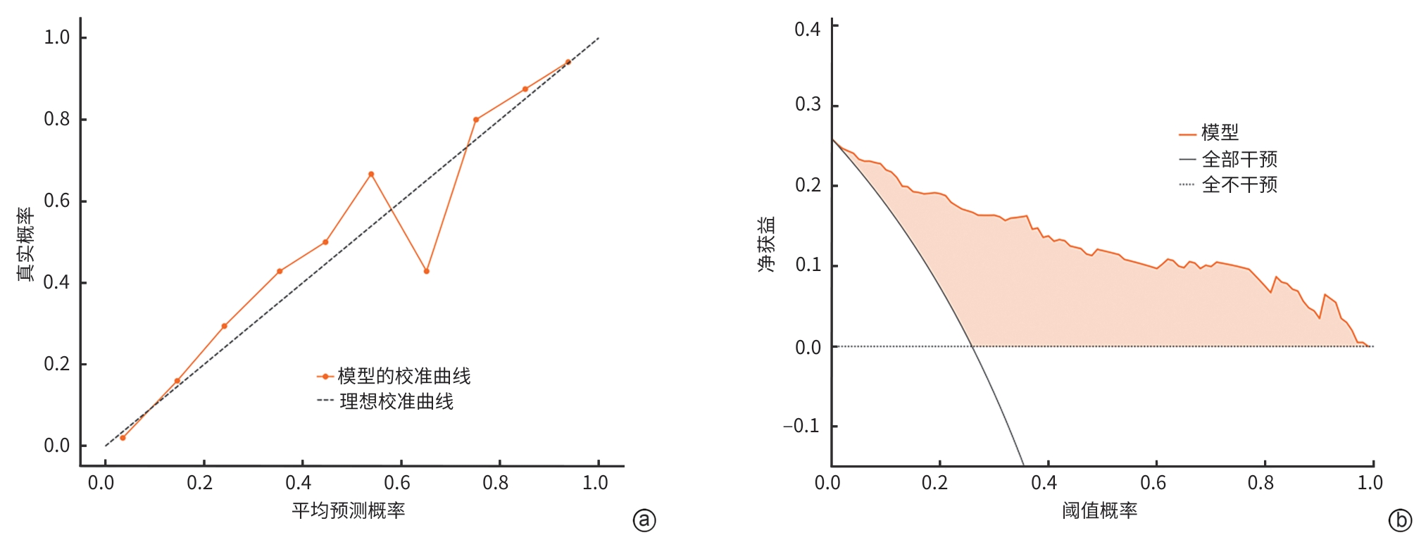
注: a,校准曲线;b,决策曲线。
图 4 校准曲线与决策曲线分析
Figure 4. Calibration curve and decision curve analysis
表 1 训练集与测试集临床资料的比较
Table 1. Comparison of clinical characteristics between the training and test sets
指标 测试集(n=87) 训练集(n=201) 统计值 P值 肿瘤[例(%)] χ2=0.137 0.711 无 67(77.0) 149(74.1) 有 20(23.0) 52(25.9) 性别[例(%)] χ2<0.001 0.999 女 37(42.5) 87(43.3) 男 50(57.5) 114(56.7) 吸烟史[例(%)] χ2=0.054 0.817 无 60(69.0) 143(71.1) 有 27(31.0) 58(28.9) 饮酒史[例(%)] χ2=1.573 0.210 无 57(65.5) 148(73.6) 有 30(34.5) 53(26.4) 2型糖尿病[例(%)] χ2=0.379 0.538 无 68(78.2) 165(82.1) 有 19(21.8) 36(17.9) 胰腺炎反复病史[例(%)] χ2=0.103 0.749 无 72(82.8) 171(85.1) 有 15(17.2) 30(14.9) 发热[例(%)] χ2=0.474 0.491 无 75(86.2) 165(82.1) 有 12(13.8) 36(17.9) 严重程度[例(%)] χ2=0.474 0.491 轻中度 75(86.2) 165(82.1) 重度 12(13.8) 36(17.9) 胆道结石[例(%)] χ2<0.001 0.999 无 57(65.5) 131(65.2) 有 30(34.5) 70(34.8) 胰管扩张[例(%)] χ2=0.264 0.608 无 71(81.6) 157(78.1) 有 16(18.4) 44(21.9) 胆管扩张[例(%)] χ2=0.379 0.538 无 68(78.2) 165(82.1) 有 19(21.8) 36(17.9) 年龄(岁) 50.00(34.50~61.00) 50.00(38.00~61.00) Z=-0.459 0.647 白细胞计数(×109/L) 9.86(6.96~13.57) 9.82(6.47~13.15) Z=0.393 0.695 中性粒细胞百分比(%) 77.40(67.60~84.45) 78.60(68.40~84.80) Z=-0.253 0.801 血红蛋白(g/L) 140.00(121.50~155.00) 139.00(125.00~156.00) Z=-0.466 0.613 淀粉酶(U/L) 182.43(86.16~537.32) 167.26(79.25~499.43) Z=0.411 0.682 脂肪酶(U/L) 297.76(109.06~1 009.25) 303.86(66.62~1 514.50) Z=0.196 0.845 血清钙(mmol/L) 2.23(2.10~2.30) 2.21(2.10~2.30) Z=0.616 0.538 空腹血糖(mmol/L) 6.64(5.33~9.20) 6.70(5.30~9.36) Z=-0.274 0.784 血肌酐(μmol/L) 58.90(50.34~72.28) 58.26(49.80~73.08) Z=0.045 0.964 白蛋白(g/L) 41.81(37.73~45.72) 40.37(37.14~44.14) Z=1.724 0.085 甘油三酯(mmol/L) 1.84(0.99~3.75) 1.65(1.05~5.45) Z=-0.032 0.975 总胆固醇(mmol/L) 4.56(3.42~5.84) 4.54(3.71~6.04) Z=-0.677 0.499 总胆红素(μmol/L) 21.88(15.62~39.20) 22.09(15.28~35.78) Z=0.079 0.937 直接胆红素(μmol/L) 8.29(5.58~13.14) 9.55(6.33~17.79) Z=-1.051 0.294 间接胆红素(μmol/L) 12.45(8.23~21.84) 12.66(8.29~20.14) Z=0.136 0.893 天冬氨酸氨基转移酶(U/L) 35.33(24.82~73.37) 37.40(27.01~80.92) Z=-0.837 0.403 丙氨酸氨基转移酶(U/L) 31.40(21.00~58.23) 33.04(20.00~95.00) Z=-0.725 0.469 碱性磷酸酶(U/L) 93.44(71.55~136.61) 98.92(74.03~151.13) Z=-0.510 0.610 降钙素原(ng/L) 0.09(0.05~0.20) 0.11(0.04~0.28) Z=-0.559 0.576 白细胞介素6(pg/L) 24.90(7.15~65.03) 22.70(7.67~69.64) Z=-0.301 0.764  下载: 导出CSV
表 2 肿瘤相关急性胰腺炎组与常规组临床资料的比较
Table 2. Comparison of clinical characteristics between the tumor-induced acute pancreatitis group and the conventional group
指标 常规组(n=216) TIAP组(n=72) 统计值 P值 性别[例(%)] χ2<0.001 0.999 女 93 (43.1) 31 (43.1) 男 123 (56.9) 41 (56.9) 吸烟史[例(%)] χ2=0.139 0.709 无 154 (71.3) 49 (68.1) 有 62 (28.7) 23 (31.9) 饮酒史[例(%)] χ2=0.683 0.409 无 157 (72.7) 48 (66.7) 有 59 (27.3) 24 (33.3) 2型糖尿病[例(%)] χ2=11.394 0.001 无 185 (85.6) 48 (66.7) 有 31 (14.4) 24 (33.3) 胰腺炎反复病史[例(%)] χ2=12.019 0.001 无 192 (88.9) 51 (70.8) 有 24 (11.1) 21 (29.2) 发热[例(%)] χ2=0.300 0.584 无 178 (82.4) 62 (86.1) 有 38 (17.6) 10 (13.9) 严重程度[例(%)] χ2=2.700 0.100 轻中度 175 (81.0) 65 (90.3) 重度 41 (19.0) 7 (9.7) 胆道结石[例(%)] χ2=3.452 0.063 无 134 (62.0) 54 (75.0) 有 82 (38.0) 18 (25.0) 胰管扩张[例(%)] χ²=79.474 <0.001 无 201 (93.1) 32 (44.4) 有 15 (6.9) 40 (55.6) 胆管扩张[例(%)] χ2=26.077 <0.001 无 190 (88.0) 43 (59.7) 有 26 (12.0) 29 (40.3) 年龄(岁) 45.00 (35.00~56.25) 59.00 (51.75~68.00) Z=-5.838 <0.001 白细胞计数(×109/L) 11.09 (7.67~13.85) 6.88 (5.25~8.94) Z=5.630 <0.001 中性粒细胞百分比(%) 80.75 (72.75~85.43) 69.90 (60.83~77.88) Z=5.269 <0.001 血红蛋白(g/L) 145.00 (128.00~161.00) 132.00 (115.00~139.50) Z=5.038 <0.001 淀粉酶(U/L) 182.91 (84.47~661.49) 156.09 (67.97~269.71) Z=2.606 0.009 脂肪酶(U/L) 330.16 (78.89~1 567.87) 192.43 (51.91~891.95) Z=2.047 0.041 血清钙(mmol/L) 2.21 (2.10~2.30) 2.22 (2.11~2.30) Z=-0.011 0.992 空腹血糖(mmol/L) 6.83 (5.32~9.94) 6.33 (5.20~8.15) Z=1.299 0.194 血肌酐(μmol/L) 59.61 (50.65~73.08) 56.74 (48.18~71.06) Z=0.938 0.349 白蛋白(g/L) 41.87 (38.16~45.46) 38.60 (33.30~41.07) Z=4.981 <0.001 甘油三酯(mmol/L) 1.85 (1.06~6.96) 1.39 (0.98~2.12) Z=2.975 0.003 总胆固醇(mmol/L) 4.87 (3.83~6.30) 3.88 (3.26~4.76) Z=4.681 <0.001 总胆红素(μmol/L) 22.64 (16.03~35.51) 18.63 (13.68~47.90) Z=0.662 0.509 直接胆红素(μmol/L) 9.83 (6.40~14.89) 7.40 (4.91~33.77) Z=0.936 0.350 间接胆红素(μmol/L) 12.74 (8.33~20.06) 12.05 (8.04~21.77) Z=0.137 0.891 天冬氨酸氨基转移酶(U/L) 37.71 (26.68~76.54) 33.88 (25.30~83.22) Z=0.600 0.549 丙氨酸氨基转移酶(U/L) 32.64 (20.00~95.00) 31.68 (20.91~59.34) Z=0.827 0.409 碱性磷酸酶(U/L) 96.73 (73.50~135.01) 103.83 (73.46~251.30) Z=-1.668 0.095 降钙素原(ng/L) 0.11 (0.04~0.26) 0.09 (0.04~0.21) Z=0.832 0.405 白细胞介素6(pg/L) 30.38 (8.96~80.60) 10.85 (4.82~28.16) Z=3.604 <0.001
下载: 导出CSV
表 3 5种机器学习算法性能的比较
Table 3. Performance comparison of five machine learning algorithms
项目 准确度 敏感度 特异度 阳性预测值 阴性预测值 F1分数 Kappa分数 训练集 RF 1.00 1.00 1.00 1.00 1.00 1.00 1.00 0.86 0.62 0.95 0.80 0.88 0.70 0.61 LR XGBoost 1.00 0.98 1.00 1.00 0.99 0.99 0.99 LightGBM 1.00 1.00 1.00 1.00 1.00 1.00 1.00 SVM 0.80 0.27 0.98 0.82 0.79 0.41 0.32 测试集 RF 0.80 0.60 0.87 0.57 0.88 0.59 0.46 0.83 0.60 0.90 0.63 0.88 0.62 0.50 LR XGBoost 0.82 0.65 0.87 0.59 0.89 0.62 0.50 LightGBM 0.83 0.65 0.88 0.62 0.89 0.63 0.52 SVM 0.78 0.30 0.93 0.55 0.82 0.39 0.27 注:RF,随机森林;LR,逻辑回归;XGBoost,极限梯度提升;LightGBM,轻量级梯度提升机;SVM,支持向量机。
下载: 导出CSV
-
[1] Li T Y, Qin C, Zhao B B, et al. Global and regional burden of pancreatitis: Epidemiological trends, risk factors, and projections to 2050 from the global burden of disease study 2021[J]. BMC Gastroenterol, 2024, 24( 1): 398. DOI: 10.1186/s12876-024-03481-8. [2] Qu Yuanxu, Cao Feng, Ding Yixuan, et al. Clinical characteristics and prognosis of acute pancreatitis with different etiologies[J]. Chin J Dig Surg, 2025, 24( 5): 623- 629. DOI: 10.3760/cma.j.cn115610-20250305-00086.曲元旭, 曹锋, 丁乙轩, 等. 不同病因急性胰腺炎的临床特征及预后情况[J]. 中华消化外科杂志, 2025, 24( 5): 623- 629. DOI: 10.3760/cma.j.cn115610-20250305-00086. [3] Chinese Pancreatic Surgery Association, Chinese Society of Surgery, Chinese Medical Association. Guidelines for diagnosis and treatment of acute pancreatitis in China(2021)[J]. Chin J Surg, 2021, 59( 7): 578- 587. DOI: 10.3760/cma.j.cn112139-20210416-00172.中华医学会外科学分会胰腺外科学组. 中国急性胰腺炎诊治指南(2021)[J]. 中华外科杂志, 2021, 59( 7): 578- 587. DOI: 10.3760/cma.j.cn112139-20210416-00172. [4] Feng Q, Li C, Zhang S, et al. Recurrence and survival after surgery for pancreatic cancer with or without acute pancreatitis[J]. World J Gastroenterol, 2019, 25( 39): 6006- 6015. DOI: 10.3748/wjg.v25.i39.6006. [5] Pitlick M M, Abeykoon J P, Dao L, et al. Non-Hodgkin lymphoma presenting as acute pancreatitis: A rare occurrence[J]. Clin Case Rep, 2018, 7( 1): 107- 109. DOI: 10.1002/ccr3.1885. [6] Tanaka H, Nakazawa T, Yoshida M, et al. Metastasis-induced acute pancreatitis in a patient with small cell carcinoma of the lungs[J]. JOP, 2009, 10( 5): 557- 561. [7] Dixon J M, Chapman R W, Berry A R. Carcinoid tumour of the Ampulla of Vater presenting as acute pancreatitis[J]. Gut, 1987, 28( 10): 1296- 1297. DOI: 10.1136/gut.28.10.1296. [8] Cuocolo R, Caruso M, Perillo T, et al. Machine learning in oncology: A clinical appraisal[J]. Cancer Lett, 2020, 481: 55- 62. DOI: 10.1016/j.canlet.2020.03.032. [9] Chung H, Ko Y, Lee I S, et al. Prognostic artificial intelligence model to predict 5 year survival at 1 year after gastric cancer surgery based on nutrition and body morphometry[J]. J Cachexia Sarcopenia Muscle, 2023, 14( 2): 847- 859. DOI: 10.1002/jcsm.13176. [10] Yin Minyue, Zhu Jinzhou, Liu Lu, et al. Research advances in machine learning models for acute pancreatitis[J]. J Clin Hepatol, 2023, 39( 12): 2978- 2984. DOI: 10.3969/j.issn.1001-5256.2023.12.034.殷民月, 朱锦舟, 刘璐, 等. 急性胰腺炎机器学习模型的研究进展[J]. 临床肝胆病杂志, 2023, 39( 12): 2978- 2984. DOI: 10.3969/j.issn.1001-5256.2023.12.034. [11] Liang Tan, Chen Zixiang, Chen Jiangming, et al. Application and prospect of artificial intelligence in the whole-process management of pancreatic cancer[J]. Chin J Dig Surg, 2024, 23( 11): 1459- 1464. DOI: 10.3760/cma.j.cn115610-20241028-00471.梁谭, 陈子祥, 陈江明, 等. 人工智能在胰腺癌全程化管理中的应用及展望[J]. 中华消化外科杂志, 2024, 23( 11): 1459- 1464. DOI: 10.3760/cma.j.cn115610-20241028-00471. [12] Ko S E, Choi I Y, Cha S H, et al. Clinical and radiologic characteristics of pancreatic head carcinoma without main pancreatic duct dilatation: Using dual-phase contrast-enhanced CT scan[J]. Clin Imag, 2016, 40( 3): 548- 552. DOI: 10.1016/j.clinimag.2015.10.008. [13] Rizvi A, Sethi A, Poneros J, et al. Does incidentally detected common bile duct dilation need evaluation?[J]. Cleve Clin J Med, 2022, 89( 6): 315- 319. DOI: 10.3949/ccjm.89a.21029. [14] Ren L J, Wang T, Sekhari Seklouli A, et al. A review on missing values for main challenges and methods[J]. Inf Syst, 2023, 119: 102268. DOI: 10.1016/j.is.2023.102268. [15] Tenner S, Vege S S, Sheth S G, et al. American college of gastroenterology guidelines: Management of acute pancreatitis[J]. Am J Gastroenterol, 2024, 119( 3): 419- 437. DOI: 10.14309/ajg.0000000000002645. [16] Mujica V R, Barkin J S, Go V L. Acute pancreatitis secondary to pancreatic carcinoma[J]. Pancreas, 2000, 21( 4): 329- 332. DOI: 10.1097/00006676-200011000-00001. [17] Li J, Zhou R, Zhang J, et al. Calcium signaling of pancreatic acinar cells in the pathogenesis of pancreatitis[J]. World J Gastroenterol, 2014, 20( 43): 16146- 16152. DOI: 10.3748/wjg.v20.i43.16146. [18] Canakis A, Baron T H. The role of underlying pancreatic ductal obstruction and risk of post-ERCP pancreatitis[J]. Gastrointest Endosc, 2025, 101( 3): 694. DOI: 10.1016/j.gie.2024.09.033. [19] Shi J Q, Nie S, Shi Z, et al. Clinical characteristics of pancreatic cancer presenting with acute pancreatitis: A propensity-matched cohort study[J]. Surg Endosc, 2025, 39( 5): 3307- 3316. DOI: 10.1007/s00464-025-11654-z. [20] Xiong Y Y, Zhao Y, Han X L, et al. Clinical characteristics and outcome of tumor-associated acute pancreatitis: A single-center cohort study[J]. Ann Transl Med, 2021, 9( 8): 639. DOI: 10.21037/atm-20-7196. [21] Ren Xia, Ye Ye, Liu Luojie, et al. Establishment of a nomogram prediction model for poor prognosis of acute pancreatitis based on inflammatory factors, lung ultrasound, and CT scores[J]. J Clin Hepatol, 2025, 41( 4): 713- 721. DOI: 10.12449/JCH250417.任夏, 叶叶, 刘罗杰, 等. 基于炎症因子、肺超声和CT评分系统的急性胰腺炎预后不良列线图预测模型的构建[J]. 临床肝胆病杂志, 2025, 41( 4): 713- 721. DOI: 10.12449/JCH250417. [22] Cho J H, Choi J S, Hwang E T, et al. Usefulness of scheduled follow-up CT in discharged patients with acute pancreatitis[J]. Pancreatology, 2015, 15( 6): 642- 646. DOI: 10.1016/j.pan.2015.09.004. [23] Zhang Huimin, Jin Meng, Qian Jiaming, et al. Differential diagnosis between pancreatic cancer with acute pancreatitis and simple acute pancreatitis cancer[J]. Chin J Pract Intern Med, 2017, 37( 2): 145- 147. DOI: 10.19538/j.nk2017020114.张慧敏, 金梦, 钱家鸣, 等. 胰腺癌合并急性胰腺炎和单纯急性胰腺炎诊断分析[J]. 中国实用内科杂志, 2017, 37( 2): 145- 147. DOI: 10.19538/j.nk2017020114. -

 本文二维码
本文二维码
计量
- 文章访问数: 4
- HTML全文浏览量: 0
- PDF下载量: 0
- 被引次数: 0