中文English
ISSN 1001-5256 (Print)
ISSN 2097-3497 (Online)
CN 22-1108/R
Volume 40 Issue 10
Oct.  2024
Turn off MathJax
Article Contents

Protective effect of folic acid against cholestatic liver injury in mice caused by bis(2-ethylhexyl) phthalate exposure

DOI: 10.12449/JCH241021
Research funding:

National Natural Science Foundation of China (82073566);

The Program of Excellent Young Talents in Universities of Anhui Province (gxyq2019014);

Anhui Public Health Clinical Center, Supported by North District Scientific Research and Cultivation Foundation of the First Affiliated Hospital of Anhui Medical University (2023YKJ14);

Anhui Public Health Clinical Center, Supported by North District Scientific Research and Cultivation Foundation of the First Affiliated Hospital of Anhui Medical University (2023YKJ06);

Anhui Public Health Clinical Center, Supported by North District Scientific Research and Cultivation Foundation of the First Affiliated Hospital of Anhui Medical University (2023YKJ11)

More Information
  • Corresponding author: WANG Jianqing, jianqingwang81@126.com (ORCID: 0000-0002-7935-9520)
  • Received Date: 2024-01-26
  • Accepted Date: 2024-03-12
  • Published Date: 2024-10-25
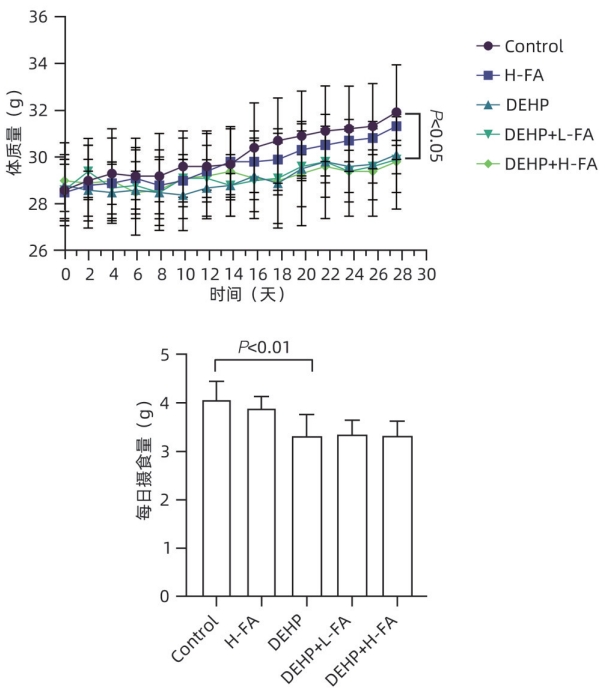
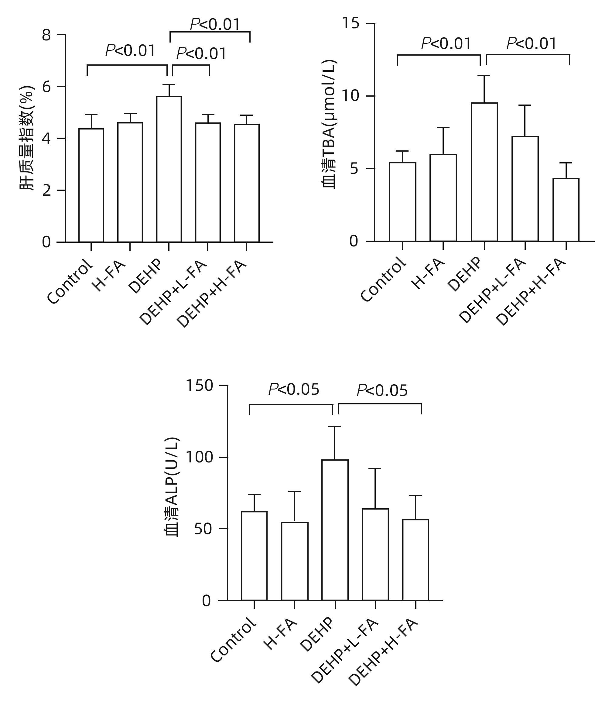
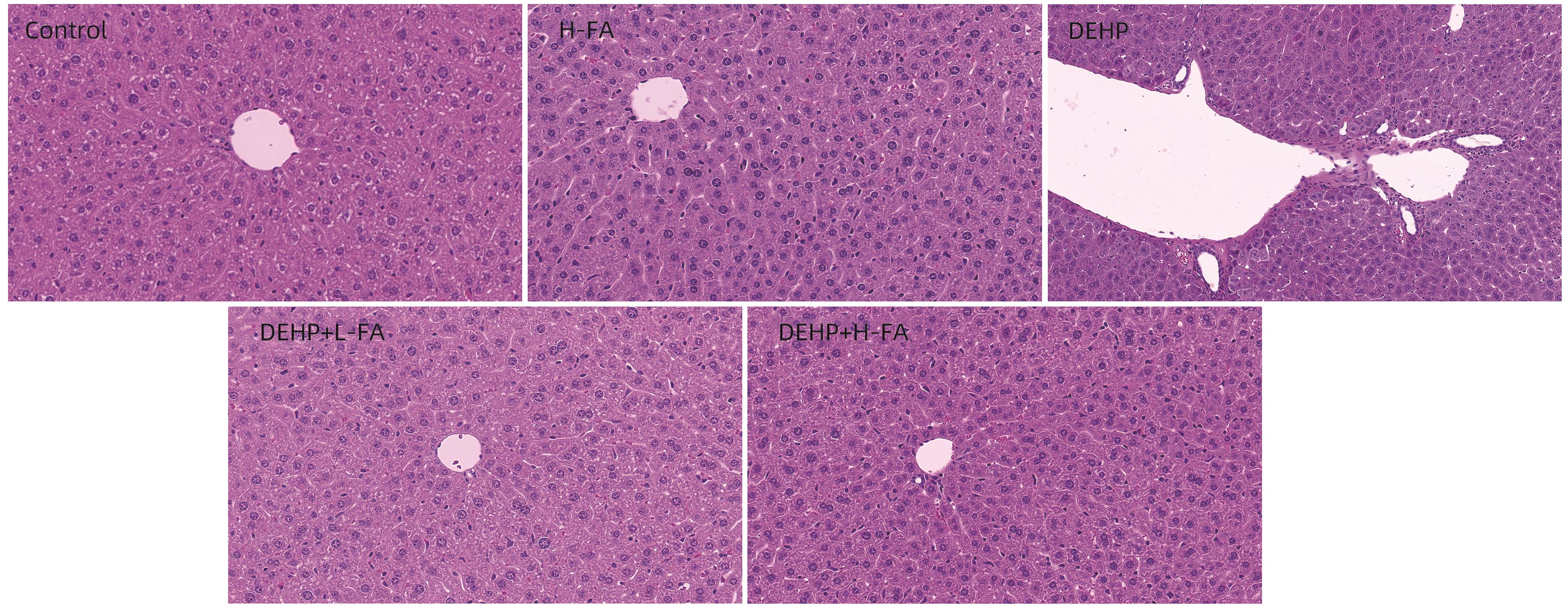
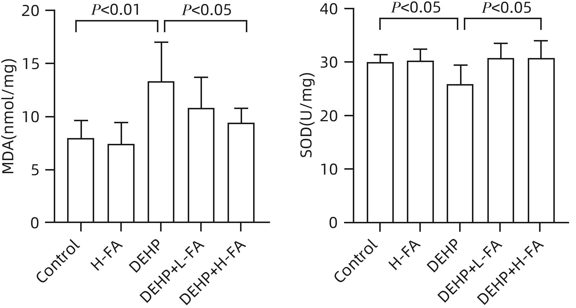
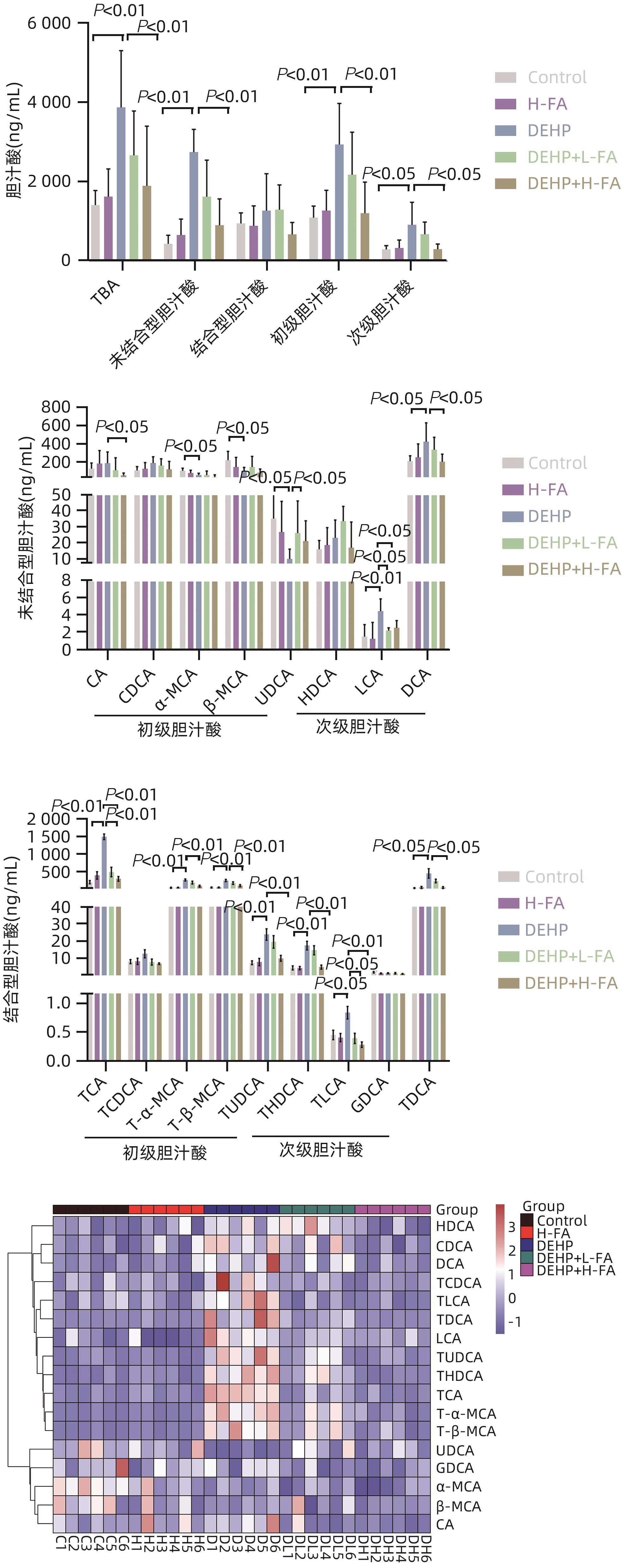
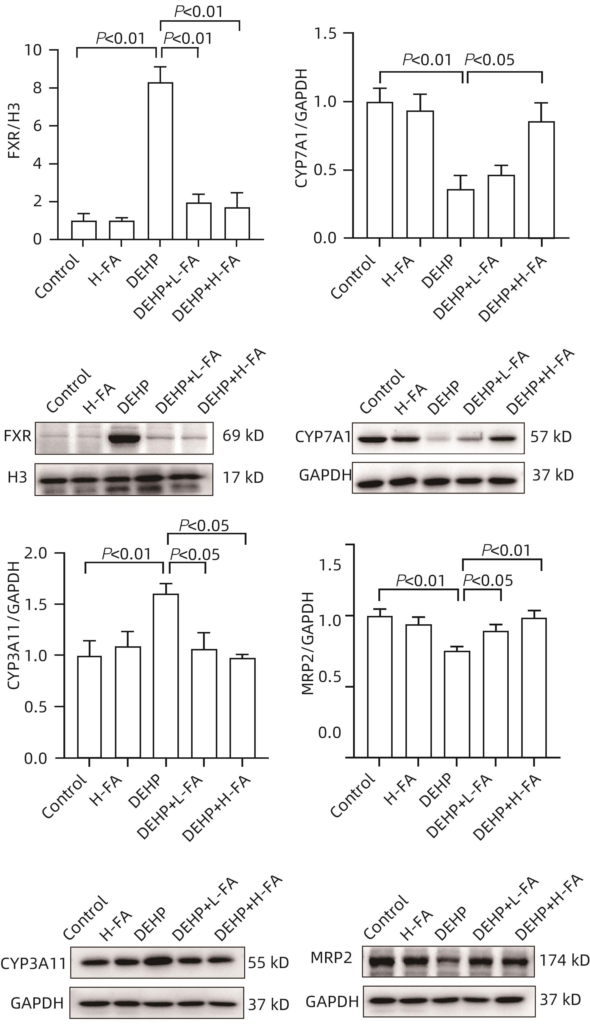
  •   Objective  To investigate the protective effect of folic acid against cholestatic liver injury in mice induced by bis(2-ethylhexyl) phthalate (DEHP) exposure and its mechanism.  Methods  ICR mice were randomly divided into control group, high-dose folic acid (H-FA) group, DEHP group, DEHP+low-dose folic acid (DEHP+L-FA) group, and DEHP+high-dose folic acid (DEHP+H-FA) group, with 6 mice in each group. The mice in the H-FA group, the DEHP+L-FA group, and the DEHP+H-FA group were given folic acid by gavage at the corresponding dose, and those in the control group and the DEHP group were given an equal volume of PBS solution by gavage. After 2 hours, the mice in the DEHP group, the DEHP+L-FA group, and the DEHP+H-FA group were given corn oil containing 200 mg/kg DEHP, and those in the control group and the H-FA group were given an equal volume of pure corn oil, by gavage for 4 weeks. Body weight and food intake were recorded every day, and blood and liver tissue samples were collected. A biochemical analyzer was used to measure the serum levels of total bile acid (TBA) and alkaline phosphatase(ALP); HE staining was used to observe the histopathological changes of liver tissue; kits were used to measure the content of malondialdehyde (MDA) and superoxide dismutase (SOD) in the liver; LC-MS/MS was used to measure serum bile acid profiles; Western blot was used to measure the expression levels of proteins associated with hepatic bile acid metabolism. A one-way analysis of variance was used for comparison of continuous data between multiple groups, and the least significant difference t-test was used for further comparison between two groups.  Results  Compared with the control group, the daily food intake of the mice in the DEHP group decreased significantly, and the body weight decreased significantly from day 10 (P<0.05), and compared with the DEHP group, the DEHP+L-FA group and the DEHP+H-FA group had basically unchanged body weight and daily food intake (P>0.05). Compared with the control group, the DEHP group had significant increases in liver weight index and the serum levels of TBA and ALP (all P<0.05), with enlarged portal area, bile duct deformity and hyperplasia, and a small amount of inflammatory cell infiltration in liver tissue; compared with the DEHP group, the DEHP+L-FA group and the DEHP+H-FA group had a significant reduction in liver weight index (P<0.01), and the DEHP+H-FA group had significant reductions in the serum levels of TBA and ALP (P<0.05), with a significant improvement in liver histomorphology and structure after folic acid intervention. Compared with the control group, the DEHP group had a significant reduction in the content of SOD (P<0.05) and a significant increase in the content of MDA in the liver (P<0.01), and compared with the DEHP group, the DEHP+H-FA group had significant reductions in the content of MDA and SOD (P<0.05). Compared with the control group, the DEHP group had significant increases in the serum levels of α-muricholic acid (α-MCA),β- muricholic acid (β-MCA),deoxycholic acid (DCA), lithocholic acid (LCA), taurocholic acid (TCA), taurodeoxycholic acid (TDCA), tauroursodeoxycholic acid (TUDCA), tauro-β-muricholic acid (T-β-MCA), tauro-α-muricholic acid (T-α-MCA), taurohyodeoxycholic acid (THDCA), and taurolithocholic acid (TLCA)P<0.05) and a significant reduction in ursodeoxycholic acid (UDCA)(P<0.05); compared with the DEHP group, the DEHP+H-FA group had significant reductions in the serum levels of DCA, LCA, TCA, TDCA, TUDCA, T-β-MCA, T-α-MCA, THDCA, and TLCA (P<0.05). Compared with the control group, the DEHP group had significant increases in the protein expression levels of FXR and CYP3A11 in the liver (P<0.01) and significant reductions in the protein expression levels of CYP7A1 and MRP2 (P<0.01); compared with the DEHP group, the DEHP+L-FA group and the DEHP+H-FA group had significant reductions in the protein expression levels of FXR and CYP3A11 in the liver (P<0.05) and a significant increase in the protein expression level of MRP2 (P<0.05), and the DEHP+H-FA group had a significant increase in the protein expression level of CYP7A1 (P<0.05).  Conclusion  Folic acid has a protective effect against cholestatic liver injury in mice induced by DEHP exposure, possibly by regulating bile acid synthesis, catabolism, and transport and maintaining bile acid homeostasis.


  • loading
  • [1]
    BAGEL S, DESSAIGNE B, BOURDEAUX D, et al. Influence of lipid type on bis(2-ethylhexyl)phthalate(DEHP) leaching from infusion line sets in parenteral nutrition[J]. JPEN J Parenter Enteral Nutr, 2011, 35( 6): 770- 775. DOI: 10.1177/0148607111414021.
    [2]
    ZHAO F, ZHANG L, QU MC, et al. Obeticholic acid alleviates intrauterine growth restriction induced by di-ethyl-hexyl phthalate in pregnant female mice by improving bile acid disorder[J]. Environ Sci Pollut Res Int, 2023, 30( 51): 110956- 110969. DOI: 10.1007/s11356-023-30149-9.
    [3]
    GALLEGO-LOPEZ MDC, OJEDA ML, ROMERO-HERRERA I, et al. Folic acid homeostasis and its pathways related to hepatic oxidation in adolescent rats exposed to binge drinking[J]. Antioxidants(Basel), 2022, 11( 2): 362. DOI: 10.3390/antiox11020362.
    [4]
    ZHANG HQ, ZUO YW, ZHAO HC, et al. Folic acid ameliorates alcohol-induced liver injury via gut-liver axis homeostasis[J]. Front Nutr, 2022, 9: 989311. DOI: 10.3389/fnut.2022.989311.
    [5]
    CHEN S, YANG MY, WANG R, et al. Suppression of high-fat-diet-induced obesity in mice by dietary folic acid supplementation is linked to changes in gut microbiota[J]. Eur J Nutr, 2022, 61( 4): 2015- 2031. DOI: 10.1007/s00394-021-02769-9.
    [6]
    JIANG L, GAI XC, NI Y, et al. Folic acid protects against tuberculosis-drug-induced liver injury in rats and its potential mechanism by metabolomics[J]. J Nutr Biochem, 2023, 112: 109214. DOI: 10.1016/j.jnutbio.2022.109214.
    [7]
    ZHANG YJ, GUO JL, XUE JC, et al. Phthalate metabolites: Characterization, toxicities, global distribution, and exposure assessment[J]. Environ Pollut, 2021, 291: 118106. DOI: 10.1016/j.envpol.2021.118106.
    [8]
    GAITANTZI H, HAKENBERG P, THEOBALD J, et al. Di(2-ethylhexyl) phthalate and its role in developing cholestasis: An in vitro study on different liver cell types[J]. J Pediatr Gastroenterol Nutr, 2018, 66( 2): e28- e35. DOI: 10.1097/MPG.0000000000001813.
    [9]
    WEI XJ, YANG DQ, ZHANG BY, et al. Di-(2-ethylhexyl) phthalate increases plasma glucose and induces lipid metabolic disorders via FoxO1 in adult mice[J]. Sci Total Environ, 2022, 842: 156815. DOI: 10.1016/j.scitotenv.2022.156815.
    [10]
    ZHOU YH, ZHOU YZ, LI YF, et al. Targeted bile acid profiles reveal the liver injury amelioration of Da-Chai-Hu Decoction against ANIT- and BDL-induced cholestasis[J]. Front Pharmacol, 2022, 13: 959074. DOI: 10.3389/fphar.2022.959074.
    [11]
    WANG GF, LI YY, SHI R, et al. Yinchenzhufu decoction protects against alpha-naphthylisothiocyanate-induced acute cholestatic liver injury in mice by ameliorating disordered bile acid homeostasis and inhibiting inflammatory responses[J]. J Ethnopharmacol, 2020, 254: 112672. DOI: 10.1016/j.jep.2020.112672.
    [12]
    LE YB, WANG KH, ZOU L. Mechanism of taurocholic acid in promoting the progression of liver cirrhosis[J]. J Clin Hepatol, 2021, 37( 11): 2658- 2662. DOI: 10.3969/j.issn.1001-5256.2021.11.037.

    乐英彪, 王昆华, 邹雷. 牛磺胆酸促进肝硬化发展的机制[J]. 临床肝胆病杂志, 2021, 37( 11): 2658- 2662. DOI: 10.3969/j.issn.1001-5256.2021.11.037.
    [13]
    LI CZ, HUANG XW, ZHANG ZP, et al. Research progress in role of gut-liver axis in occurrence and development of atherosclerosis[J]. J Jilin Univ Med Ed, 2023, 49( 6): 1669- 1676. DOI: 10.13481/j.1671-587X.20230636.

    李朝政, 黄晓巍, 张泽鹏, 等. 肠-肝轴在动脉粥样硬化发生发展中作用的研究进展[J]. 吉林大学学报(医学版), 2023, 49( 6): 1669- 1676. DOI: 10.13481/j.1671-587X.20230636.
    [14]
    TRAUNER M, FUCHS CD. Novel therapeutic targets for cholestatic and fatty liver disease[J]. Gut, 2022, 71( 1): 194- 209. DOI: 10.1136/gutjnl-2021-324305.
    [15]
    HAN X, LIN C, LIU H, et al. Allocholic acid protects against α‍- naphthylisothiocyanate-induced cholestasis in mice by ameliorating disordered bile acid homeostasis[J]. J Appl Toxicol, 2024, 44( 4): 582- 594. DOI: 10.1002/jat.4562.
    [16]
    CHAI J, CAI SY, LIU XC, et al. Canalicular membrane MRP2/ABCC2 internalization is determined by Ezrin Thr567 phosphorylation in human obstructive cholestasis[J]. J Hepatol, 2015, 63( 6): 1440- 1448. DOI: 10.1016/j.jhep.2015.07.016.
    [17]
    PAULUSMA CC, KOTHE MJ, BAKKER CT, et al. Zonal down-regulation and redistribution of the multidrug resistance protein 2 during bile duct ligation in rat liver[J]. Hepatology, 2000, 31( 3): 684- 693. DOI: 10.1002/hep.510310319.
    [18]
    ZU Y, LIU YN, LAN LL, et al. Consecutive baicalin treatment relieves its accumulation in rats with intrahepatic cholestasis by increasing MRP2 expression[J]. Heliyon, 2023, 9( 1): e12689. DOI: 10.1016/j.heliyon.2022.e12689.
    [19]
    RAZORI MV, MAIDAGAN PM, CIRIACI N, et al. Anticholestatic mechanisms of ursodeoxycholic acid in lipopolysaccharide-induced cholestasis[J]. Biochem Pharmacol, 2019, 168: 48- 56. DOI: 10.1016/j.bcp.2019.06.009.
    [20]
    HINOSHITA E, TAGUCHI K, INOKUCHI A, et al. Decreased expression of an ATP-binding cassette transporter, MRP2, in human livers with hepatitis C virus infection[J]. J Hepatol, 2001, 35( 6): 765- 773. DOI: 10.1016/s0168-8278(01)00216-1.
  • 加载中

Catalog

    通讯作者: 陈斌, bchen63@163.com
    • 1. 

      沈阳化工大学材料科学与工程学院 沈阳 110142

    1. 本站搜索
    2. 百度学术搜索
    3. 万方数据库搜索
    4. CNKI搜索

    Figures(6)

    Article Metrics

    Article views (112) PDF downloads(23) Cited by()
    Proportional views
    Related

    /

    DownLoad:  Full-Size Img  PowerPoint
    Return
    Return