2026,
42(4):
908-917.
DOI: 10.12449/JCH260420
Abstract:
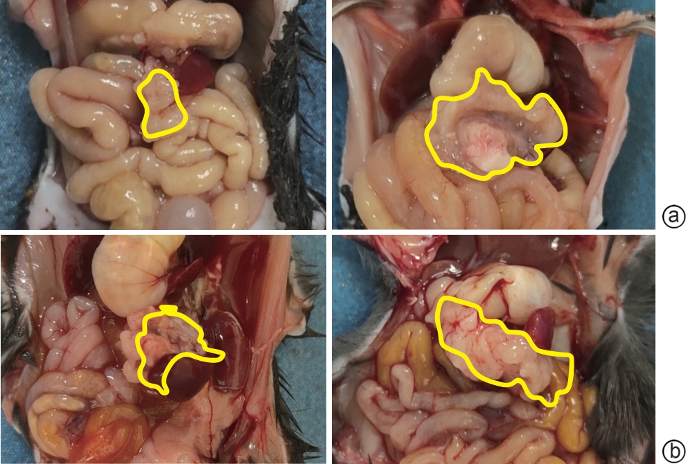
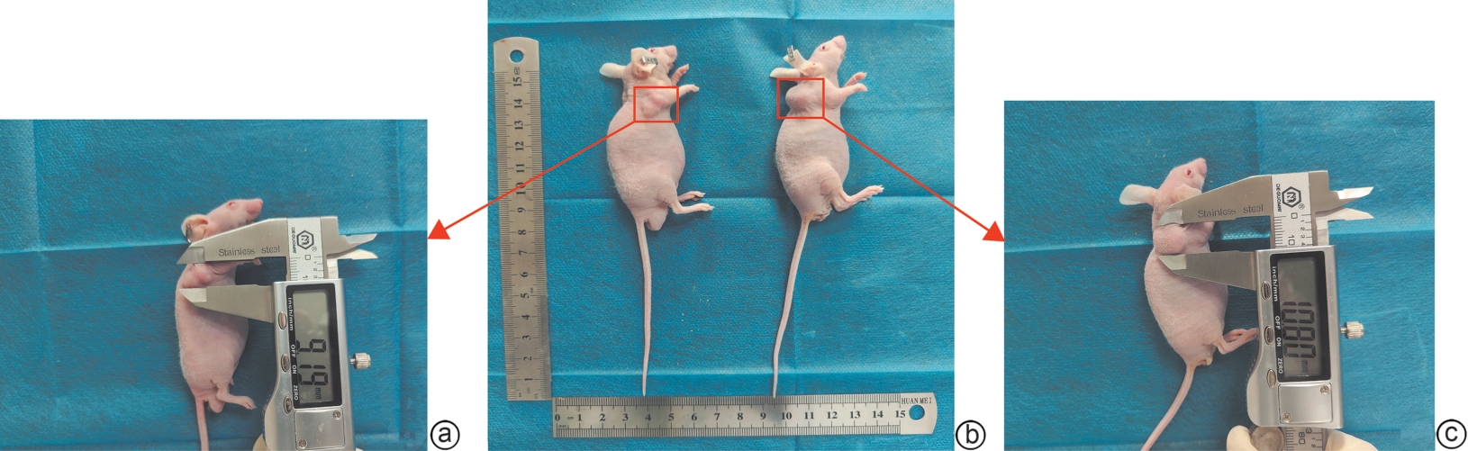
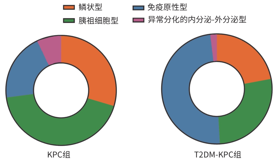
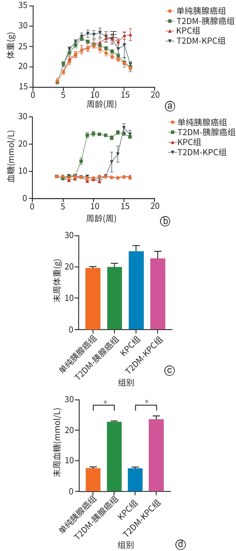
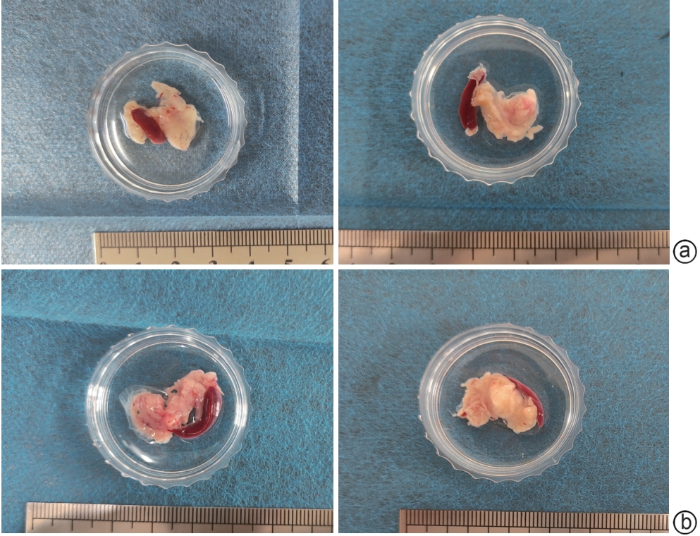
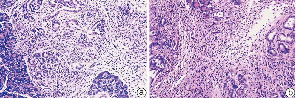
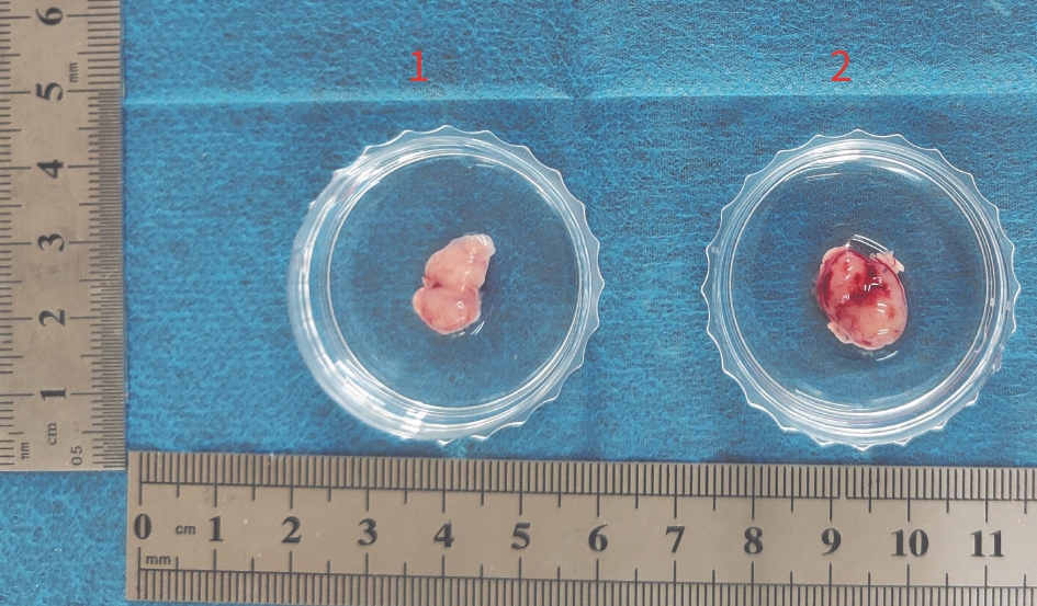
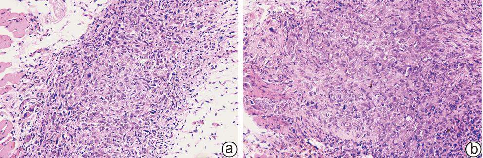
Objective To construct a novel KPC mouse model of type 2 diabetes mellitus (T2DM) comorbid with spontaneous pancreatic cancer based on the gene editing-metabolic intervention dual-driven strategy, and to compare it with traditional models. Methods A total of 14 male KPC mice were randomly divided into novel model group (T2DM-KPC group with 7 mice) and control group (KPC group with 7 mice), and 14 male BALB/c-nu nude mice were randomly divided into traditional model group (T2DM-pancreatic cancer group with 7 mice) and control group (pancreatic cancer group with 7 mice). The mice in the KPC group and the pancreatic cancer group were fed with normal diet, and those in the T2DM-KPC group and the T2DM-pancreatic cancer group were fed with a high-fat diet. After 4 weeks, the mice in the T2DM-KPC group and the T2DM-pancreatic cancer group were given intraperitoneal injection of streptozotocin. Subsequently, the mice in the KPC group and the T2DM-KPC group developed primary pancreatic tumor spontaneously over time, while those in the T2DM-pancreatic cancer group and the pancreatic cancer group were inoculated with tumor cells to form subcutaneous tumor xenograft at 2 weeks after stabilization of blood glucose. The 4 groups were observed in terms of tumor formation rate, tumor formation time, body weight, and the change in blood glucose; RNA sequencing was performed for tumors from the KPC group and the T2DM-KPC group, and then molecular subtyping was performed; HE staining, Masson staining, and immunohistochemical staining were used to assess the histopathological features and tumor microenvironment of pancreatic tumor from the T2DM-KPC group, which were compared with those of the T2DM-pancreatic cancer group. A one-way analysis of variance was used for comparison of continuous data between multiple groups, and the least significant difference t-test was used for further comparison between two groups; the Fisher’s exact test was used for comparison of categorical data between multiple groups. Results The T2DM-KPC group had a tumor formation rate of 85.71% and a tumor formation time of 104.40±2.87 days, while the T2DM-pancreatic cancer group had a tumor formation rate of 71.43% and a tumor formation time of 95.20±9.47 days, and there were no significant differences between the two groups in tumor formation rate, tumor formation time, body weight, and blood glucose (all P>0.05). Molecular subtyping showed that the model in the KPC group highly resembled the pancreatic progenitor subtype of human pancreatic ductal adenocarcinoma (PDAC), and the model in the T2DM-KPC group highly resembled the immunogenic subtype of PDAC. HE staining showed that tumor cells in the T2DM-KPC group were arranged into glandular tubular structures of varying shapes, exhibiting significant cellular atypia, and this model faithfully recapitulated the pathological features of primary pancreatic cancer and showed greater invasiveness than the KPC group. Immunohistochemical staining and Masson staining showed that compared with the T2DM-pancreatic cancer group, the T2DM-KPC group had significantly higher degrees of tumor proliferation (assessed by Ki-67 expression) and fibrosis (assessed by α-SMA and Masson) (all P<0.05), suggesting that the mouse model in the T2DM-KPC group could better recapitulate the features of hyperproliferation and pronounced desmoplasia in human pancreatic cancer. Conclusion A novel KPC mouse model of T2DM comorbid with spontaneous pancreatic cancer is successfully constructed in this study. This model can accurately mimic the histopathological architecture and stromal microenvironment of T2DM comorbid with pancreatic cancer, realize the longitudinal simulation of the progression of pancreatic tissue from intraepithelial neoplasia to invasive carcinoma and metastasis in the presence of T2DM, and support the translational research on immunotherapy, thereby providing a novel experimental carrier for in vivo studies on spontaneous pancreatic cancer in T2DM.
HUANG XH, ZHAO CH, XU YN, et al. Construction and evaluation of a novel KPC mouse model of type 2diabetes mellitus comorbid with spontaneous pancreatic cancer[J]. J Clin Hepatol, 2026, 42(4): 908-917.. doi: 10.12449/JCH260420.

 Abstract
 HTML
HTML PDF (529KB)