中文English
ISSN 1001-5256 (Print)
ISSN 2097-3497 (Online)
CN 22-1108/R
Volume 40 Issue 12
Dec.  2024
Turn off MathJax
Article Contents

Influencing factors for rebleeding after endoscopic therapy in patients with liver cirrhosis receiving secondary prevention of gastroesophageal varices

DOI: 10.12449/JCH241213
Research funding:

National Natural Science Foundation of China (81900467);

Shandong Medical and Health Science and Technology Project (202103031040)

More Information
  • Corresponding author: LIU Xiaofeng, liuxf0531@126.com (ORCID: 0000-0002-0531-8187)
  • Received Date: 2024-04-17
  • Accepted Date: 2024-06-21
  • Published Date: 2024-12-25
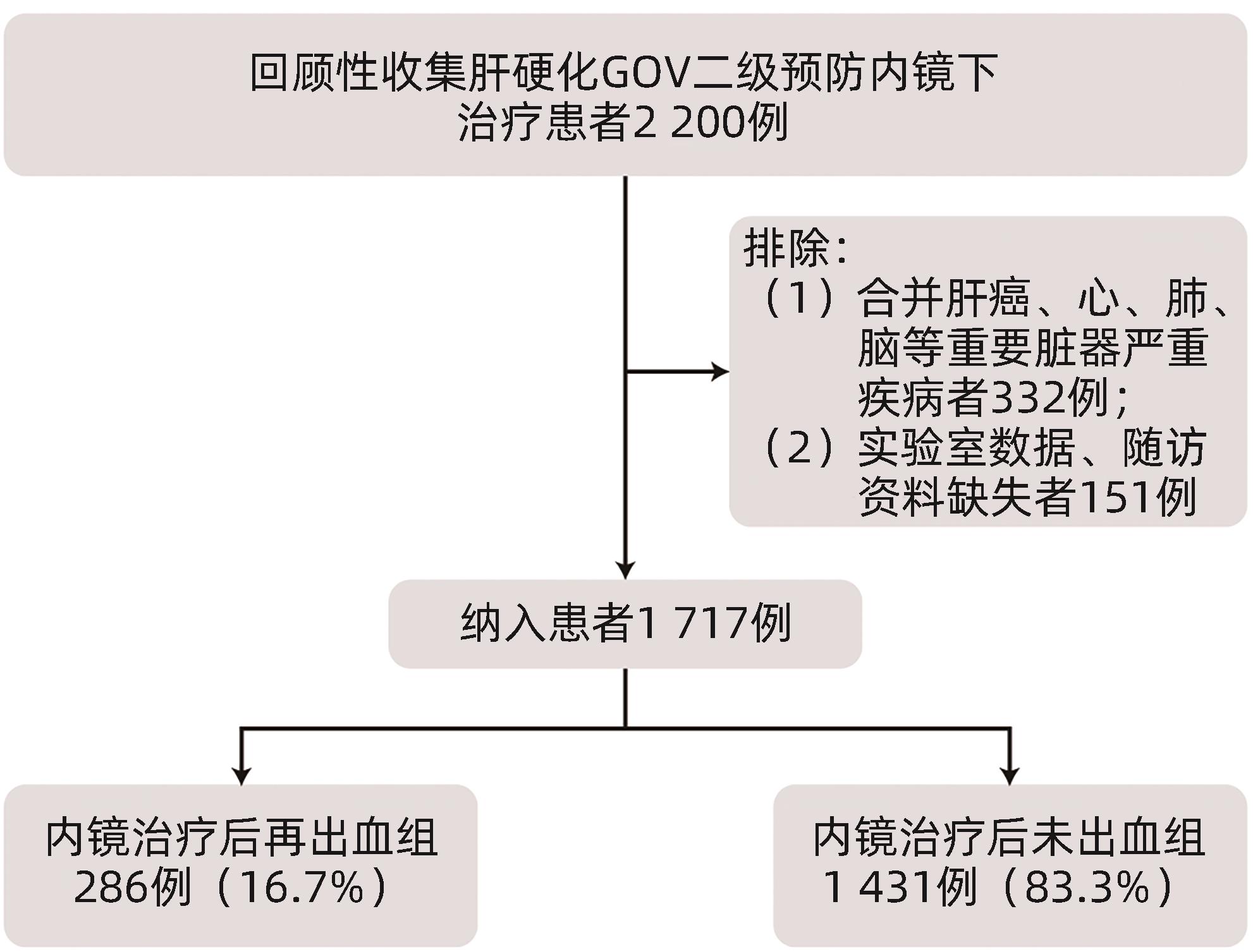
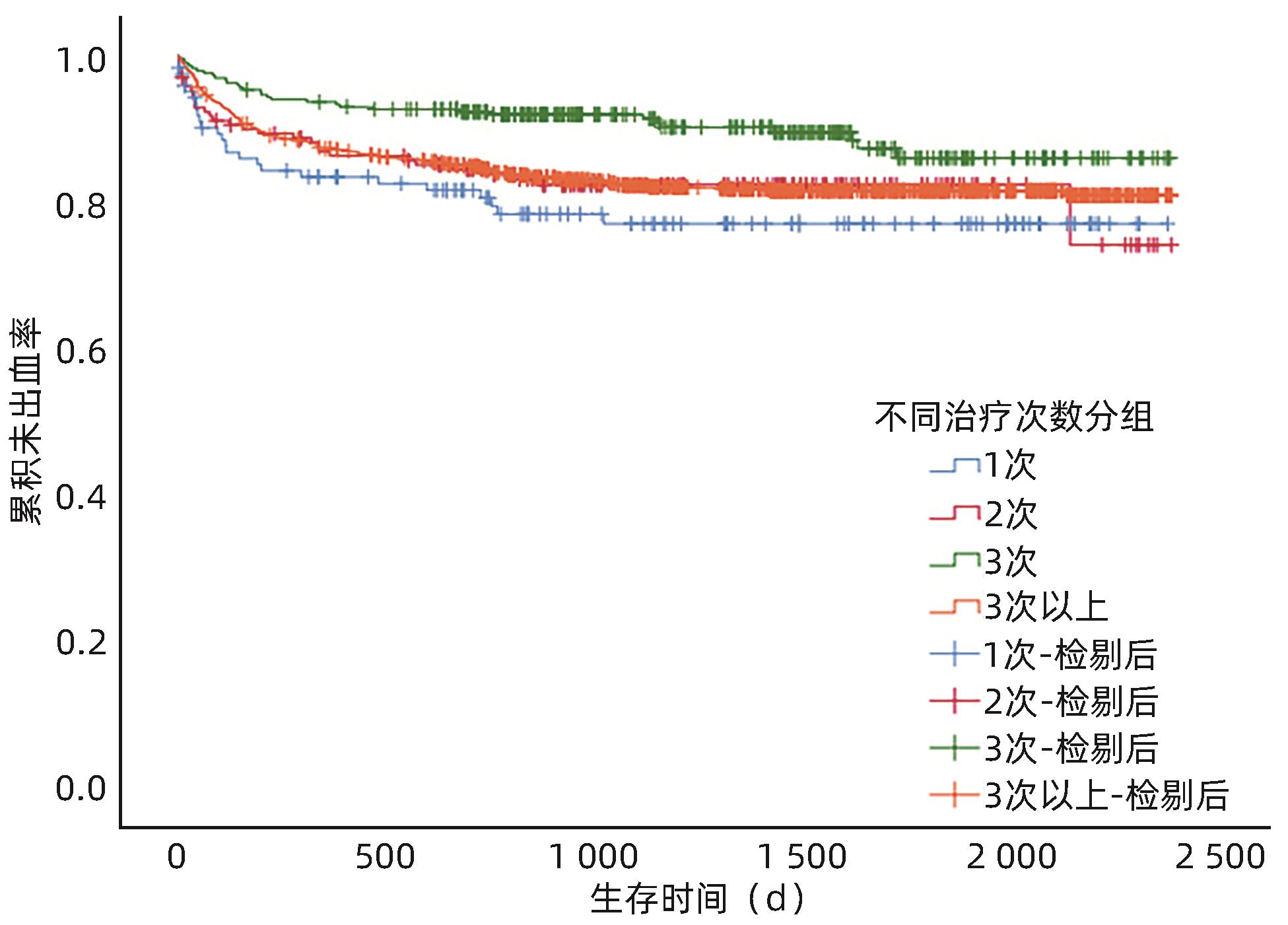
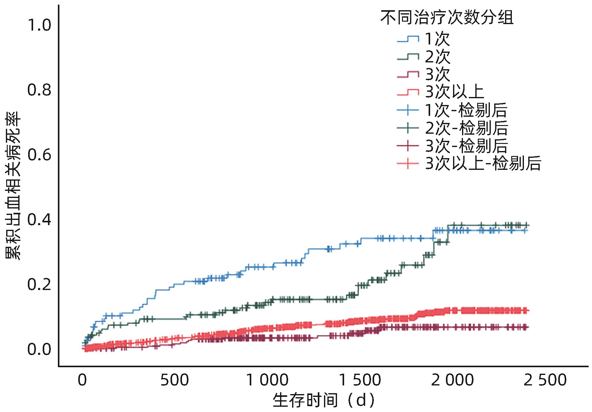
  •   Objective  To investigate the influencing factors for rebleeding after endoscopic therapy and the effect of the number of sequential treatment sessions on postoperative rebleeding in patients with liver cirrhosis receiving secondary prevention of gastroesophageal varices (GOV).  Methods  A total of 1 717 patients with liver cirrhosis who received secondary prevention of GOV and attended The 960th Hospital of the PLA Joint Logistice Support Force from January 2017 to December 2021 were enrolled, and according to the presence or absence of bleeding after endoscopic therapy, they were divided into non-bleeding group and rebleeding group. The influencing factors for rebleeding were analyzed, as well as the association between the number of endoscopic treatment sessions and rebleeding. The chi-square test was used for comparison of categorical data between groups; the independent-samples t test or the Mann-Whitney U test was used for comparison of continuous data between the two groups; the Kruskal-Wallis H test was used for comparison bertween multiple groups, and the Wilcoxon test was used for further comparison between two groups. The Cox regression model was used to investigate the influencing factors for rebleeding, and the Kaplan-Meier method was used to plot survival curves, while the Log-rank test was used for comparison between groups.  Results  Of all patients, 286 (16.7%) experienced rebleeding after endoscopic therapy, and 1 431 (83.3%) did not experience bleeding. There were significant differences between the two groups in history of smoking and drinking, etiology of liver cirrhosis, hemoglobin (Hb), prothrombin time (PT), prothrombin activity (PTA), international normalized ratio (INR), albumin (Alb), fasting blood glucose, blood urea nitrogen, Child-Pugh class, aspartate aminotransferase-to-platelet ratio index (APRI) score, albumin-bilirubin (ALBI) score, use of non-selective beta-blocker (NSBB) before surgery, treatment modality, type of varices, and maximal varicose vein diameter (all P<0.05). The univariate Cox regression analysis showed that in the patients with liver cirrhosis who received secondary prevention of GOV, rebleeding was associated with history of smoking and drinking, etiology of liver cirrhosis, use of NSBB before surgery, treatment modality, maximal varicose vein diameter, Hb, platelet count, PT, PTA, INR, Alb, total bilirubin (TBil), alkaline phosphatase (ALP), gamma-glutamyl transpeptidase, blood glucose, Child-Pugh class, and ALBI score (all P<0.05). The multivariate Cox regression analysis showed that Hb (hazard ratio [HR]=0.989, 95% confidence interval [CI]: 0.983‍ ‍—‍ ‍0.994, P<0.001), TBil (HR=1.020, 95%CI: 1.006‍ ‍—‍ ‍1.034, P=0.005), Alb (HR=0.868, 95%CI: 0.758‍ ‍—‍ ‍0.994, P=0.041), treatment modality (sclerosing agent: HR=2.158, 95%CI: 1.342‍ ‍—‍ ‍3.470, P=0.002; tissue adhesive: HR=2.709, 95%CI: 1.343‍ ‍—‍ ‍5.462, P=0.005; ligation+sclerosing agent: HR=3.181, 95%CI: 1.522‍ ‍—‍ ‍6.645, P=0.002; sclerosing agent+tissue adhesive: HR=1.851, 95%CI: 1.100‍ ‍—‍ ‍3.113, P=0.020), ALP (HR=1.003, 95%CI: 1.001‍ ‍—‍ ‍1.004, P=0.002), and maximal varicose vein diameter (HR=1.346, 95%CI: 1.119‍ ‍—‍ ‍1.618, P=0.002) were independent influencing factors for rebleeding after endoscopic therapy. Comparison of rebleeding rate after different numbers of sequential treatment sessions showed that the patients treated for three sessions had a significantly lower rebleeding rate than those treated for one or two sessions (χ2=8.643 and 5.277, P=0.003 and 0.022). The survival analysis showed that with the increase in the number of treatment sessions, there was a significantly longer interval between rebleeding (P=0.006) and a significantly lower mortality rate (P<0.001).  Conclusion  The levels of TBil, ALP, Hb, and Alb on admission, endoscopic treatment modality, and maximal varicose vein diameter were the main predictive factors for rebleeding after endoscopic therapy for GOV in liver cirrhosis, and such predictive factors should be closely monitored in clinical practice. Regular endoscopic therapy can reduce the rebleeding and mortality rates of patients with liver cirrhosis and GOV and prolonmg the interval between rebleeding.


  • loading
  • [1]
    IWAKIRI Y, TREBICKA J. Portal hypertension in cirrhosis: Pathophysiological mechanisms and therapy[J]. JHEP Rep, 2021, 3( 4): 100316. DOI: 10.1016/j.jhepr.2021.100316.
    [2]
    LUNOVA M, FRANKOVA S, GOTTFRIEDOVA H, et al. Portal hypertension is the main driver of liver stiffness in advanced liver cirrhosis[J]. Physiol Res, 2021, 70( 4): 563- 577. DOI: 10.33549/physiolres.934626.
    [3]
    KIM KR, JUN CH, CHO KM, et al. Can proton pump inhibitors reduce rebleeding following Histoacryl sclerotherapy for gastric variceal hemorrhage?[J]. Korean J Intern Med, 2015, 30( 5): 593- 601. DOI: 10.3904/kjim.2015.30.5.593.
    [4]
    ZHAO Y. Application value of endoscopic ultrasonography in precise devascularization of esophageal and gastric varices caused by portal hypertension[D]. Kaifeng: Henan University, 2020.

    赵一. 超声内镜在门脉高压所致食管胃静脉曲张精准断流术中的应用价值[D]. 开封: 河南大学, 2020.
    [5]
    MA L, TSENG Y, LUO T, et al. Risk stratification for secondary prophylaxis of gastric varices due to portal hypertension[J]. Dig Liver Dis, 2019, 51( 12): 1678- 1684. DOI: 10.1016/j.dld.2019.05.020.
    [6]
    LIU LM, ZHANG CQ. Advances in high risk factors of rebleeding after endoscopic treatment of esophagogastric variceal bleeding[J/CD]. Chin J Dig Med Imageology Electron Ed, 2022, 12( 4): 236- 240. DOI: 10.3877/cma.j.issn.2095-2015.2022.04.010.

    刘黎明, 张春清. 食管-胃底静脉曲张破裂出血内镜治疗后再出血高危因素研究进展[J/CD]. 中华消化病与影像杂志(电子版), 2022, 12( 4): 236- 240. DOI: 10.3877/cma.j.issn.2095-2015.2022.04.010.
    [7]
    GUO YL, XU BH, LIU X, et al. Risk factors analysis of early rebleeding after endoscopic treatment of esophageal varices[J]. Chin J Dig Endosc, 2018, 35( 2): 89- 93. DOI: 10.3760/cma.j.issn.1007-5232.2018.02.003.

    郭雅丽, 徐宝宏, 刘贤, 等. 食管静脉曲张内镜治疗术后早期再出血的危险因素分析[J]. 中华消化内镜杂志, 2018, 35( 2): 89- 93. DOI: 10.3760/cma.j.issn.1007-5232.2018.02.003.
    [8]
    GIRI S, SUNDARAM S, JEARTH V, et al. Predictors of early bleeding after endoscopic variceal ligation for esophageal varices: A systematic review and meta-analysis[J]. Clin Exp Hepatol, 2022, 8( 4): 267- 277. DOI: 10.5114/ceh.2022.123096.
    [9]
    ZHANG N, PENG CY, ZHANG F, et al. Clinical value of regular endoscopic treatment for esophageal and gastric variceal bleeding in patients with liver cirrhosis[J]. Chin J Dig Endosc, 2022, 39( 5): 384- 387. DOI: 10.3760/cma.j.cn321463-20210113-00760.

    张娜, 彭春艳, 张峰, 等. 肝硬化食管胃静脉曲张破裂出血行规律内镜下治疗的临床价值[J]. 中华消化内镜杂志, 2022, 39( 5): 384- 387. DOI: 10.3760/cma.j.cn321463-20210113-00760.
    [10]
    Chinese Society of Hepatology, Chinese Society of Gastroenterology, Chinese Society of Digestive Endoscopology of Chinese Medical Association. Guidelines on the management of esophagogastric variceal bleeding in cirrhotic portal hypertension[J]. J Clin Hepatol, 2023, 39( 3): 527- 538.

    中华医学会肝病学分会, 中华医学会消化病学分会, 中华医学会消化内镜学分会. 肝硬化门静脉高压食管胃静脉曲张出血的防治指南[J]. 临床肝胆病杂志, 2023, 39( 3): 527- 538.
    [11]
    Esophagageal and Gastric Varices Group of Digestive Endoscopy Branch of Chinese Medical Association. Tentative guidelines for endoscopic diagnosis and treatment of varicosity and variceal bleeding in digestive tract(2009)[J]. Chin J Dig Endosc, 2010, 27( 1): 1- 4. DOI: 10.3760/cma.j.issn.1007-5232.2010.01.001.

    中华医学会消化内镜学分会食管胃静脉曲张学组. 消化道静脉曲张及出血的内镜诊断和治疗规范试行方案(2009年)[J]. 中华消化内镜杂志, 2010, 27( 1): 1- 4. DOI: 10.3760/cma.j.issn.1007-5232.2010.01.001.
    [12]
    MOSTAFA EF, MOHAMMAD AN. Incidence and predictors of rebleeding after band ligation of oesophageal varices[J]. Arab J Gastroenterol, 2014, 15( 3-4): 135- 141. DOI: 10.1016/j.ajg.2014.10.002.
    [13]
    HUANG C, SU N, WU SL, et al. Risk factors of early rebleeding after endoscopic variceal ligation[J]. J Clin Intern Med, 2019, 36( 4): 264- 266. DOI: 10.3969/j.issn.1001-9057.2019.04.014.

    黄超, 苏宁, 吴胜兰, 等. 内镜下食管静脉曲张套扎术后早期再出血的危险因素分析[J]. 临床内科杂志, 2019, 36( 4): 264- 266. DOI: 10.3969/j.issn.1001-9057.2019.04.014.
    [14]
    ZHANG Y. Meta-analysis of risk factors of early rebleeding after endoscopic variceal ligation[D]. Changchun: Jilin University, 2023.

    张赟. 食管静脉曲张内镜下套扎术后早期再出血危险因素的Meta分析[D]. 长春: 吉林大学, 2023.
    [15]
    DROLZ A, SCHRAMM C, SEIZ O, et al. Risk factors associated with bleeding after prophylactic endoscopic variceal ligation in cirrhosis[J]. Endoscopy, 2021, 53( 3): 226- 234. DOI: 10.1055/a-1214-5355.
    [16]
    JIMÉNEZ ROSALES R, MARTÍNEZ-CARA JG, VADILLO-CALLES F, et al. Analysis of rebleeding in cases of an upper gastrointestinal bleed in a single center series[J]. Rev Esp Enferm Dig, 2019, 111( 3): 189- 192. DOI: 10.17235/reed.2018.5702/2018.
    [17]
    ZHONG BH, WANG JP, LIN JH, et al. Risk assessment and prevention of gastroesophageal varices and variceal hemorrhage in cirrhosis[J]. Chin J Pract Intern Med, 2013, 33( 9): 690- 693.

    钟碧慧, 王锦萍, 林菁华, 等. 肝硬化食管胃底静脉曲张破裂出血风险评估及预防策略[J]. 中国实用内科杂志, 2013, 33( 9): 690- 693.
    [18]
    ZHAO MY, WU XR, LI HX, et al. Study on correlation between serum 25-hydroxyvitamin D3 level and esophageal variceal bleeding in cirrhotic patients[J]. Chin J Hepatol, 2019, 27( 5): 358- 362. DOI: 10.3760/cma.j.issn.1007-3418.2019.05.007.

    赵明远, 武希润, 李红霞, 等. 血清25羟维生素D3的水平与肝硬化食管静脉曲张破裂出血的相关性[J]. 中华肝脏病杂志, 2019, 27( 5): 358- 362. DOI: 10.3760/cma.j.issn.1007-3418.2019.05.007.
    [19]
    ZHANG Q, SHI K, WANG XB. Developing the prediction model of esophagogastric variceal rebleeding in patients with liver cirrhosis based on artificial neural network[J]. J Clin Hepatol, 2022, 38( 11): 2493- 2498. DOI: 10.3969/j.issn.1001-5256.2022.11.011.

    张群, 时克, 王宪波. 肝硬化食管胃底静脉曲张破裂出血患者再出血预测模型的建立[J]. 临床肝胆病杂志, 2022, 38( 11): 2493- 2498. DOI: 10.3969/j.issn.1001-5256.2022.11.011.
    [20]
    YUAN XY, HUANG YQ, JIANG M. Influence of hyponatremia on the condition and prognosis of patients with liver cirrhosis[J]. J China Med Univ, 2019, 48( 11): 1003- 1006. DOI: 10.12007/j.issn.0258-4646.2019.11.010.

    袁晓艳, 黄颖秋, 姜敏. 低钠血症对肝硬化患者病情和预后的影响[J]. 中国医科大学学报, 2019, 48( 11): 1003- 1006. DOI: 10.12007/j.issn.0258-4646.2019.11.010.
    [21]
    ZHANG XB, LI HP, LU CL, et al. Relationship between serum sodium level and liver function and complications in patients with decompensated cirrhosis[J]. Anhui Med J, 2019, 40( 6): 673- 676. DOI: 10.3969/j.issn.1000-0399.2019.06.022.

    张小宝, 李华平, 陆春雷, 等. 失代偿期肝硬化患者血清钠水平与肝功能及并发症的关系[J]. 安徽医学, 2019, 40( 6): 673- 676. DOI: 10.3969/j.issn.1000-0399.2019.06.022.
    [22]
    GUO H, ZHANG M, ZHANG N, et al. Number of endoscopic sessions to eradicate varices identifies high risk of rebleeding in cirrhotic patients[J]. BMC Gastroenterol, 2022, 22( 1): 213. DOI: 10.1186/s12876-022-02283-0.
    [23]
    RÍOS E, SIERRALTA A, ABARZÚA M, et al. Comparison of band ligation with sclerotherapy for the treatment of bleeding esophageal varices[J]. Rev Med Chil, 2012, 140( 6): 713- 718. DOI: 10.4067/s0034-98872012000600003.
    [24]
    YANG YJ. Comparative analysis of ligation, sclerosing agent and drug therapy in patients with esophageal varices bleeding[J]. Guide China Med, 2011, 9( 19): 288- 289.

    杨悦军. 食管静脉曲张破裂出血患者套扎术、硬化剂、药物治疗方法比较分析[J]. 中国医药指南, 2011, 9( 19): 288- 289.
    [25]
    XIANG Y, WANG X, MEI XC, et al. Comparative analysis of endoscopic ligation and tissue adhesive injection for tortuous gastric varices[J]. Chin J Dig Endosc, 2021, 38( 11): 901- 906. DOI: 10.3760/cma.j.cn321463-20210223-00123.

    项艺, 王曦, 梅雪灿, 等. 蔓状隆起型胃底静脉曲张内镜下套扎与组织胶注射治疗的对比分析[J]. 中华消化内镜杂志, 2021, 38( 11): 901- 906. DOI: 10.3760/cma.j.cn321463-20210223-00123.
    [26]
    HUANG XQ, WU L, JIANG SY, et al. Clinical evaluation of different modalities in emergency endoscopic treatment of esophageal variceal bleeding[J]. J Chin Physician, 2022, 24( 5): 653- 657. DOI: 10.3760/cma.j.cn431274-20220419-00361.

    黄晓铨, 吴玲, 姜思雨, 等. 食管静脉曲张出血的急诊内镜治疗选择与疗效评价[J]. 中国医师杂志, 2022, 24( 5): 653- 657. DOI: 10.3760/cma.j.cn431274-20220419-00361.
    [27]
    SONG ZM, CHEN YM, YANG M. Comparison of clinical evaluation of endoscopic therapy for esophageal variceal bleeding[J]. Chin J Curr Adv Gen Surg, 2010, 13( 3): 199- 201. DOI: 10.3969/j.issn.1009-9905.2010.03.009.

    宋振梅, 陈艳明, 杨猛. 内镜下硬化与套扎治疗食管静脉曲张破裂出血疗效比较[J]. 中国现代普通外科进展, 2010, 13( 3): 199- 201. DOI: 10.3969/j.issn.1009-9905.2010.03.009.
  • 加载中

Catalog

    通讯作者: 陈斌, bchen63@163.com
    • 1. 

      沈阳化工大学材料科学与工程学院 沈阳 110142

    1. 本站搜索
    2. 百度学术搜索
    3. 万方数据库搜索
    4. CNKI搜索

    Figures(3)  / Tables(5)

    Article Metrics

    Article views (119) PDF downloads(18) Cited by()
    Proportional views
    Related

    /

    DownLoad:  Full-Size Img  PowerPoint
    Return
    Return