中文|English
ISSN 1001-5256 (Print)
ISSN 2097-3497 (Online)
CN 22-1108/R
Volume 42 Issue 9
Sep.  2026
Turn off MathJax
Article Contents

Protective effect and mechanism of trophoblast-derived mesenchymal stem cells against liver injury in a mouse model of autoimmune hepatitis

DOI: 10.12449/JCH260916
Research funding:

National Natural Science Foundation of China (82270626);

China Mega-Project for Infectious Diseases (2017ZX10203202);

China Mega-Project for Infectious Diseases (2013ZX10002005);

Project of Beijing Science and Technology Committee (Z191100007619037);

Sanming Project of Medicine in Shenzhen (SZSM202311033);

Peking University Medicine Sailing Program for Young Scholars’ Scientific & Technological Innovation (BMU2025YFJHPY010)

More Information
  • Corresponding author: Wang Guiqiang, john131212@126.com (ORCID: 0000-0003-0515-7974)
  • Received Date: 2026-03-22
  • Accepted Date: 2026-04-16
  • Published Date: 2026-09-25
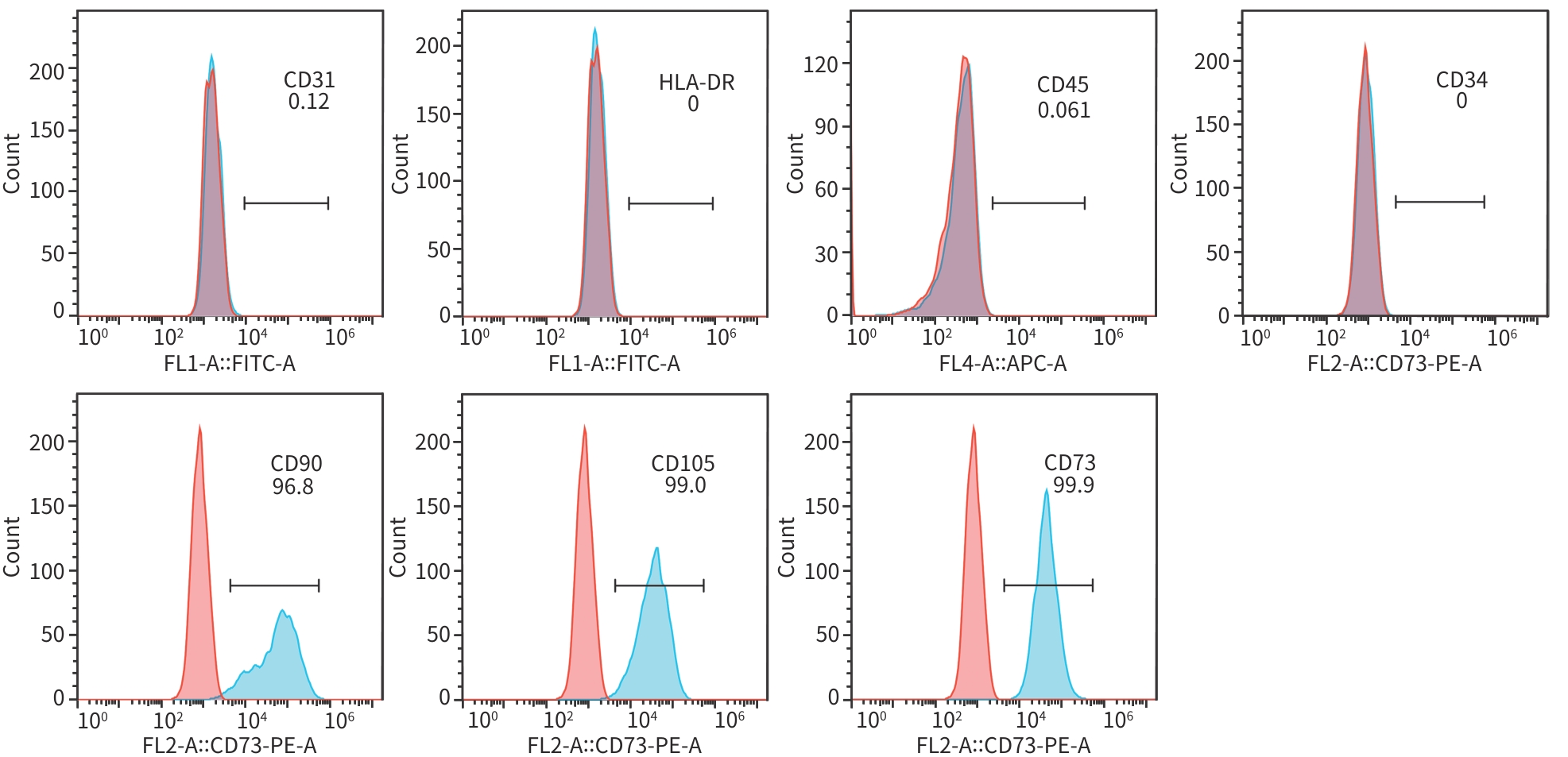
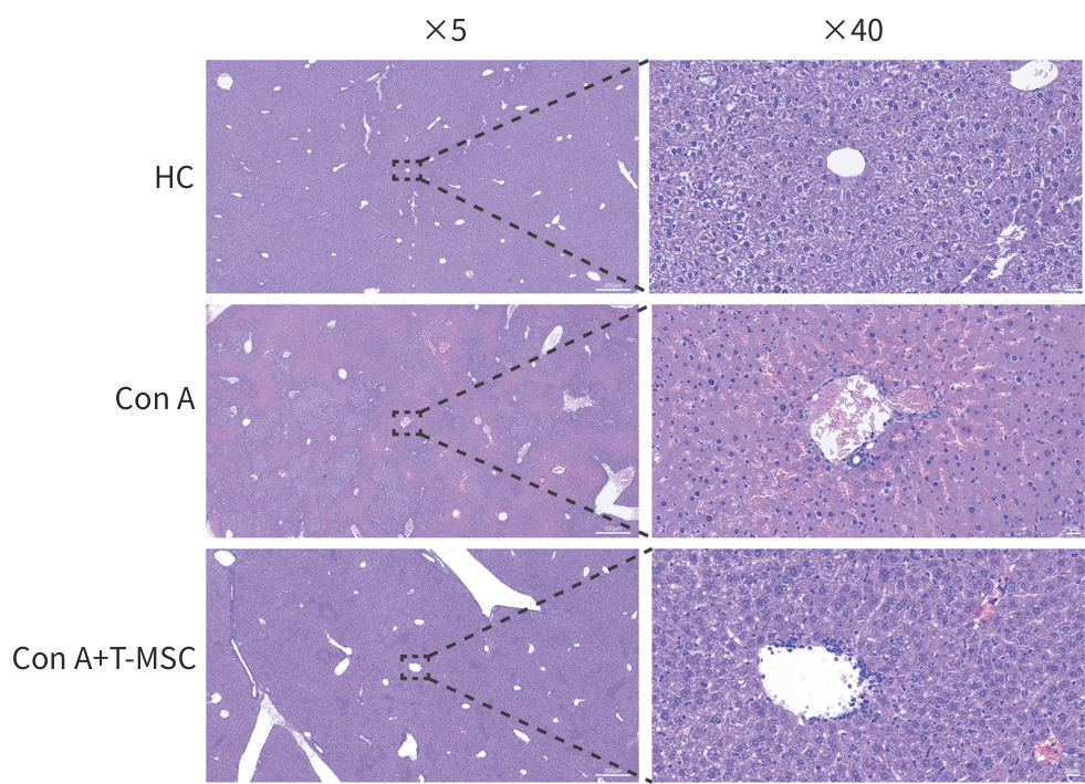
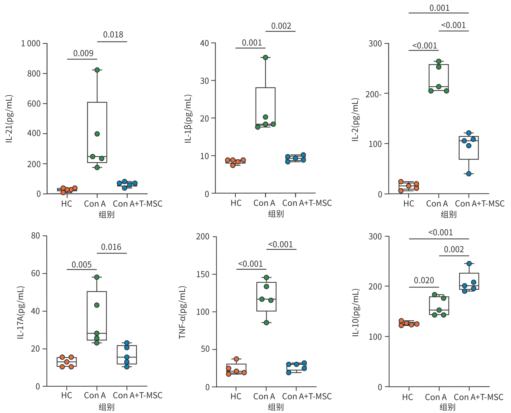
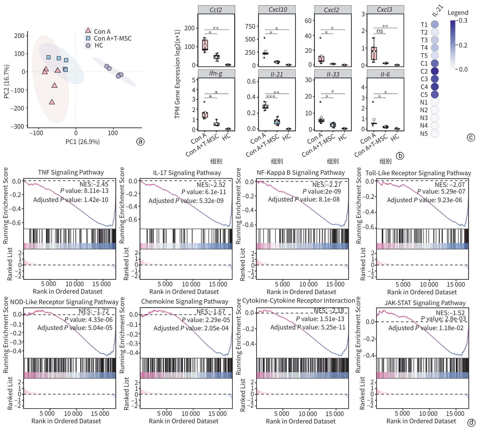
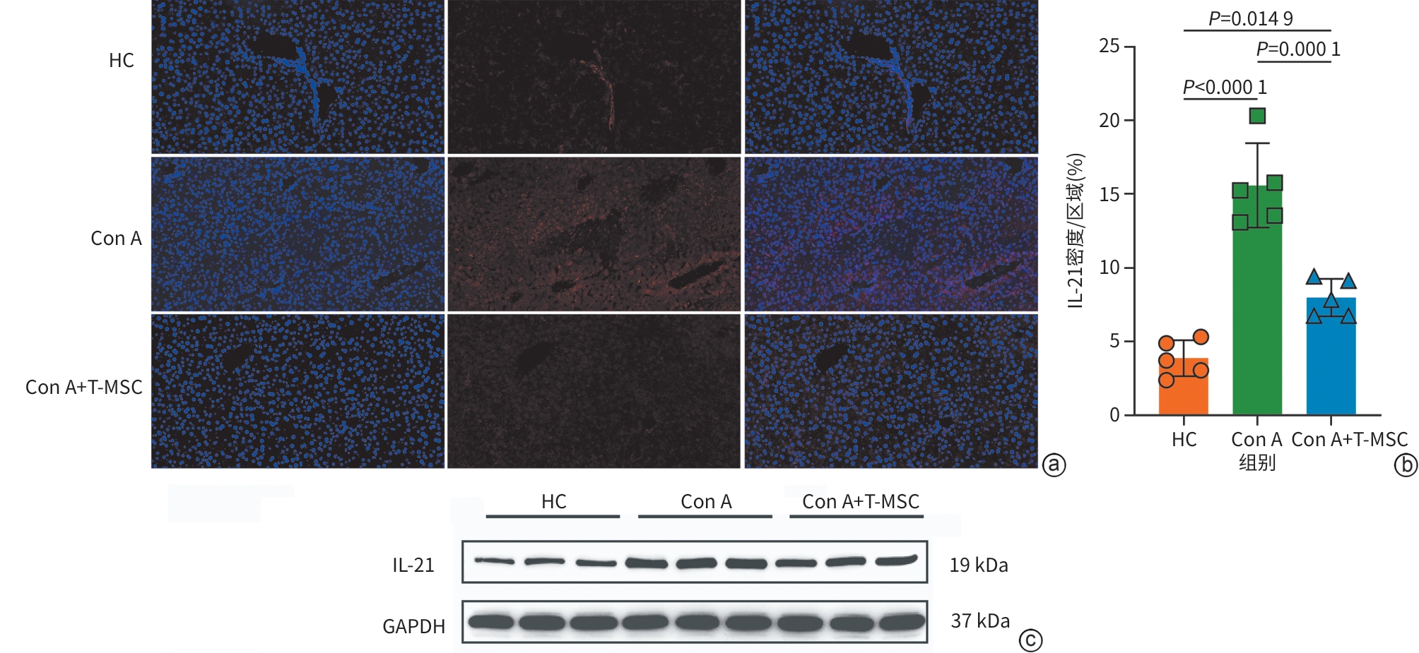
  •   Objective  To investigate the effect of trophoblast-derived mesenchymal stem cell (T-MSC) on concanavalin A (Con A)-induced immune-mediated liver injury in mice, as well as its association with the changes in inflammatory response.  Methods  Morphological assessment, flow cytometry, and tri-lineage differentiation assay were used to observe the biological characteristics of T-MSC. In the pilot experiment, mice were divided into PBS group, T-MSC monotherapy group, and Con A groups at a concentration of 10, 15, or 20 mg/kg to evaluate the safety of T-MSC monotherapy on liver function and determine the appropriate dose for modeling. In the formal experiment, mice were divided into healthy control group (HC group), Con A group, and Con A+T-MSC group, with 5 mice in each group. The mice were given injection of Con A (15 mg/kg) via the caudal vein to establish a mouse model of immune-mediated liver injury, while the mice in the T-MSC monotherapy group were given transfusion of 1×10⁶ T-MSCs via the caudal vein at 3 hours after modeling, and samples were collected 12 hours later. The serum levels of alanine aminotransferase (ALT), aspartate aminotransferase (AST), and lactate dehydrogenase (LDH) were measured; liver histopathological changes were observed, and Ishak inflammatory activity score and inflammatory necrosis area were assessed; the serum levels of inflammatory cytokines were measured; transcriptome sequencing, immunofluorescence assay, and Western blot were used to measure the changes in inflammation-related molecules in liver tissue. The Shapiro-Wilk test was used to assess the normality of continuous data, and the homogeneity of variance test was also performed; a one-way analysis of variance was used for comparison of normally distributed data with homogeneity of variance between multiple groups, and the Tukey’s post-hoc test was used for further comparison between two groups; the Kruskal-Wallis H test was used for comparison of non-normally distributed data with heterogeneity of variance between multiple groups, and the Wilcoxon test was used for further comparison between two groups.  Results  T-MSC had a long spindle shape, showed the characteristics of fibroblast-like morphology and adherent growth, and possessed the osteogenic, chondrogenic, and adipogenic differentiation abilities. Flow cytometry showed high expression levels of CD90, CD105, and CD73 in T-MSC, with a positive rate of 96.8%, 99.0%, and 99.9%, respectively, while there was low or no expression of CD31, human leukocyte antigen DR, CD45, and CD34, with a positive rate of 0.120%, 0, 0.061%, and 0, respectively. Administration of T-MSC alone did not cause obvious abnormalities in ALT, AST, or LDH. Con A at the concentrations of 10, 15, and 20 mg/kg all increased the serum levels of biochemical parameters for liver injury in a dose-dependent manner, with mortality observed in the 20 mg/kg group. In the formal experiment, compared with the HC group, the Con A group had significant increases in the serum levels of ALT, AST, and LDH (P<0.01), and after T-MSC treatment, there were significant reductions in these markers compared with the Con A group (P<0.01). HE staining showed that the Con A group had disordered structure of the liver, accompanied by marked inflammatory cell infiltration and focal/patchy necrosis, and these pathological changes were alleviated after T-MSC treatment. Compared with the HC group, the Con A group had significant increases in Ishak score and the percentage of inflammatory necrosis area (P<0.001), while there were significant reductions in these indicators after T-MSC treatment compared with the Con A group (P<0.01). ELISA showed that compared with the HC group, the Con A group had significant increases in the serum levels of interleukin-21 (IL-21), interleukin-1β (IL-1β), interleukin-2 (IL-2), interleukin-17A (IL-17A), and tumor necrosis factor-α (TNF-α) (P<0.05), and there were significant reductions in these markers after T-MSC treatment compared with the Con A group (P<0.05), with no significant differences in IL-21, IL-1β, IL-17A, and TNF-α compared with the HC group (P>0.05); although there was a significant reduction in the level of IL-2, the Con A+T-MSC group still had a significantly higher level of IL-2 than the HC group (P=0.001). In contrast, compared with the HC group, the Con A group had a significant increase in IL-10 (P<0.05), with a further increase after T-MSC treatment (P<0.05). The transcriptome analysis showed that T-MSC treatment downregulated the expression of the inflammation- and chemokine-related genes including Ccl2, Cxcl10, Il-21, and Il-6 and inhibited the inflammation-related pathways such as TNF, IL-17, nuclear factor-κB, and JAK-STAT. Immunofluorescence assay showed that compared with the HC group, the Con A group had a significant increase in the expression of IL-21 in the liver (P<0.001), and there was a significant reduction in IL-21 after T-MSC treatment compared with the Con A group (P<0.001); the results of Western blot showed the same trend.  Conclusion  T-MSC can alleviate Con A-induced immune-mediated liver injury in mice, as evidenced by the improvement in liver biochemical parameters, the alleviation of histopathological damage, and the overall downregulation of inflammation-related molecules. T-MSC exerts marked anti-inflammatory and immunomodulatory effects in this model, and IL-21 shows a significant change in inflammatory response and can be used as a candidate molecule for further investigation.


  • loading
  • [1]
    European Association for the Study of the Liver. EASL Clinical Practice Guidelines on the management of autoimmune hepatitis[J]. J Hepatol, 2025, 83( 2): 453- 501. DOI: 10.1016/j.jhep.2025.03.017.
    [2]
    Mack C L, Adams D, Assis D N, et al. Diagnosis and management of autoimmune hepatitis in adults and children: 2019 practice guidance and guidelines from the American Association for the Study of Liver Diseases[J]. Hepatology, 2020, 72( 2): 671- 722. DOI: 10.1002/hep.31065.
    [3]
    Wang G Q, Tanaka A, Zhao H, et al. The Asian Pacific Association for the Study of the Liver clinical practice guidance: The diagnosis and management of patients with autoimmune hepatitis[J]. Hepatol Int, 2021, 15( 2): 223- 257. DOI: 10.1007/s12072-021-10170-1.
    [4]
    Gleeson D, Bornand R, Brownlee A, et al. British Society of Gastroenterology guidelines for diagnosis and management of autoimmune hepatitis[J]. Gut, 2025, 74( 9): 1364- 1409. DOI: 10.1136/gutjnl-2024-333171.
    [5]
    Korean Association for the Study of the Liver(KASL). KASL clinical practice guidelines for management of autoimmune hepatitis 2022[J]. Clin Mol Hepatol, 2023, 29( 3): 542- 592. DOI: 10.3350/cmh.2023.0087.
    [6]
    Ohira H, Takahashi A, Zeniya M, et al. Clinical practice guidelines for autoimmune hepatitis[J]. Hepatol Res, 2022, 52( 7): 571- 585. DOI: 10.1111/hepr.13776.
    [7]
    Zhou Y M, Tang R Q, Gershwin M E, et al. Autoimmune hepatitis: Pathophysiology[J]. Clin Liver Dis, 2024, 28( 1): 15- 35. DOI: 10.1016/j.cld.2023.06.003.
    [8]
    Dalekos G N, Gatselis N K. Autoimmune serology testing in clinical practice: An updated roadmap for the diagnosis of autoimmune hepatitis[J]. Eur J Intern Med, 2023, 108: 9- 17. DOI: 10.1016/j.ejim.2022.11.013.
    [9]
    Song N, Scholtemeijer M, Shah K. Mesenchymal stem cell immunomodulation: Mechanisms and therapeutic potential[J]. Trends Pharmacol Sci, 2020, 41( 9): 653- 664. DOI: 10.1016/j.tips.2020.06.009.
    [10]
    Chen Y, Chen S, Liu L Y, et al. Mesenchymal stem cells ameliorate experimental autoimmune hepatitis by activation of the programmed death 1 pathway[J]. Immunol Lett, 2014, 162( 2 Pt B): 222- 228. DOI: 10.1016/j.imlet.2014.10.021.
    [11]
    Wang H X, Liu M, Weng S Y, et al. Immune mechanisms of Concanavalin A model of autoimmune hepatitis[J]. World J Gastroenterol, 2012, 18( 2): 119- 125. DOI: 10.3748/wjg.v18.i2.119.
    [12]
    Tiegs G, Hentschel J, Wendel A. A T cell-dependent experimental liver injury in mice inducible by concanavalin A[J]. J Clin Invest, 1992, 90( 1): 196- 203. DOI: 10.1172/JCI115836.
    [13]
    Fei M M, Xie Q, Zou Y, et al. Alpha-lipoic acid protects mice against concanavalin A-induced hepatitis by modulating cytokine secretion and reducing reactive oxygen species generation[J]. Int Immunopharmacol, 2016, 35: 53- 60. DOI: 10.1016/j.intimp.2016.03.023.
    [14]
    He R, Wang L P, Zhu J L, et al. Methane-rich saline protects against concanavalin A-induced autoimmune hepatitis in mice through anti-inflammatory and anti-oxidative pathways[J]. Biochem Biophys Res Commun, 2016, 470( 1): 22- 28. DOI: 10.1016/j.bbrc.2015.12.080.
    [15]
    Kimura N, Yamagiwa S, Sugano T, et al. Possible involvement of chemokine C-C receptor 7- programmed cell death-1+ follicular helper T-cell subset in the pathogenesis of autoimmune hepatitis[J]. J Gastroenterol Hepatol, 2018, 33( 1): 298- 306. DOI: 10.1111/jgh.13844.
    [16]
    Biewenga M, Heidt S, Vergunst M, et al. B-cell activating factor and IL-21 levels predict treatment response in autoimmune hepatitis[J]. JHEP Rep, 2022, 4( 5): 100460. DOI: 10.1016/j.jhepr.2022.100460.
    [17]
    Aoki N, Kido M, Iwamoto S, et al. Dysregulated generation of follicular helper T cells in the spleen triggers fatal autoimmune hepatitis in mice[J]. Gastroenterology, 2011, 140( 4): 1322-1333.e1-5. DOI: 10.1053/j.gastro.2011.01.002.
    [18]
    Thomas-Dupont P, Remes-Troche J M, Izaguirre-Hernández I Y, et al. Elevated circulating levels of IL-21 and IL-22 define a cytokine signature profile in type 2 autoimmune hepatitis patients[J]. Ann Hepatol, 2016, 15( 4): 550- 558. DOI: 10.5604/16652681.1203152.
    [19]
    Spolski R, Leonard W J. Interleukin-21: Basic biology and implications for cancer and autoimmunity[J]. Annu Rev Immunol, 2008, 26: 57- 79. DOI: 10.1146/annurev.immunol.26.021607.090316.
  • 加载中

Catalog

    通讯作者: 陈斌, bchen63@163.com
    • 1. 

      沈阳化工大学材料科学与工程学院 沈阳 110142

    1. 本站搜索
    2. 百度学术搜索
    3. 万方数据库搜索
    4. CNKI搜索

    Figures(6)  / Tables(2)

    Article Metrics

    Article views (105) PDF downloads(14) Cited by()
    Proportional views
    Related

    /

    DownLoad:  Full-Size Img  PowerPoint
    Return
    Return