中文English
ISSN 1001-5256 (Print)
ISSN 2097-3497 (Online)
CN 22-1108/R

Research advances in exosomes in diagnosis of pancreatic cancer

DOI: 10.3969/j.issn.1001-5256.2019.05.053
Research funding:


  • Received Date: 2019-02-12
  • Published Date: 2019-05-20
  • Pancreatic cancer is a digestive system malignancy with high mortality. Exosomes are membranous vesicles with a diameter of 30-200 nm, which are released by various cells into the extracellular matrix. Exosomes can carry various molecules such as RNA, DNA, proteins, and lipids and bind to target cells to mediate the communication between cells and regulate the physiological function of cells. Recent studies have shown that exosomes play an important role in the development and progression of pancreatic cancer and can help with the early diagnosis of pancreatic cancer, and exosomes are expected to become a new biomarker for the diagnosis of pancreatic cancer. This article reviews the research advances in the role of exosomes in the early diagnosis of pancreatic cancer.


  • 肝细胞癌(HCC)是我国最常见的恶性肿瘤之一。国家癌症中心最新数据显示,我国每年新发肝癌约37万人,约32.6万人死于肝癌,其中94.7%与慢性HBV感染有关[1-2]。早期发现、早期诊断和早期治疗是改善原发性肝癌预后的关键。然而目前尚缺乏原发性肝癌高敏感度和高特异度的诊断指标。自噬是细胞自我吞噬胞内长寿蛋白或受损细胞器并通过溶酶体途径进行的分解代谢过程,从而维持细胞稳态[3]。近年研究[4-7]显示自噬在HBV感染和HCC的发生发展起重要作用。但目前关于自噬相关蛋白在HBV相关HCC(HBV-HCC)患者血清中的表达及意义鲜有报道。本研究通过分析HBV-HCC、非HBV相关HCC(nonHBV-HCC)、慢性乙型肝炎(CHB)和健康对照者血清自噬相关蛋白7(ATG7)的表达水平,探讨ATG7对HBV-HCC的诊断价值。

    选取2018年6月—2020年12月于本院住院的HCC患者89例。HCC的诊断标准符合《原发性肝癌诊疗规范(2017年版)》[8]并经病理确认。排除标准如下:病理类型为肝内胆管癌或HCC-肝内胆管癌混合型者;已接受过肝移植术、局部消融、肝动脉化疗栓塞术、放化疗等抗肿瘤治疗者;继发性HCC患者;同时伴有其他肿瘤患者。CHB诊断符合《慢性乙型肝炎防治指南(2019年版)》[9]。根据血清是否检出HBsAg和/或HBV DNA分为HBV-HCC组及nonHBV-HCC组。选取同期50例CHB患者(CHB组)和20例健康体检者作为对照组(HC组)。

    收集研究对象性别和年龄,总蛋白(TP)、白蛋白(Alb)、TBil、DBil、ALT、AST、GGT和ALP等实验室指标。另收集HBV-HCC组患者的BCLC分期和肿瘤直径等病理资料。

    所有研究对象清晨空腹肘静脉采血,采血后1 h内送实验室,3000 r/min离心10 min分离血清,-80 ℃冰箱保存。ATG7检测前4 h室温下复溶。使用上海江莱生物技术有限公司人ATG7酶联免疫吸附测定试剂盒(批号:Aug 2020)进行检测,严格按试剂盒说明书操作,在加入终止液15 min内用PHOMO酶标仪(安图生物,合肥)在450 nm波长测定各孔光密度(OD值),用WPS表格以标准品的浓度为纵坐标,OD值为横坐标绘制标准曲线并取得多项式公式,根据多项式公式计算每个样本的ATG7浓度。

    本研究方案经由福建医科大学孟超肝胆医院伦理委员会审批,批号:科审2020-029-01。

    采用SPSS 22.0进行统计分析。非正态分布的计量资料以M(P25~P75)表示,多组间比较采用Kruskal-Wallis H检验,2组间比较采用Mann-Whitney U检验;计数资料组间比较采用χ2检验;采用Spearman进行相关性分析。运用GraphPad Prism7.0绘制散点图,采用Medcalc18.11.3绘制受试者工作特征曲线(ROC曲线)并比较不同指标曲线下面积(AUC)。P<0.05为差异有统计学意义。

    HCC、CHB及HC 3组比较,男女比例差异无统计学意义,其余指标差异均有统计学意义(P值均<0.01)(表 1)。89例HCC患者中,67例(75.28%)血清HBsAg和/或HBV DNA阳性,纳入HBV-HCC组;剩余22例(24.72%)纳入nonHBV-HCC组,包括5例酒精性肝硬化,8例非酒精性脂肪肝,1例慢性丙型肝炎,2例肝硬化(病因未明),6例慢性肝炎(病因未明)。HBV-HCC组血清TBil水平及具有肝硬化背景病例数高于nonHBV-HCC组,而nonHBV-HCC组的血清AST水平和最大肿瘤直径高于HBV-HCC组,差异均有统计学意义(P值均<0.05),其余参数差异均无统计学意义(P值均>0.05)(表 2)。

    表  1  3组人口学和实验室特征比较
    参数 HCC组(n=89) CHB组(n=50) HC组(n=20) χ2 P
    年龄(岁) 56.0(48.0~66.5) 42.0(32.8~52.5) 37.5(30.0~48.8) 38.807 <0.001
    男性[例(%)] 77(86.52) 38(76.00) 13(65.00) 5.480 0.076
    TBil(μmol/L) 19.30(12.65~26.10) 19.55(12.78~29.28) 12.15(9.48~18.63) 10.478 0.005
    DBil(μmol/L) 7.60(4.70~10.85) 4.45(2.20~7.05) 5.30(4.15~6.35) 17.168 <0.001
    TP(g/L) 61.0(54.5~67.5) 66.5(60.8~73.3) 72.0(70.0~77.0) 34.168 <0.001
    Alb(g/L) 34.0(31.0~38.5) 37.5(34.0~41.0) 44.0(41.0~47.0) 42.516 <0.001
    ALT(U/L) 115.0(62.0~218.5) 87.5(26.5~206.8) 19.0(12.3~26.5) 37.212 <0.001
    AST(U/L) 149.0(54.0~286.0) 57.0(31.5~110.5) 17.0(14.3~20.0) 54.345 <0.001
    GGT(U/L) 52.0(34.0~110.5) 68.5(29.3~112.0) 17.0(14.3~24.5) 31.527 <0.001
    ALP(U/L) 81.0(60.5~103.0) 102.0(82.5~117.0) 66.5(52.8~88.3) 21.160 <0.001
    AFP(ng/mL) 37.90(5.00~712.32) 7.20(3.05~241.27) 2.85(2.18~4.48) 33.048 <0.001
    ATG7(ng/mL) 21.11(17.76~22.73) 19.21(16.65~20.82) 13.82(8.70~17.82) 33.134 <0.001
    下载: 导出CSV 
    | 显示表格
    表  2  HBV-HCC组与nonHBV-HCC组生化和病理特征比较
    参数 HBV-HCC组(n=67) nonHBV-HCC组(n=22) 统计值 P
    年龄(岁) 55.0(46.0~ 65.0) 63.5(50.8~71.8) U=535.000 0.055
    男/女(例) 58/9 19/3 χ2=0.001 0.981
    TBil(μmol/L) 20.60(13.30~27.40) 16.00(11.15~21.48) U=524.500 0.043
    DBil(μmol/L) 7.70(4.80~10.90) 7.15(3.80~9.65) U=602.500 0.201
    TP(g/L) 61.0(50.0~67.0) 58.5(51.3~68.3) U=638.500 0.348
    Alb(g/L) 34.0(31.0~38.0) 33.0(30.3~39.0) U=709.500 0.793
    ALT(U/L) 95.0(54.0~198.0) 152.0(89.8~268.0) U=557.000 0.087
    AST(U/L) 136.0(37.0~240.0) 236.5(93.8~348.8) U=523.000 0.042
    GGT(U/L) 51.0(30.0~111.0) 58.0(35.0~113.3) U=658.000 0.452
    ALP(U/L) 76.0(60.0~99.0) 92.0(62.8~127.8) U=575.500 0.124
    AFP(ng/mL) 58.00(7.10~945.80) 18.28(2.48~405.33) U=536.500 0.057
    BCLC(A/B/C/D,例) 51/4/12/0 12/5/5/0 χ2=5.229 0.073
    最大肿瘤直径(cm) 3.95(2.50~7.70) 8.00(3.70~11.00) U=520.000 0.047
    肝硬化[例(%)] 55(82.09) 9(40.91) χ2=13.904 <0.001
    下载: 导出CSV 
    | 显示表格

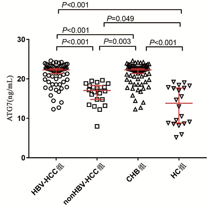
    HBV- HCC组、nonHBV-HCC组、CHB组和HC组血清ATG7水平分别为22.88(19.79~23.04)ng/mL、17.06(14.45~19.40)ng/mL、19.21(16.65~20.82)ng/mL和13.82(8.70~17.82)ng/mL,差异有统计学意义(χ2=65.144,P<0.001)。两两比较显示HBV-HCC组血清ATG7不仅高于CHB组(U=758.5,P<0.001)和HC组(U=94.0,P<0.001),也高于nonHBV-HCC组(U=142.0,P<0.001);而nonHBV-HCC组血清ATG7水平不仅低于HBV-HCC组,也低于CHB组(U=311.0,P=0.003),仅稍高于HC组(U=142.0,P=0.049)(图 1)。此外,在67例HBV-HCC患者中,有肝硬化背景和无肝硬化背景患者的血清ATG7水平分别为22.32 (19.79~22.99)ng/mL和20.89(19.40~23.37)ng/mL,差异无统计学意义(P>0.05)。

    图  1  4组血清ATG7表达水平比较

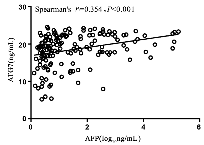
    Spearman相关性分析显示,血清ATG7表达水平和血清AFP水平呈正相关,但相关性较弱(r=0.354,95%CI:0.205~0.486,P<0.001)(图 2)。

    图  2  血清ATG7和AFP相关性分析

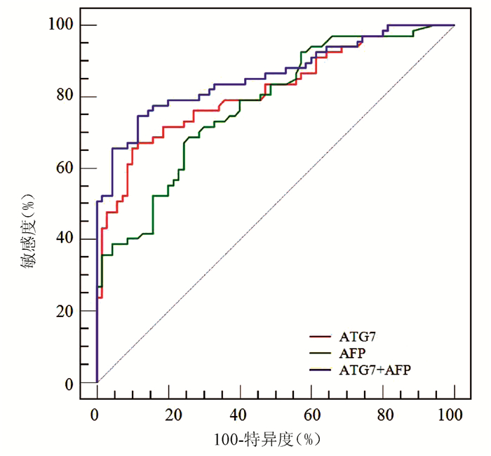
    ROC曲线分析显示,ATG7诊断HBV-HCC的AUC为0.818(95% CI:0.743~0.879),稍高于AFP(AUC=0.777,95%CI:0.698~0.843),但二者差异无统计学意义(Z=0.852,P=0.394)(图 3);ATG7诊断HBV-HCC的cut-off值为20.08 ng/mL,敏感度和特异度分别为71.64%和77.14%,均略高于AFP(68.66%和74.29%)。ATG7联合AFP的二元logistic回归预测概率的AUC为0.859 (95%CI:0.790~0.913),显著高于ATG7(Z=2.192,P=0.028)和AFP(Z=2.076,P=0.038)(图 3, 表 3)。

    图  3  ATG7、AFP及二者联合检测的ROC曲线
    表  3  ATG7、AFP及其联合检测诊断HBV-HCC的性能
    项目 AUC cut-off值 敏感度(%) 特异度(%) 阳性预测值(%) 阴性预测值(%)
    AFP 0.777 12.20 ng/mL 68.66 74.29 71.9 71.2
    ATG7 0.818 20.08 ng/mL 71.64 77.14 75.0 74.0
    ATG7+AFP 0.859 0.56 74.63 88.57 86.2 78.5
    下载: 导出CSV 
    | 显示表格

    本研究通过ELISA技术检测HCC患者血清ATG7表达水平,结果显示HBV-HCC患者血清ATG7显著升高,血清ATG7是诊断HBV-HCC的良好标志物,ATG7与AFP联合诊断可显著提高HBV-HCC的诊断效率。

    ATG7作为一种泛素样修饰物激活酶,通过激活ATG8和ATG12调节自噬体的延伸,是自噬体形成过程的关键蛋白之一[10]。近年研究[11-12]提示ATG7及自噬在HBV-HCC发生发展中发挥重要作用。首先,HBV感染或HBV病毒蛋白的表达可诱导ATG7表达及自噬反应。本研究中CHB组血清ATG7显著高于健康对照组,印证了自噬与HBV感染有关。其次,多项研究[13-14]显示包括ATG7在内的多种自噬相关蛋白在HCC组织表达显著提高。近年的分子细胞学试验进一步揭示多种分子可通过ATG7调节自噬促进HCC发生发展[15-17]。研究[15]显示长链非编码RNA HOX转录反义RNA(HOTAIR)可通过上调ATG7表达激活自噬促进HCC细胞系增殖。癌性锚蛋白重复序列可直接与胞质中的ATG7蛋白相互作用诱导自噬从而促进HCC细胞进展[16]。肿瘤坏死因子α诱导蛋白8(TNFAIP8)则通过与ATG3-ATG7复合物相互作用调节自噬促进HCC细胞增殖[17]。相反,靶向敲减ATG7可抑制自噬,从而促进细胞凋亡、延缓细胞周期及抑制HCC细胞增殖[13]。上述研究结果表明ATG7与HBV-HCC密切相关。本研究数据显示HBV-HCC患者血清ATG7显著高于CHB和健康对照组,进一步证实了自噬与HBV-HCC的相关性,提示血清ATG7是一种潜在HBV-HCC标志物。而且HBV-HCC患者血清ATG7也显著高于nonHBV-HCC,提示不同病因的HCC发病机制不同,其血清标志物也可不同。

    虽然AFP已在临床应用数十年,但诊断敏感度不高。最近的一项系统综述[18]显示,AFP诊断HCC的合并敏感度仅为63.9%。与此相符,本研究中AFP诊断HBV-HCC的敏感度、特异度和AUC分别为68.66%、74.29%和0.777。虽然ATG7的敏感度与AFP相仿,但其特异度较高,总体诊断性能较好。此外,ATG7和AFP相关性较弱,提示二者联合检测可有效提高诊断性能。早前有学者[19]提出应用logistic回归分析有助于提高多指标联合诊断效率。本研究结果显示ATG7联合AFP的二元logistic回归预测模型对HBV-HCC的整体诊断性能较好,AUC及诊断敏感度和特异度均显著高于ATG7及AFP。

    总之,本研究显示ATG7是HBV-HCC的良好标志物,与AFP有较好的互补性,二者联合检测可显著提高HBV-HCC的诊断效率。然而本研究为单中心横断面研究,纳入研究病例有限,因此其诊断性能仍有待进一步扩大样本的多中心临床验证。

  • [1] TESFAYE AA, AZMI AS, PHILIP PA. miRNA and gene expression in pancreatic ductal adenocarcinoma[J]. Am J Pathol, 2019, 189 (1) :58-70.
    [2] STROBEL O, NEOPTOLEMOS J, JÃGER D, et al. Optimizing the outcomes of pancreatic cancer surgery[J]. Nat Rev Clin Oncol, 2019, 16 (1) :11-26.
    [3] KAMISAWA T, WOOD LD, ITOI T, et al. Pancreatic cancer[J]. Lancet, 2016, 388 (10039) :73-85.
    [4] de LIGUORI CARINO N, O'REILLY DA, SIRIWARDENA AK, et al. Irreversible Electroporation in pancreatic ductal adenocarcinoma:Is there a role in conjunction with conventional treatment[J]. Eur J Surg Oncol, 2018, 44 (10) :1486-1493.
    [5] HERMANN PC, SAINZ B Jr. Pancreatic cancer stem cells:A state or an entity[J]. Semin Cancer Biol, 2018, 53:223-231.
    [6] LIAO W, DU Y, ZHANG C, et al. Exosomes:The next generation of endogenous nanomaterials for advanced drug delivery and therapy[J]. Acta Biomater, 2018, 86:1-14.
    [7] XU H, HAN H, SONG S, et al. Exosome-transmitted PSMA3and PSMA3-AS1 promotes proteasome inhibitors resistance in multiple myeloma[J]. Clin Cancer Res, 2019, 25 (6) :2363.
    [8] MATHIEU M, MARTIN-JAULAR L, LAVIEU G, et al. Specificities of secretion and uptake of exosomes and other extracellular vesicles for cell-to-cell communication[J]. Nat Cell Biol, 2019, 21 (1) :9-17.
    [9] CONIGLIARO A, CICCHINI C. Exosome-mediated signaling in epithelial to mesenchymal transition and tumor progression[J]. J Clin Med, 2018, 8 (1) :e26.
    [10] PAN BT, TENG K, WU C, et al. Electron microscopic evidence for externalization of the transferrin receptor in vesicular form in sheep reticulocytes[J]. J Cell Biol, 1985, 101 (3) :942-948.
    [11] ZHOU Y, REN H, DAI B, et al. Hepatocellular carcinomaderived exosomal miRNA-21 contributes to tumor progression by converting hepatocyte stellate cells to cancer-associated fibroblasts[J]. J Exp Clin Cancer Res, 2018, 37 (1) :324.
    [12] KHARE D, OR R, RESNICK I, et al. Mesenchymal stromal cell-derived exosomes affect mRNA expression and function of B-lymphocytes[J]. Front Immunol, 2018, 9:3053.
    [13] FENG M, ZHAO J, WANG L, et al. Upregulated expression of serum exosomal microRNAs as diagnostic biomarkers of lung adenocarcinoma[J]. Ann Clin Lab Sci, 2018, 48 (6) :712-718.
    [14] MASSOUMI RL, HINES OJ, EIBL G, et al. Emerging evidence for the clinical relevance of pancreatic cancer exosomes[J].Pancreas, 2019, 48 (1) :1-8.
    [15] MAZIVEYI M, DONG S, BARANWAL S, et al. Exosomes from Nischarin-expressing cells reduce breast cancer cell motility and tumor growth[J]. Cancer Res, 2019.[Epub ahead of print]
    [16] KAJIWARA C, FUMOTO K, KIMURA H, et al. p63-dependent Dickkopf3 expression promotes esophageal cancer cell proliferation via CKAP4[J]. Cancer Res, 2018, 78 (21) :6107-6120.
    [17] CHEN L, YOU C, JIN X, et al. Cytoskeleton-associated protein 4 is a novel serodiagnostic marker for esophageal squamous-cell carcinoma[J]. Onco Targets Ther, 2018, 11:8221-8226.
    [18] IGBINIGIE E, GUO F, JIANG SW, et al. Dkk1 involvement and its potential as a biomarker in pancreatic ductal adenocarcinoma[J]. Clin Chim Acta, 2019, 488:226-234.
    [19] KIKUCHI A, FUMOTO K, KIMURA H. The Dickkopf1-cytoskeleton-associated protein 4 axis creates a novel signalling pathway and may represent a molecular target for cancer therapy[J]. Br J Pharmacol, 2017, 174 (24) :4651-4665.
    [20] KIMURA H, YAMAMOTO H, HARADA T, et al. CKAP4, a DKK1receptor, is a biomarker in exosomes derived from pancreatic cancer and a molecular target for therapy[J]. Clin Cancer Res, 2019, 25 (6) :1936-1947.
    [21] KITAGAWA T, TANIUCHI K, TSUBOI M, et al. Circulating pancreatic cancer exosomal RNAs for detection of pancreatic cancer[J]. Mol Oncol, 2019, 13 (2) :212-227.
    [22] KINOSHITA-KAWADA M, HASEGAWA H, HONGU T, et al.A crucial role for Arf6 in the response of commissural axons to Slit[J]. Development, 2019, 146 (3) :dev172106.
    [23] LIN JS, JEON JS, FAN Q, et al. ARF6 mediates nephrin tyrosine phosphorylation-induced podocyte cellular dynamics[J]. PLo S One, 2017, 12 (9) :e0184575.
    [24] LI R, PENG C, ZHANG X, et al. Roles of Arf6 in cancer cell invasion, metastasis and proliferation[J]. Life Sci, 2017, 182:80-84.
    [25] LIANG C, QIN Y, ZHANG B, et al. ARF6, induced by mutant Kras, promotes proliferation and Warburg effect in pancreatic cancer[J]. Cancer Lett, 2017, 388:303-311.
    [26] MELO SA, LUECKE LB, KAHLERT C, et al. Glypican-1 identifies cancer exosomes and detects early pancreatic cancer[J]. Nature, 2015, 523 (7559) :177-182.
    [27] DIAMANDIS EP, PLEBANI M. Glypican-1 as a highly sensitive and specific pancreatic cancer biomarker[J]. Clin Chem Lab Med, 2016, 54 (1) :e1-e2.
    [28] ZHOU CY, DONG YP, SUN X, et al. High levels of serum glypican-1 indicate poor prognosis in pancreatic ductal adenocarcinoma[J]. Cancer Med, 2018, 7 (11) :5525-5533.
    [29] LAI X, WANG M, McELYEA SD, et al. A microRNA signature in circulating exosomes is superior to exosomal glypican-1levels for diagnosing pancreatic cancer[J]. Cancer Lett, 2017, 393:86-93.
    [30] OSTEIKOETXEA X, BENKE M, RODRIGUEZ M, et al. Detection and proteomic characterization of extracellular vesicles in human pancreatic juice[J]. Biochem Biophys Res Commun, 2018, 499 (1) :37-43.
    [31] ZHANG Y, HUANG S, LI P, et al. Pancreatic cancer-derived exosomes suppress the production of GIP and GLP-1 from STC-1 cells in vitro by down-regulating the PCSK1/3[J].Cancer Lett, 2018, 431:190-200.
    [32] Pancreatic Cancer Committee of Chinese Anti-Cancer Association. Comprehensive guidelines for the diagnosis and treatment of pancreatic cancer (2018 version) [J]. J Clin Hepatol, 2018, 34 (10) :2109-2120. (in Chinese) 中国抗癌协会胰腺癌专业委员会.胰腺癌综合诊治指南 (2018版) [J].临床肝胆病杂志, 2018, 34 (10) :2109-2120.
    [33] ZHAO X, REN Y, CUI N, et al. Identification of key microRNAs and their targets in exosomes of pancreatic cancer using bioinformatics analysis[J]. Medicine (Baltimore) , 2018, 97 (39) :e12632.
    [34] LI N, ZHAO X, YOU S. Identification of key regulators of pancreatic ductal adenocarcinoma using bioinformatics analysis of microarray data[J]. Medicine (Baltimore) , 2019, 98 (2) :e14074.
  • Relative Articles

    [1]Lixian WU, Weiqiang ZHENG, Huanqin HAN. Effect of different antiviral drugs in reducing the risk of hepatitis B virus-related hepatocellular carcinoma[J]. Journal of Clinical Hepatology, 2022, 38(5): 1165-1168. doi: 10.3969/j.issn.1001-5256.2022.05.040
    [2]Mingyang FENG, Hui WANG. Selection of antiviral drugs for patients with hepatitis B virus-associated hepatocellular carcinoma[J]. Journal of Clinical Hepatology, 2022, 38(11): 2452-2456. doi: 10.3969/j.issn.1001-5256.2022.11.004
    [3]LIU YuWei, JIN JingLan, REN TianYi, GAO XiuZhu, LI Jie, ZHU Qing, NIU JunQi. Effect of direct-acting antiviral on the recurrence hepatitis C virus-related hepatocellular carcinoma after curative treatment: A Meta-analysis[J]. Journal of Clinical Hepatology, 2020, 36(12): 2714-2719. doi: 10.3969/j.issn.1001-5256.2020.12.015
    [4]Ning HuiBin, Liu JunPing, Jin HuiMing, Li Kuan, Xiao ErHui, Shang Jia. Direct-acting antiviral durgs for hepatitis C in children: A case report[J]. Journal of Clinical Hepatology, 2019, 35(1): 164-165. doi: 10.3969/j.issn.1001-5256.2019.01.032
    [5]Zhang JieBing, Guo HongHua. Research advances in hepatitis B cirrhosis with renal injury and the application of antiviral drugs[J]. Journal of Clinical Hepatology, 2019, 35(1): 191-196. doi: 10.3969/j.issn.1001-5256.2019.01.042
    [6]Zhang YaoDi, Zhang YueRong, Wang Hui, Lin Yan, Zhou Ning, Wu LiYang, Wei ShiFang, Li XiangLin. Clinical features of hepatitis C patients with failure or recurrence after treatment with pegylated interferon-α combined with ribavirin and the clinical effect of direct-acting antiviral agents[J]. Journal of Clinical Hepatology, 2019, 35(11): 2456-2460. doi: 10.3969/j.issn.1001-5256.2019.11.014
    [7]Wang MengLan, Tang Hong. Drug-drug interaction of direct-acting antivirals in treatment of chronic hepatitis C[J]. Journal of Clinical Hepatology, 2018, 34(2): 229-232. doi: 10.3969/j.issn.1001-5256.2018.02.002
    [8]Zhang Xi, Li YongGuo. Current status of the application of direct-acting antiviral agents in treatment of chronic hepatitis C and existing problems[J]. Journal of Clinical Hepatology, 2018, 34(4): 853-857. doi: 10.3969/j.issn.1001-5256.2018.04.034
    [9]Li XiaoLou, Zhuo HaiYan, Liu ZhiQiang, Chen RuanQin, Chen Li. Clinical features of HBV-related liver failure associated with recurrence after withdrawal of nucleos ( t) ide analogues and influencing factors for prognosis[J]. Journal of Clinical Hepatology, 2018, 34(7): 1423-1427. doi: 10.3969/j.issn.1001-5256.2018.07.012
    [10]Bian DanDan, Zheng SuJun. Influencing factors for the therapeutic effect of direct-acting antiviral agents in hepatitis C[J]. Journal of Clinical Hepatology, 2017, 33(11): 2205-2208. doi: 10.3969/j.issn.1001-5256.2017.11.034
    [11]Chen XinYue, Liu YaLi, Ren Shan. Value of direct-acting antivirals combined with PR regimen (PEG-IFN combined with ribavirin) in the new era of antiviral therapy for hepatitis C[J]. Journal of Clinical Hepatology, 2017, 33(6): 1063-1066. doi: 10.3969/j.issn.1001-5256.2017.06.009
    [12]Ceng Qun, Jie ShengHua. Research advances in antiviral drugs and their treatment regimens in chronic hepatitis C[J]. Journal of Clinical Hepatology, 2017, 33(11): 2200-2204. doi: 10.3969/j.issn.1001-5256.2017.11.033
    [13]Wen XiaoYu, Niu JunQi. Mechanism of action of direct-acting antiviral agents in treatment of chronic hepatitis C[J]. Journal of Clinical Hepatology, 2016, 32(9): 1699-1705. doi: 10.3969/j.issn.1001-5256.2016.09.013
    [14]Zhang Jing, Liu XueMin, Liu ZhengWen, Lu: Yi. Application of direct-acting antiviral agents in perioperative period of liver transplantation for patients with hepatitis C[J]. Journal of Clinical Hepatology, 2015, 31(12): 2084-2087. doi: 10.3969/j.issn.1001-5256.2015.12.023
    [15]Ren Shan, Chen XinYue. Current research on hepatitis C virus resistance to direct-acting antiviral agents[J]. Journal of Clinical Hepatology, 2015, 31(11): 1807-1812. doi: 10.3969/j.issn.1001-5256.2015.11.010
    [16]Liu HongLing, Zhang Min, Zang Hong, Zhou Xia, Yao Hong, Gao YinJie, Zhou ShuangNan, Liu ZhenWen. Clinical efficacy of direct-acting antiviral agents in treatment of patients with hepatitis C recurrence after liver transplantation[J]. Journal of Clinical Hepatology, 2015, 31(12): 2039-2041. doi: 10.3969/j.issn.1001-5256.2015.12.012
    [17]Xie ZhiWei, Zhou FuYuan. Influence of quantitative changes in HBeAg on the therapeutic efficacy of antiviral drugs [J]. Journal of Clinical Hepatology, 2013, 29(2): 150-152+157.
    [18]Deng Le, Wen ZhiLi. Review of recent research progress in correlating HBV genotypes with efficacy of antiviral treatments[J]. Journal of Clinical Hepatology, 2012, 28(6): 469-473.
    [19]Zhou YouQian, Yin FengMing, Feng JingHua. Regularity of relapse after antiviral treatment in patients with chronic hepatitis C[J]. Journal of Clinical Hepatology, 2012, 28(8): 609-611.
    [20]Liang YanXiu, Jiang JianNing, Su MingHua, Guo WenWen, Huang XiaoHong, Liu ZhiHong, Xie Rong, Fu WuDao, He LiXia, Zhong ShaoHua. Influence of HBeAg seroconversion on virological relapse in chronic hepatitis B patients on withdrawal of nucleos (t) ide analogues therapy[J]. Journal of Clinical Hepatology, 2011, 27(12): 1275-1277+1290.
  • Cited by

    Periodical cited type(2)

    1. 郭佳佳,张文果,王文秀,张晓丽,马徜徉. 血清Nox2、ATG7水平对新生儿窒息心肌损伤的评估价值. 安徽医药. 2024(09): 1791-1795 .
    2. 蔡卓玮,胡刚峰,章波. miR-203在乙型肝炎病毒相关肝细胞癌诊断及预后中的意义. 广西医科大学学报. 2022(11): 1773-1780 .

    Other cited types(0)

  • 加载中
    Created with Highcharts 5.0.7Chart context menuAccess Class DistributionFULLTEXT: 4.5 %FULLTEXT: 4.5 %META: 90.6 %META: 90.6 %PDF: 5.0 %PDF: 5.0 %FULLTEXTMETAPDF
    Created with Highcharts 5.0.7Chart context menuAccess Area Distribution其他: 5.5 %其他: 5.5 %其他: 0.8 %其他: 0.8 %India: 0.3 %India: 0.3 %上海: 0.8 %上海: 0.8 %丽水: 0.3 %丽水: 0.3 %北京: 7.6 %北京: 7.6 %南京: 0.3 %南京: 0.3 %南宁: 0.5 %南宁: 0.5 %台州: 1.6 %台州: 1.6 %吉林: 1.0 %吉林: 1.0 %孝感: 0.3 %孝感: 0.3 %张家口: 5.2 %张家口: 5.2 %成都: 0.5 %成都: 0.5 %昆明: 0.3 %昆明: 0.3 %杭州: 1.3 %杭州: 1.3 %格兰特县: 0.3 %格兰特县: 0.3 %济南: 0.5 %济南: 0.5 %深圳: 0.3 %深圳: 0.3 %石家庄: 0.3 %石家庄: 0.3 %罗奥尔凯埃: 0.8 %罗奥尔凯埃: 0.8 %芒廷维尤: 41.9 %芒廷维尤: 41.9 %苏州: 0.3 %苏州: 0.3 %莫斯科: 0.8 %莫斯科: 0.8 %西宁: 26.2 %西宁: 26.2 %重庆: 0.3 %重庆: 0.3 %长春: 1.3 %长春: 1.3 %长沙: 0.8 %长沙: 0.8 %长治: 0.3 %长治: 0.3 %其他其他India上海丽水北京南京南宁台州吉林孝感张家口成都昆明杭州格兰特县济南深圳石家庄罗奥尔凯埃芒廷维尤苏州莫斯科西宁重庆长春长沙长治

Catalog

    通讯作者: 陈斌, bchen63@163.com
    • 1. 

      沈阳化工大学材料科学与工程学院 沈阳 110142

    1. 本站搜索
    2. 百度学术搜索
    3. 万方数据库搜索
    4. CNKI搜索

    Article Metrics

    Article views (1726) PDF downloads(335) Cited by(2)
    Proportional views
    Related

    /

    DownLoad:  Full-Size Img  PowerPoint
    Return
    Return