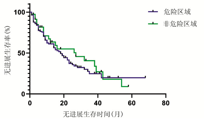
| [1] |
Bureau of Medical AdministrationNational Health Commission of the People's Republic of China. Guidelines for diagnosis and treatment of primary liver cancer in China (2019 edition)[J]. J Clin Hepatol, 2020, 36(2): 277-292. DOI: 10.3969/j.issn.1001-5256.2020.02.007. |
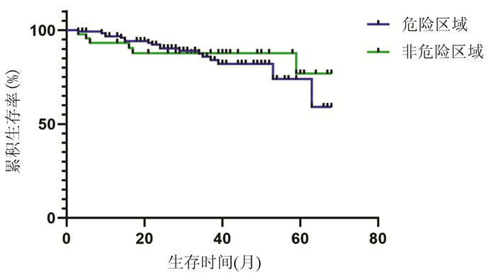
| [2] |
LEE WC, JENG LB, CHEN MF. Estimation of prognosis after hepatectomy for hepatocellular carcinoma[J]. Br J Surg, 2002, 89(3): 311-316. DOI: 10.1046/j.0007-1323.2001.02034.x. |
| [3] |
Bureau of Medical Administration National Health Commission of the People's Republic of China. Guidelines for diagnosis and treatment of primary liver cancer in China (2019 edition)[J]. J Clin Hepatol, 2020, 36(2): 277-292. DOI: 10.3969/j.issn.1001-5256.2020.02.007. |
| [4] |
TOSHIMORI J, NOUSO K, NAKAMURA S, et al. Local recurrence and complications after percutaneous radiofrequency ablation of hepatocellular carcinoma: A retrospective cohort study focused on tumor location[J]. Acta Med Okayama, 2015, 69(4): 219-226. DOI: 10.18926/AMO/53558. |
| [5] |
LEE CH, CHEN WT, LIN CC, et al. Radiofrequency ablation assisted by real-time virtual sonography for hepatocellular carcinoma inconspicuous under sonography and high-risk locations[J]. Kaohsiung J Med Sci, 2015, 31(8): 413-419. DOI: 10.1016/j.kjms.2015.06.002. |
| [6] |
NAKAZAWA T, KOKUBU S, SHIBUYA A, et al. Radiofrequency ablation of hepatocellular carcinoma: Correlation between local tumor progression after ablation and ablative margin[J]. AJR Am J Roentgenol, 2007, 188(2): 480-488. DOI: 10.2214/AJR.05.2079. |
| [7] |
JIANG C, LIU B, CHEN S, et al. Safety margin after radiofrequency ablation of hepatocellular carcinoma: Precise assessment with a three-dimensional reconstruction technique using CT imaging[J]. Int J Hyperthermia, 2018, 34(8): 1135-1141. DOI: 10.1080/02656736.2017.1411981. |
| [8] |
Chinese Society of Liver Cancer, Chinese Anti-Cancer Association; Chinese Society of Clinical Oncology, Chinese Anti-Cancer Association; Liver Cancer Study Group, Chinese Society of Hepatology, Chinese Medical Association. Expert consensus on the norms of local ablation therapy for hepatocellular carcinoma[J]. J Clin Hepatol, 2011, 27(3): 236-238, 244. http://lcgdbzz.org/article/id/LCGD201103004 |
| [9] |
LENCIONI R, LLOVET JM. Modified RECIST (mRECIST) assessment for hepatocellular carcinoma[J]. Semin Liver Dis, 2010, 30(1): 52-60. DOI: 10.1055/s-0030-1247132. |
| [10] |
BENSON AB 3rd, D'ANGELICA MI, ABBOTT DE, et al. NCCN guidelines insights: Hepatobiliary cancers, version 1.2017[J]. J Natl Compr Canc Netw, 2017, 15(5): 563-573. DOI: 10.6004/jnccn.2017.0059. |
| [11] |
ZHU P, WANG YM. Updated key points and clinical pathway for NCCN clinical practice guidelines in oncology: Hepatobiliary cancers (Version2. 2016)[J]. J Clin Hepatol, 2016, 32(9): 1644-1652. DOI: 10.3969/j.issn.1001-5256.2016.09.003. |
| [12] |
YANG BS, YUAN M, HOU YB, et al. TACE combined with precision radiofrequency ablation by using multiple imaging guidance technology for HCC located at exceptionalsites[J]. J Intervent Radiol, 2018, 27(12): 1193-1198. DOI: 10.3969/j.issn.1008-794X.2018.12.018. |
| [13] |
HAN HY, JING X, DING JM, et al. Safety and efficacy of microwave ablation for hepatocellular carcinoma at dangerous locations[J]. Chin J Interv Imaging Ther, 2017, 14(4): 205-209. DOI: 10.13929/j.1672-8475.201611009. |
| [14] |
TERATANI T, YOSHIDA H, SHⅡNA S, et al. Radiofrequency ablation for hepatocellular carcinoma in so-called high-risk locations[J]. Hepatology, 2006, 43(5): 1101-1108. DOI: 10.1002/hep.21164. |
| [15] |
HUANG H, LIANG P, YU XL, et al. Safety assessment and therapeutic efficacy of percutaneous microwave ablation therapy combined with percutaneous ethanol injection for hepatocellular carcinoma adjacent to the gallbladder[J]. Int J Hyperthermia, 2015, 31(1): 40-47. DOI: 10.3109/02656736.2014.999017. |
| [16] |
ZHANG M, LIANG P, CHENG ZG, et al. Efficacy and safety of artificial ascites in assisting percutaneous microwave ablation of hepatic tumours adjacent to the gastrointestinal tract[J]. Int J Hyperthermia, 2014, 30(2): 134-141. DOI: 10.3109/02656736.2014.891765. |
| [17] |
WONG SN, LIN CJ, LIN CC, et al. Combined percutaneous radiofrequency ablation and ethanol injection for hepatocellular carcinoma in high-risk locations[J]. AJR Am J Roentgenol, 2008, 190(3): w187-w195. DOI: 10.2214/AJR.07.2537. |
| [18] |
LIU A, XU W, XU H, et al. Effects of ultrasound-guided low-power microwave ablation for the treatment of liver cancer in special sites[J]. J Pract Radiol, 2018, 34(12): 1925-1928. DOI: 10.3969/j.issn.1002-1671.2018.12.028. |
| [19] |
ZHANG NN, CHENG XJ, LIU JY, et al. High-powered microwave ablation in treating patients with hepatocellular carcinoma and the risk fac-tors of recurrence[J]. J Prac Hepatol, 2015, 18(3): 249-253. DOI: 10.3969/j.issn.1672-5069.2015.02.008. |


 DownLoad: