中文English
ISSN 1001-5256 (Print)
ISSN 2097-3497 (Online)
CN 22-1108/R
Volume 38 Issue 4
Apr.  2022
Turn off MathJax
Article Contents

Inhibitory effect of 6-paradol on the proliferation, migration, and invasion of intrahepatic cholangiocarcinoma cells and its mechanism

DOI: 10.3969/j.issn.1001-5256.2022.04.022
Research funding:

National Natural Science Foundation of China (81871965)

More Information
  • Corresponding author: JIANG Jianixn, rm002979@whu.edu.cn(ORCID: 0000-0001-7939-9082)
  • Received Date: 2021-08-24
  • Published Date: 2022-04-20
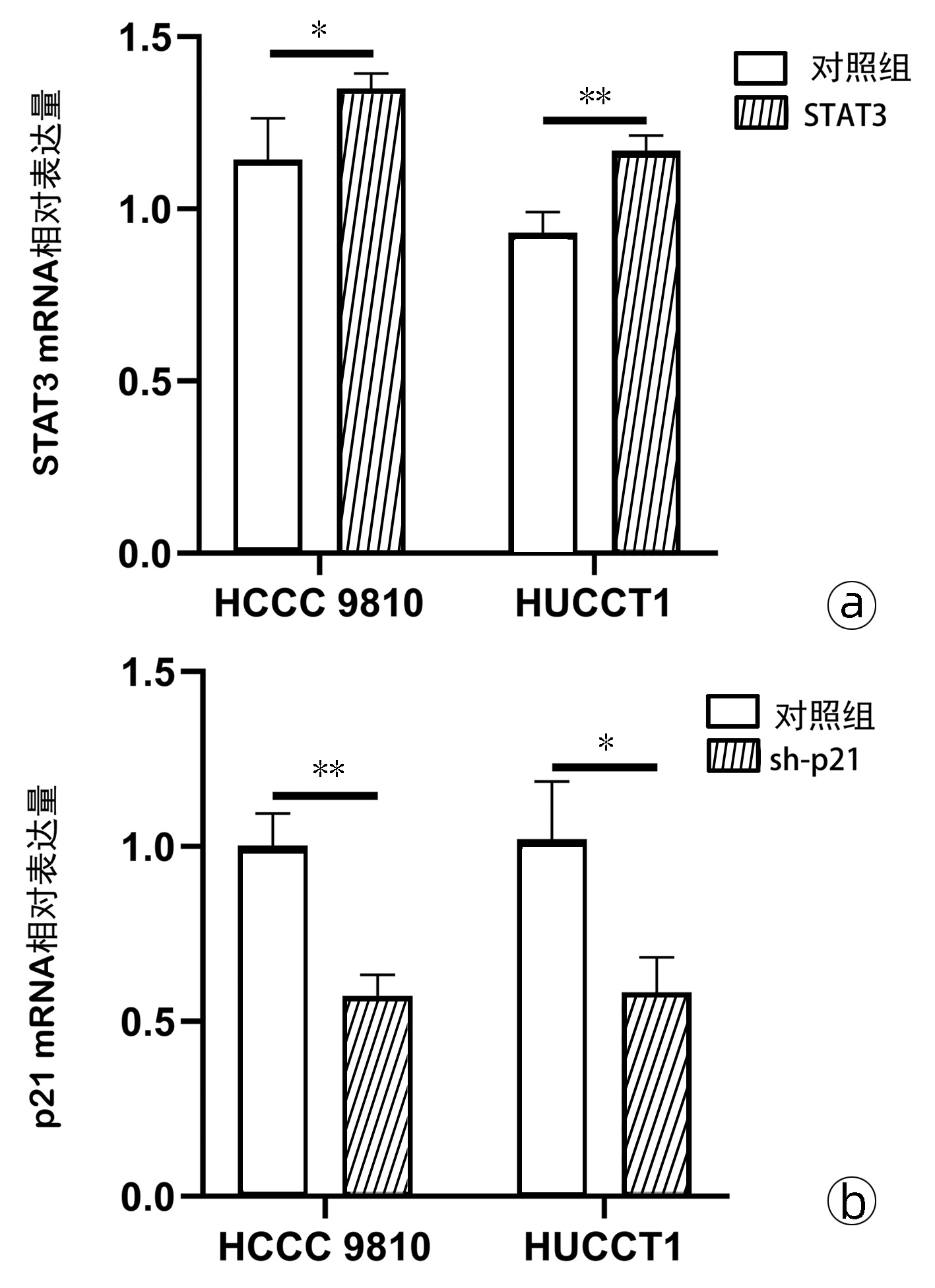
  •   Objective  To investigate the effect of 6-paradol on the proliferation, migration, and invasion of human intrahepatic cholangiocarcinoma cells and its mechanism.  Methods  Human intrahepatic cholangiocarcinoma cell lines HCCC 9810 and HUCCT1 were treated with different concentrations of 6-paradol or an equal volume of DMSO (control group), and then CCK-8 assay, plate colony formation assay, wound healing assay, and Transwell assay were used to measure cell proliferation, migration, and invasion. The bioinformatics software Swiss Target Prediction was used to predict the protein targets of 6-paradol, and Western blot was used to measure the protein expression levels of STAT3, p-STAT3, SRC, p-mTOR, p21, Bcl-2, and p53; Drug Affinity Responsive Target Stability (DARTS) assay was used to investigate the interaction between 6-paradol and STAT3. After cholangiocarcinoma HCCC 9810 and HUCCT1 cells were transfected with STAT3 overexpression plasmid or sh-p21 plasmid, quantitative real-time PCR was used to measure the mRNA expression levels of STAT3 and p21, and Western blot was used to measure the protein expression levels of STAT3 and p21; CCK-8 assay, wound healing assay, and Transwell assay were used to measure cell proliferation, migration, and invasion. The t-test was used for comparison of data between two groups; an analysis of variance was used for comparison between multiple groups, and the least significant difference t-test was used for further comparison between two groups.  Results  Compared with the control group, the 6-paradol treatment groups had significant reductions in cell proliferation, migration, and invasion (P < 0.05). Compared with the control group, the 6-paradol treatment groups had significant reductions in the expression levels of STAT3 and p-STAT3 (all P < 0.05) and a significant increase in the expression level of p21 (all P < 0.05), while there were no significant changes in the expression levels of Bcl-2, SRC, and p-mTOR (all P > 0.05). In the 6-paradol treatment groups, the proportion of STAT3 hydrolyzed by protease was reduced by 48.66% and 45.33%, respectively (t=16.64 and 8.76, both P < 0.05); after transfection with STAT3 overexpression plasmid or p21-silencing plasmid in cholangiocarcinoma cells, there was a significant increase in the mRNA expression level of STAT3 (tHCCC 9810=2.82, tHUCCT1=5.60, both P < 0.05) and a significant reduction in the mRNA expression level of p21 (tHCCC 9810=6.84, tHUCCT1=3.91, both P < 0.05). CCK-8 assay showed that for HCCC 9810 and HUCCT1 cells treated with 6-paradol for 48 and 72 hours, the STAT3 overexpression group had a significantly higher proliferation rate than the single administration group, and the p21 silencing group also had a significantly higher proliferation rate than the single administration group (P < 0.05). The wound healing assay showed that the HCCC 9810 and HUCCT1 cells with STAT3 overexpression or p21 silencing had a significantly higher wound healing rate than the single administration group (all P < 0.05). Transwell assay showed that the HCCC 9810 and HUCCT1 cells with STAT3 overexpression or p21 silencing had significant increases in migration rate and invasion rate compared with the single administration group (all P < 0.05).  Conclusion  6-Paradol inhibits the proliferation, migration, and invasion of cholangiocarcinoma cells by targeting the STAT3-p21 pathway.


  • loading
  • [1]
    RAZUMILAVA N, GORES GJ. Cholangiocarcinoma[J]. Lancet, 2014, 383(9935): 2168-2179. DOI: 10.1016/s0140-6736(13)61903-0.
    [2]
    MOEINI A, SIA D, BARDEESY N, et al. Molecular pathogenesis and targeted therapies for intrahepatic cholangiocarcinoma[J]. Clin Cancer Res, 2016, 22(2): 291-300. DOI: 10.1158/1078-0432.CCR-14-3296.
    [3]
    ANDERSEN JB. Molecular pathogenesis of intrahepatic cholangiocarcinoma[J]. J Hepatobiliary Pancreat Sci, 2015, 22(2): 101-113. DOI: 10.1002/jhbp.155.
    [4]
    KIM HJ, KIM IS, REHMAN SU, et al. Effects of 6-paradol, an unsaturated ketone from gingers, on cytochrome P450-mediated drug metabolism[J]. Bioorg Med Chem Lett, 2017, 27(8): 1826-1830. DOI: 10.1016/j.bmcl.2017.02.047.
    [5]
    SURH YJ, PARK KK, CHUN KS, et al. Anti-tumor-promoting activities of selected pungent phenolic substances present in ginger[J]. J Environ Pathol Toxicol Oncol, 1999, 18(2): 131-139.
    [6]
    MARIADOSS AV, KATHIRESAN S, MUTHUSAMY R, et al. Protective effects of[6]-paradol on histological lesions and immunohistochemical gene expression in DMBA induced hamster buccal pouch carcinogenesis[J]. Asian Pac J Cancer Prev, 2013, 14(5): 3123-3129. DOI: 10.7314/apjcp.2013.14.5.3123.
    [7]
    HUYNH J, CHAND A, GOUGH D, et al. Therapeutically exploiting STAT3 activity in cancer - using tissue repair as a road map[J]. Nat Rev Cancer, 2019, 19(2): 82-96. DOI: 10.1038/s41568-018-0090-8.
    [8]
    HARPER JW, ADAMI GR, WEI N, et al. The p21 Cdk-interacting protein Cip1 is a potent inhibitor of G1 cyclin-dependent kinases[J]. Cell, 1993, 75(4): 805-816. DOI: 10.1016/0092-8674(93)90499-g.
    [9]
    LECHNER JF, STONER GD. Gingers and their purified components as cancer chemopreventative agents[J]. Molecules, 2019, 24(16): 2859. DOI: 10.3390/molecules24162859.
    [10]
    LI Q, WANG R, WANG LP, et al. Paradol inhibits proliferation and migration of human hepatocellular carcinoma cells[J]. Sci Adv Mater, 2019, 11(10): 1467-1473. DOI: 10.1166/sam.2019.3555.
    [11]
    YU H, JOVE R. The STATs of cancer-new molecular targets come of age[J]. Nat Rev Cancer, 2004, 4(2): 97-105. DOI: 10.1038/nrc1275.
    [12]
    HE G, KARIN M. NF-κB and STAT3 - key players in liver inflammation and cancer[J]. Cell Res, 2011, 21(1): 159-168. DOI: 10.1038/cr.2010.183.
    [13]
    FUKUDA A, WANG SC, MORRIS JPT, et al. Stat3 and MMP7 contribute to pancreatic ductal adenocarcinoma initiation and progression[J]. Cancer Cell, 2011, 19(4): 441-455. DOI: 10.1016/j.ccr.2011.03.002.
    [14]
    SAINI U, NAIDU S, ELNAGGAR AC, et al. Elevated STAT3 expression in ovarian cancer ascites promotes invasion and metastasis: A potential therapeutic target[J]. Oncogene, 2017, 36(2): 168-181. DOI: 10.1038/onc.2016.197.
    [15]
    GARTEL AL, RADHAKRISHNAN SK. Lost in transcription: p21 repression, mechanisms, and consequences[J]. Cancer Res, 2005, 65(10): 3980-3985. DOI: 10.1158/0008-5472.CAN-04-3995.
    [16]
    SHAMLOO B, USLUER S. p21 in cancer research[J]. Cancers (Basel), 2019, 11(8): 1178. DOI: 10.3390/cancers11081178.
    [17]
    GEORGAKILAS AG, MARTIN OA, BONNER WM. p21: A two- faced genome guardian[J]. Trends Mol Med, 2017, 23(4): 310-319. DOI: 10.1016/j.molmed.2017.02.001.
  • 加载中

Catalog

    通讯作者: 陈斌, bchen63@163.com
    • 1. 

      沈阳化工大学材料科学与工程学院 沈阳 110142

    1. 本站搜索
    2. 百度学术搜索
    3. 万方数据库搜索
    4. CNKI搜索

    Figures(7)

    Article Metrics

    Article views (980) PDF downloads(83) Cited by()
    Proportional views
    Related

    /

    DownLoad:  Full-Size Img  PowerPoint
    Return
    Return