中文English
ISSN 1001-5256 (Print)
ISSN 2097-3497 (Online)
CN 22-1108/R
Volume 39 Issue 1
Jan.  2023
Turn off MathJax
Article Contents

Role of glutathione transferase in nonalcoholic fatty liver disease: An analysis based on gene expression profile

DOI: 10.3969/j.issn.1001-5256.2023.01.014
Research funding:

The Three-year Action Plan of Shanghai TCM Development (ZY-(2018-2020)-CCCX-5001);

Science and Technology Planning Project of Shanghai Science and Technology Commission (20Z21900100);

Shanghai Key Specialty of Traditional Chinese Clinical Medicine (shslczdzk01201)

More Information
  • Corresponding author: TAO yanyan, taoyanyan1023@126.com (ORCID: 0000-0002-8962-3137); LIU Chenghai, chenghailiu@hotmail.com (ORCID: 0000-0002-2033-0934)
  • Received Date: 2022-06-24
  • Accepted Date: 2022-09-27
  • Published Date: 2023-01-20
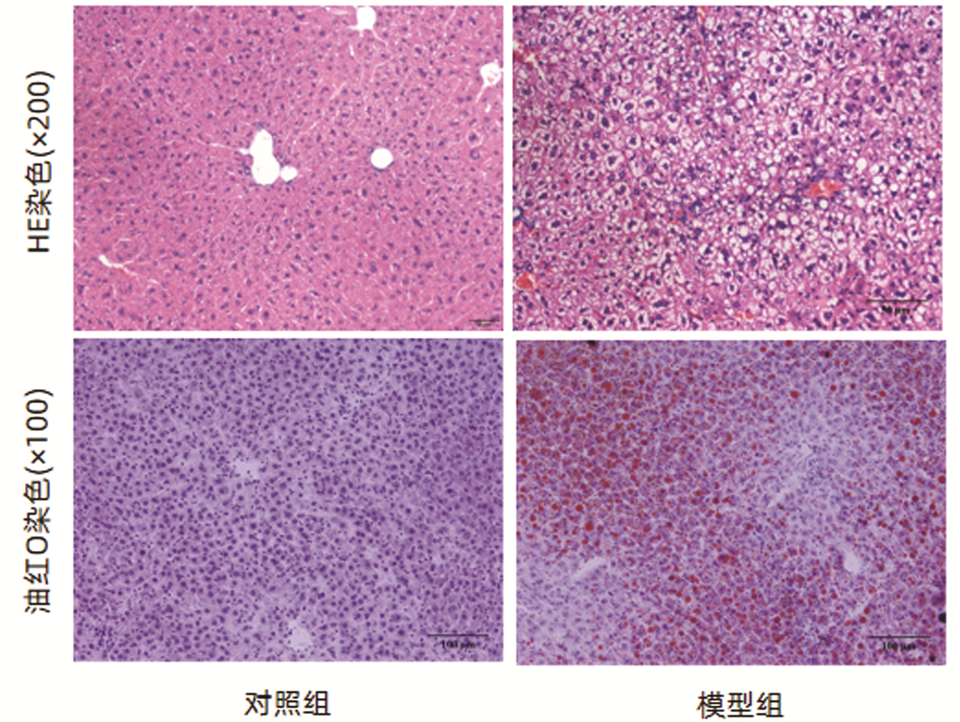
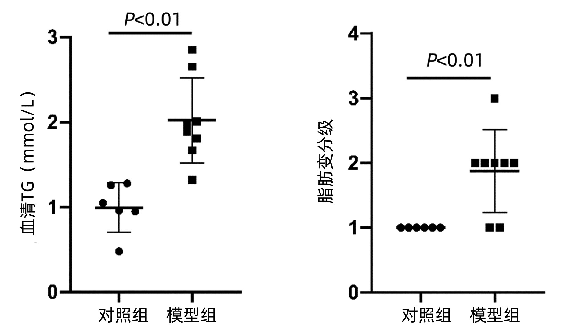
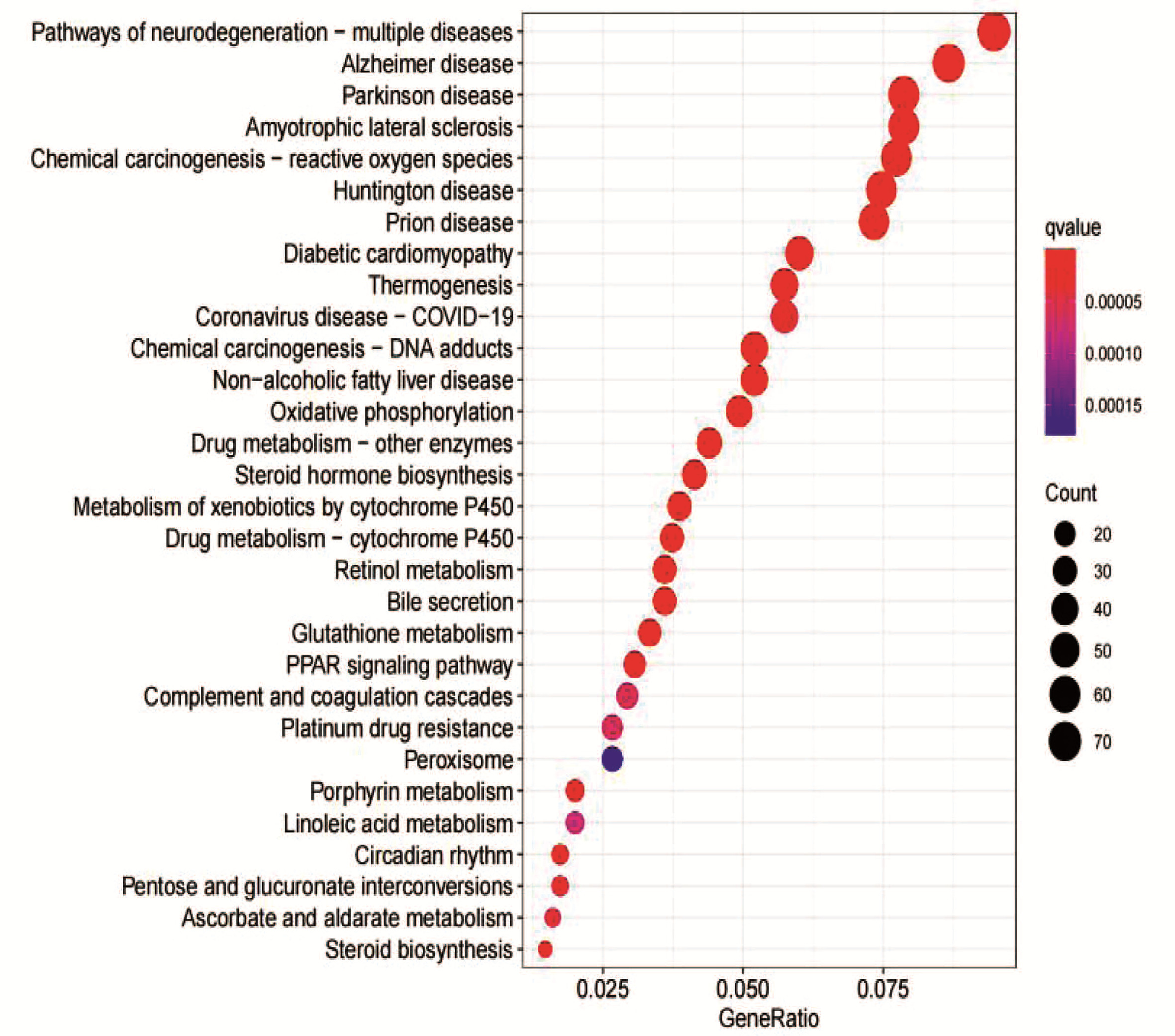
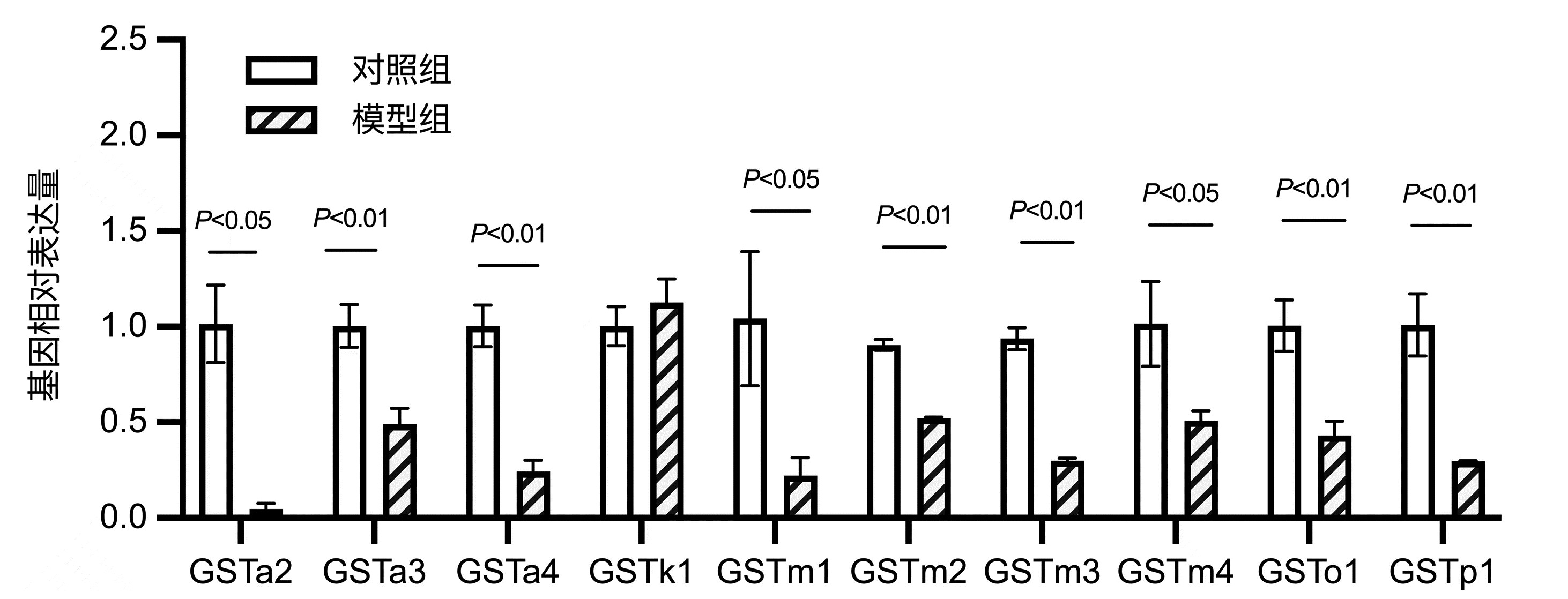
  •   Objective  To investigate the role of glutathione transferase in nonalcoholic fatty liver disease (NAFLD) induced by high-fat diet using the RNA-Seq technique in combination with gene ontology (GO) and Kyoto Encyclopedia of Genes and Genomes (KEGG) enrichment analyses of differentially expressed genes.  Methods  A total of 14 male C57BL/6J mice were divided into control group with 6 mice and model group with 8 mice by random sampling. The mice in the control group were fed with normal diet, and those in the model group were fed with high-fat diet for 7 consecutive weeks to establish a model of NAFLD. Kits were used to measure the activities of serum alanine aminotransferase (ALT) and aspartate aminotransferase (AST) and the level of triglyceride (TG), and HE staining and oil red staining were used to observe liver pathology and deposition of lipid droplets. Liver tissue RNA was extracted for RNA-Seq, and genes with a fold change of ≥2.0 and a P value of < 0.05 were defined as differentially expressed genes; after differentially expressed genes were screened out between the control group and the model group, GO and KEGG enrichment analyses were performed, and qRT-PCR was used to validate the expression of the differentially expressed genes. The independent samples t-test was used for comparison of normally distributed continuous data between two groups.  Results  There were no significant differences between the two groups in body weight and the serum levels of ALT and AST (all P > 0.05). Compared with the control group, the model group had a significantly higher serum level of TG (2.02±0.50 mmol/L vs 1.00±0.29 mmol/L, t=-4.45, P=0.001). HE staining showed diffuse steatosis and ballooning degeneration in the model group, and oil red staining showed that the model group had a significant increase in orange-red lipid droplets in the cytoplasm of hepatocytes and a significantly higher grade of hepatocyte steatosis than the control group (1.88±0.64 vs 1.00±0.00, t=-3.86, P=0.006). RNA-seq results showed a total of 1367 differentially expressed genes between the two groups, among which there were 608 upregulated genes and 759 downregulated genes, and there were 17 differentially expressed GST genes between the two groups. The top 10 GST genes in terms of fold change were validated, and compared with the control group, the model group had downregulated expression of GSTa2, GSTa3, GSTa4, GSTm1, GSTm2, GSTm3, GSTm4, GSTp1, and GSTo1 and upregulated expression of GSTk1. The results of qRT-PCR were consistent with the results of sequencing.  Conclusion  GST affects lipid metabolism by participating in various biological processes such as steroid metabolism, fatty acid metabolism, and cholesterol metabolism and is closely associated with the pathogenesis of NAFLD.


  • loading
  • [1]
    National Workshop on Fatty Liver and Alcoholic Liver Disease, Chinese Society of Hepatology, Chinese Medical Association, Fatty Liver Expert Committee, Chinese Medical Doctor Association. Guidelines of prevention and treatment for nonalcoholic fatty liver disease: A 2018 update[J]. J Clin Hepatol, 2018, 34(5): 947-957. DOI: 10.3969/j.issn.1001-5256.2018.05.007.

    中华医学会肝病学分会脂肪肝和酒精性肝病学组, 中国医师协会脂肪性肝病专家委员会. 非酒精性脂肪性肝病防治指南(2018年更新版)[J]. 临床肝胆病杂志, 2018, 34(5): 947-957. DOI: 10.3969/j.issn.1001-5256.2018.05.007.
    [2]
    LAN T, HU Y, HU F, et al. Hepatocyte glutathione S-transferase mu 2 prevents non-alcoholic steatohepatitis by suppressing ASK1 signaling[J]. J Hepatol, 2022, 76(2): 407-419. DOI: 10.1016/j.jhep.2021.09.040.
    [3]
    HAYES PC, BOUCHIER IA, BECKETT GJ. Glutathione S-transferase in humans in health and disease[J]. Gut, 1991, 32(7): 813-818. DOI: 10.1136/gut.32.7.813.
    [4]
    CAI QF, WU Q. Research progress of glutathione transferase[J]. J Hanan Med Coll, 2011, 17(12): 1735-1738. DOI: 46-1049/R.20111011.1408.007.

    蔡群芳, 邬强. 谷胱甘肽转移酶的研究进展[J]. 海南医学院学报, 2011, 17(12): 1735-1738. DOI: 46-1049/R.20111011.1408.007.
    [5]
    MORRIS MJ, LIU D, WEAVER LM, et al. A structural basis for cellular uptake of GST-fold proteins[J]. PLoS One, 2011, 6(3): e17864. DOI: 10.1371/journal.pone.0017864.
    [6]
    MOREL F, ANINAT C. The glutathione transferase kappa family[J]. Drug Metab Rev, 2011, 43(2): 281-291. DOI: 10.3109/03602532.2011.556122.
    [7]
    National Workshop on Fatty Liver and Alcoholic Liver Disease. Guidelines for diagnosis and treatment of nonalcoholic fatty liver disease (revised in February 2006)[J]. Mod Digest Interv, 2007, 12(4): 266-268. DOI: 10.3969/j.issn.1672-2159.2007.04.020.

    中华医学会肝脏病学分会脂肪肝和酒精性肝病学组. 非酒精性脂肪性肝病诊疗指南(2006年2月修订)[J]. 现代消化及介入诊疗, 2007, 12(4): 266-268. DOI: 10.3969/j.issn.1672-2159.2007.04.020.
    [8]
    ZHU GR, WANG J, HUANG K, et al. A transcriptomic analysis of acute hepatotoxicity induced by aristolochic acidⅠin mice[J]. J Clin Hepatol, 2021, 37(10): 2389-2394. DOI: 10.3969/j.issn.1001-5256.2021.10.026.

    朱哿瑞, 王静, 黄恺, 等. 马兜铃酸Ⅰ致小鼠急性肝毒性的转录组学分析[J]. 临床肝胆病杂志, 2021, 37(10): 2389-2394. DOI: 10.3969/j.issn.1001-5256.2021.10.026.
    [9]
    PRYSYAZHNYUK V, SYDORCHUK L, SYDORCHUK R, et al. Glutathione-S-transferases genes-promising predictors of hepatic dysfunction[J]. World J Hepatol, 2021, 13(6): 620-633. DOI: 10.4254/wjh.v13.i6.620.
    [10]
    HAYES JD, FLANAGAN JU, JOWSEY IR. Glutathione transferases[J]. Annu Rev Pharmacol Toxicol, 2005, 45: 51-88. DOI: 10.1146/annurev.pharmtox.45.120403.095857.
    [11]
    NEBERT DW, VASILIOU V. Analysis of the glutathione S-transferase (GST) gene family[J]. Hum Genomics, 2004, 1(6): 460-464. DOI: 10.1186/1479-7364-1-6-460.
    [12]
    PRYSYAZHNYUK VP, ROSSOKHA ZI, GOROVENKO NG. Variation in particular biochemical indicators, cytokine and adipokine profiles of the blood, and the structural and functional parameters of the liver in patients with nonalcoholic fatty liver disease and different genotypes by the polymorphic locus A313G of the GSTP1 gene[J]. Cytol Genet, 2017, 6: 50-57. DOI: 10.3103/S0095452717060111.
    [13]
    HASHEMI M, ESKANDARI-NASAB E, FAZAELI A, et al. Association of genetic polymorphisms of glutathione-S-transferase genes (GSTT1, GSTM1, and GSTP1) and susceptibility to nonalcoholic fatty liver disease in Zahedan, Southeast Iran[J]. DNA Cell Biol, 2012, 31(5): 672-677. DOI: 10.1089/dna.2011.1343.
    [14]
    KASER S, MOSCHEN A, KASER A, et al. Circulating adiponectin reflects severity of liver disease but not insulin sensitivity in liver cirrhosis[J]. J Intern Med, 2005, 258(3): 274-280. DOI: 10.1111/j.1365-2796.2005.01543.x.
    [15]
    PRYSYAZHNYUK V, VOLOSHYN O, PRYSIAZHNIUK I, et al. Glutathione S-transferase T1 and M1 null genotype distribution among non-alcoholic fatty liver disease patients and its association with cytokine and adipokine profiles[J]. Clin Exp Hepatol, 2020, 6(2): 142-149. DOI: 10.5114/ceh.2020.95678.
    [16]
    RONIS M, MERCER K, ENGI B, et al. Global deletion of glutathione S-transferase A4 exacerbates developmental nonalcoholic steatohepatitis[J]. Am J Pathol, 2017, 187(2): 418-430. DOI: 10.1016/j.ajpath.2016.10.022.
    [17]
    LI N, SUN YR, HE LB, et al. Amelioration by idesia polycarpa maxim. var. vestita diels. of oleic acid-induced nonalcoholic fatty liver in HepG2 cells through antioxidant and modulation of lipid metabolism[J]. Oxid Med Cell Longev, 2020, 2020: 1208726. DOI: 10.1155/2020/1208726.
    [18]
    ZHU YL, ZHANG SJ, YANG C, et al. Quantitative analysis of differential proteins in liver tissues of patients with non-alcoholic steatohepatitis using iTRAQ technology[J]. J South Med Univ, 2021, 41(9): 1381-1387. DOI: 10.12122/j.issn.1673-4254.2021.09.13.

    朱雅莉, 章述军, 阳成, 等. 非酒精性脂肪性肝病患者的肝组织差异蛋白的定量分析: 基于iTRAQ技术[J]. 南方医科大学学报, 2021, 41(9): 1381-1387. DOI: 10.12122/j.issn.1673-4254.2021.09.13.
  • 加载中

Catalog

    通讯作者: 陈斌, bchen63@163.com
    • 1. 

      沈阳化工大学材料科学与工程学院 沈阳 110142

    1. 本站搜索
    2. 百度学术搜索
    3. 万方数据库搜索
    4. CNKI搜索

    Figures(5)  / Tables(2)

    Article Metrics

    Article views (1971) PDF downloads(112) Cited by()
    Proportional views
    Related

    /

    DownLoad:  Full-Size Img  PowerPoint
    Return
    Return