中文English
ISSN 1001-5256 (Print)
ISSN 2097-3497 (Online)
CN 22-1108/R
Issue 10
Oct.  2016

Role of circulating microRNAs in development and progression of liver fibrosis

DOI: 10.3969/j.issn.1001-5256.2016.10.042
  • Received Date: 2016-05-11
  • Published Date: 2016-10-20
  • Recent studies have found that microRNAs( miRNAs) can be secreted via exosomes,stay outside the cells,and keep stable in circulating blood. Various studies have shown that the changes in circulating miRNAs are associated with different pathological states,suggesting that the circulating miRNAs can be used as potential molecular targets for the diagnosis and treament of diseases. Research has shown that miRNAs are closely associated with the development and progression of liver fibrosis. This article reviews the studies on the differences in the expression of circulating miRNAs in the course of liver fibrosis and points out that circulating microRNAs can provide a new research direction for early noninvasive diagnosis and treatment of liver fibrosis.


  • 原发性肝癌作为临床最为常见的恶性肿瘤之一,具有较高的发病率和死亡率,是最为常见的癌症相关性死亡原因之一[1-2]。由于原发性肝癌发病隐匿,患者缺乏早期典型的临床症状,大部分患者在临床确诊时已经处于疾病的中晚期阶段,能够进行根治性手术切除的患者比例甚至不足20%。经肝动脉化疗栓塞术(transcatheter arterial chemoembolization, TACE)是中晚期原发性肝癌患者治疗的主要手段,可有效改善患者临床预后[3-5]。然而,当前仍缺乏评价中晚期肝癌TACE术后预后的有效方式。研究[6-7]显示,表皮生长因子受体(epidermal growth factor receptor, EGFR)信号通路是原发性肝癌发生发展的关键。KRAS基因属于Ras原癌基因家族中的一员,不仅是抗EGFR靶向治疗选择的重要依据,同时也可能成为患者预后的预测因子。本研究探讨KRAS基因状态对TACE治疗中晚期肝癌患者预后的预测价值。

    选择2017年4月—2020年5月在海南省第三人民医院接受TACE治疗的中晚期肝癌患者为研究对象。纳入标准:(1)患者均经穿刺活检,临床病理诊断为原发性肝癌患者;(2)心、肺、肾等机体主要器官功能无明显障碍患者;(3)巴塞罗那肝癌临床分期(BCLC)B期或C期患者;(4)肝功能Child-Pugh分级为A级或B级患者;(5)无法接受外科手术治疗切除患者;(6)卡氏(KPS)评分>60分;(7)术前未接受其他治疗。排除标准:(1)合并严重凝血功能障碍患者;(2)大量腹水或顽固性腹水患者;(3)有TACE治疗禁忌证患者;(4)合并远处转移患者。

    使用Seldinger法穿刺患者股动脉,插管直至肝总动脉进行造影,以明确肿瘤大小、数目、位置。以微导管超选择插管至支配肝癌病灶的主要供血动脉内,将化疗药物(氟尿嘧啶750 mg/m2,奥沙利铂60 mg/m2)稀释液缓慢注入,然后使用15~25 mL 40%碘化油栓塞肿瘤末梢血管。若有必要可使用明胶海绵颗粒,以尽可能完全阻断患者肿瘤血供。根据患者肿瘤大小、数量以及患者肝功能具体情况确定碘化油及栓塞剂的使用剂量。此外,根据患者情况决定TACE治疗次数,本研究患者治疗2~3次。

    TACE手术前,在超声引导下对肝癌组织进行穿刺取材,取5 g左右肿瘤组织,按照QIAamp DNAFFPF Kit试剂盒(Qiagen公司)步骤提取肿瘤组织DNA,使用紫外分光光度计检测提取DNA浓度,然后调节浓度至100 μg/mL,-20 ℃保存待测。扩增KRAS基因12/13号密码子特需特异性引物,根据文献[8]报道,引物序列,正义:5′-CTGTATCAAAGAATGGTCCTGCAC-3′,反义:5′-CTGTATCAAAGAATGGTCCTGCAC-3′。引物及Premix PCR试剂盒购于宝生物工程(大连)有限公司,PCR反应体系50 μL。PCR反应条件:94 ℃ 5 min,94 ℃ 30 s、60 ℃ 30 s、70 ℃ 40 s,共完成30个循环,使用1.5%琼脂糖凝胶电泳分析PCR产物,选取PCR阳性凝胶条带作为目的片段,使用QIAquick PCR Purification Kit(Qiagen公司)对PCR产物回收,然后送至上海基因技术有限公司进行测序分析。

    所有患者在接受TACE术后,每月复查肝功能、AFP、肝脏增强CT或MRI,以评估患者肿瘤控制情况以及术后复发情况,记录患者无进展生存期(PFS)以及总体生存期(OS)。PFS指的是从患者TACE术后到观察到疾病进展或发生死亡(由于任何原因)之间的时间;OS指的是从患者TACE术后到患者死亡(由于任何原因)之间的时间。

    采用统计学软件SPSS 25.0处理数据。计量资料以x±s表示,两组间比较采用t检验,计数资料两组间比较采用χ2检验,生存分析绘制Kaplan-Meier生存曲线,生存曲线比较采用Log-rank检验,对可能影响患者预后的各因素进行Cox回归分析。P<0.05为差异具有统计学意义。

    97例患者中男61例、女36例,年龄30~78岁,平均(56.48±12.31)岁。肿瘤最大径2.89~9.74 cm,平均(7.19±2.08)cm;肿瘤单个89例、≥2个18例。在本组97例晚期肝癌患者中,共检出KRAS基因突变患者34例(35.05%),其中检出12号密码子突变患者21例(61.76%),13号密码子突变患者13例(38.24%)。在检出KRAS基因突变患者中,均为单个碱基的点突变,而并未检出两个或两个以上碱基突变或其他形式的突变(图 1)。

    图  1  KRAS基因12、13密码子序列突变图谱
    注:a, KRAS基因12、13密码子野生型基因序列;b, KRAS基因12密码子GGT→GAT突变;c, KRAS基因12密码子GGT→GTT突变;d, KRAS基因13密码子GGC→GAC突变。
    Figure  1.  Mutation map of KRAS gene codons 12 and 13

    KRAS基因突变型与野生型比较,肝内转移、肿瘤数目的差异均有统计学意义(χ2值分别为3.965、6.593,P值均<0.05)(表 1)。

    表  1  KRAS基因突变状态与患者临床特征的关系
    Table  1.  The relationship between KRAS gene mutation status and clinical characteristics of patients
    临床资料 KRAS突变型
    (n=34)
    KRAS野生型
    (n=63)
    统计值 P
    性别(例) χ2=0.370 0.543
    20 41
    14 22
    年龄(岁) 55.12±14.21 56.93±10.04 t=0.730 0.468
    乙型肝炎病史(例) χ2=0.057 0.811
    阴性 30 56
    阳性 4 7
    ALT(U/L) 49.85±14.20 51.24±15.22 t=0.439 0.662
    凝血酶原时间(s) 12.83±1.84 13.02±2.10 t=0.443 0.659
    总胆红素(mmol/L) 17.94±3.73 18.05±3.16 t=0.153 0.878
    白蛋白(g/L) 40.27±6.49 40.53±5.98 t=0.198 0.843
    AFP(ng/L) 438.95±109.32 409.64±97.58 t=1.353 0.179
    肿瘤大小(cm) 7.39±2.30 7.02±2.15 t=0.789 0.432
    肝硬化(例) χ2=0.035 0.852
    27 49
    7 14
    肝内转移(例) χ2=3.965 0.047
    16 17
    18 46
    肿瘤数目(例) χ2=6.593 0.010
    单个 23 56
    多个 11 7
    腹水(例) χ2=0.057 0.811
    31 55
    3 8
    Child-Pugh分级(例) χ2=0.056 0.812
    A 24 43
    B 10 20
    BCLC分期(例) χ2=0.074 0.785
    B 22 39
    C 12 24
    下载: 导出CSV 
    | 显示表格

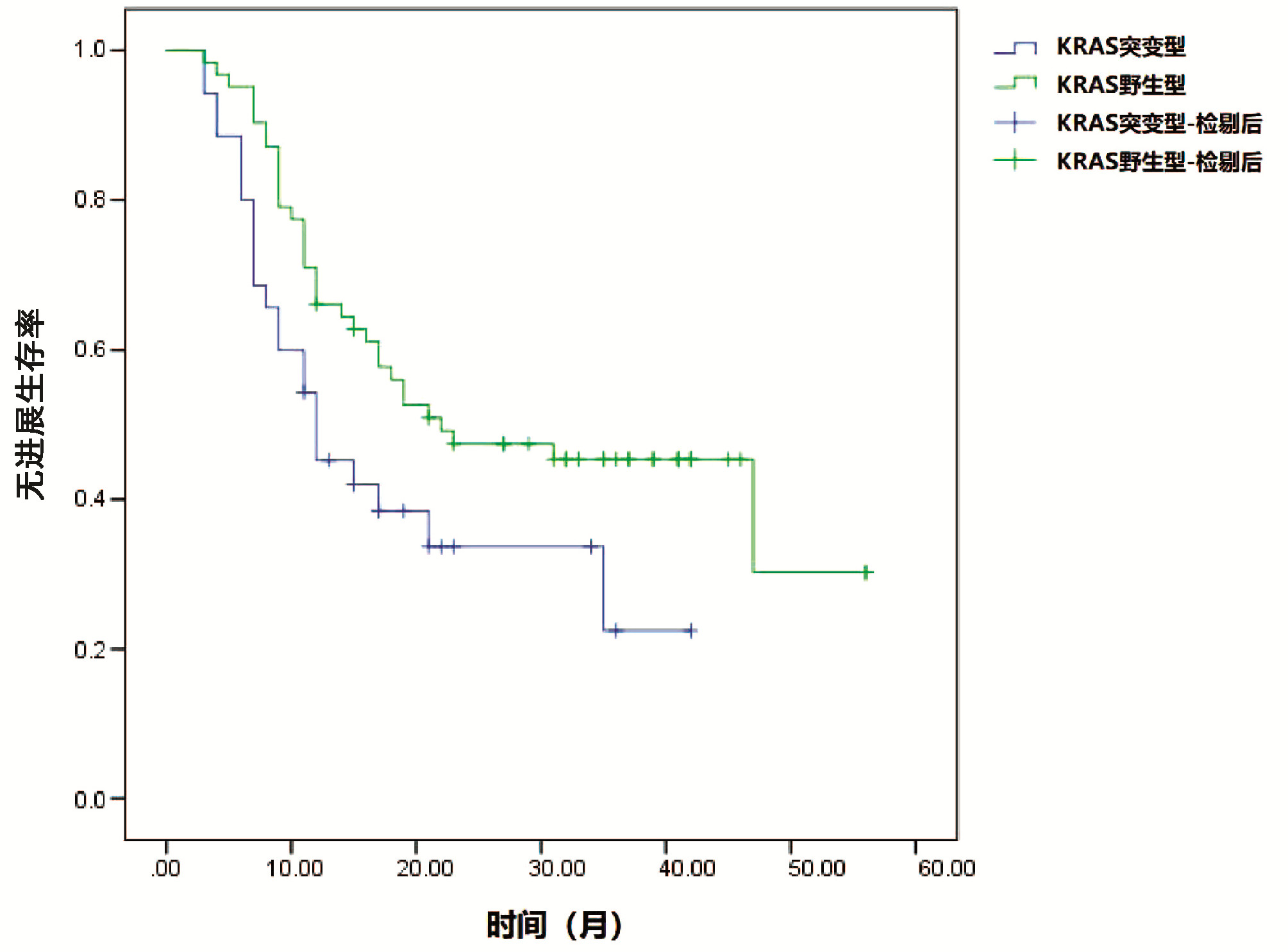
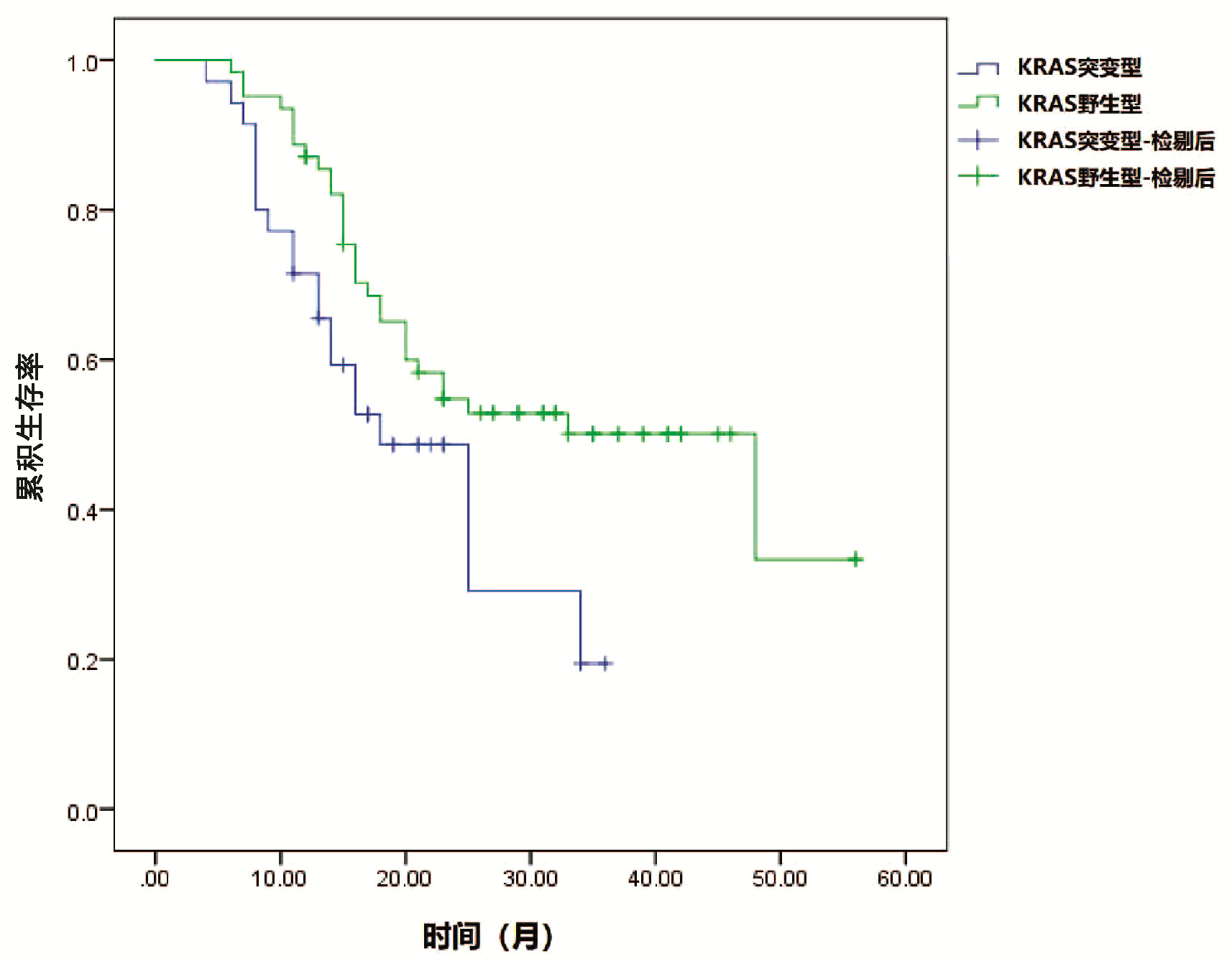
    对本组97例患者进行追踪随访显示,KRAS基因野生型患者平均PFS为11.35个月,突变型患者平均PFS为19.65个月;KRAS基因野生型患者平均OS为19.18个月,突变型患者平均OS为26.54个月。Kaplan-Meier生存分析结果显示,KRAS基因野生型患者PFS及OS均显著优于KRAS突变型(P值均<0.05)(图 23)。

    图  2  KRAS基因状态与患者PFS的关系
    Figure  2.  Relationship between KRAS gene status and progression-free survival analysis of patients
    图  3  KRAS基因状态与患者OS的关系
    Figure  3.  Relationship between KRAS gene status and overall survival analysis of patients

    对可能影响患者总体生存预后的各因素进行Cox分析,结果显示,KRAS基因状态(RR=18.273, 95%CI:5.584~98.305)、肝内转移(RR=11.475, 95%CI:3.029~56.490)、肿瘤数目(RR= 10.038, 95%CI:2.973~19.328)、BCLC分期(RR=12.384, 95%CI:2.385~29.305)与患者预后密切相关,为影响患者总体生存预后的重要因素(P值均<0.05)(表 2)。

    表  2  影响患者预后的Cox分析
    Table  2.  Cox analysis affecting patient outcomes
    自变量 RR 95%CI P
    性别(男性=0,女性=1) 2.734 0.983~7.935 0.283
    年龄(≥60岁=0,<60岁=1) 3.038 0.627~10.293 0.276
    KRAS基因状态(突变型=0,野生型=1) 18.273 5.584~98.305 0.001
    乙型肝炎病史(有=0,无=1) 5.484 0.719~9.380 0.193
    ALT(≥40 U/L=0,<40 U/L=1) 2.079 0.417~12.953 0.389
    凝血酶原时间(≥12 s=0,<12 s=1) 1.092 0.271~9.182 0.849
    总胆红素(≥18 nmol/L=0,<18
    nmol/L=1)
    1.684 0.495~7.293 0.711
    白蛋白(≥40 g/L=0,<40 g/L=1) 2.087 0.408~11.235 0.419
    AFP(≥400 ng/L=0,<400 ng/L=1) 2.193 0.602~11.293 0.619
    肿瘤大小(≥7 cm=0,<7 cm=1) 2.183 0.485~7.304 0.280
    肝硬化(有=0,无=1) 1.804 0.198~10.237 0.695
    肝内转移(有=0,无=1) 11.475 3.029~56.490 0.018
    肿瘤数目(单发=0,多发=1) 10.038 2.973~19.328 0.021
    腹水(有=0,无=1) 2.013 0.421~8.106 0.174
    Child-Pugh分级(A=0,B=1) 2.189 0.569~7.491 0.209
    BCLC分期(B=0,C=1) 12.384 2.385~29.305 0.011
    下载: 导出CSV 
    | 显示表格

    目前,TACE已经成为无法外科切除中晚期肝癌患者的首选治疗方式。肝癌的主要供血为肝动脉供血。经肝动脉碘油注射后,碘油可在肝癌的组织间隙、肝窦以及细小血管内选择性停滞,从而导致癌细胞失去血液供应而导致肿瘤组织缺血坏死[9-10];在碘油中混合化疗药物,从而使化疗药物在肿瘤组织中缓慢释放,延长了化疗药物与肿瘤细胞之间的接触时间,进而显著提高化疗药物治疗效果,同时可显著减小化疗药物对患者全身的不良影响,并阻断肿瘤血供,引起肿瘤组织发生缺血性坏死,进一步增加抗肿瘤疗效[11-12]。研究提出[13-14],采取非手术多模式治疗,尤其对于原发性肝癌患者采取TACE治疗,可有效延长患者生存期,在中晚期肝癌的治疗中具有重要作用。

    原发性肝癌是多基因、多分子、多通路间相互作用的结果,因此检测原发性肝癌患者基因突变状况,对于患者治疗反应以及生存预后具有预测价值,从而有助于实现精准化治疗的目的[15-16]。KARS基因突变在原发性肝癌患者中具有着较高的发生率。研究[17-18]显示,原发性肝癌的发生是一种多步骤过程,而在肿瘤恶性发生的整个过程中,均与KRAS基因突变有关。有研究[19-20]认为,KRAS基因突变被认为是原发性肝癌发生的启动性因子,尤其G-A碱基突变,可能在多种恶性肿瘤发生中具有重要作用。

    本研究结果显示,在本组97例中晚期肝癌患者中,共检出KRAS基因突变患者34例(35.05%),其中检出12号密码子突变患者21例(61.76%),13号密码子突变患者13例(38.24%),且检出的患者均为单个碱基突变。本组结果与相关研究报道结果基本一致[21]。KRAS基因突变率接近35%,是一种较为常见的基因突变形式。此外,KRAS基因突变与患者肝硬化、肝内转移、肿瘤数目有关。提示KRAS基因突变的存在可能与肝癌疾病的发生发展具有一定的内在联系,从而为KRAS突变可能成为预测患者治疗预后的因素提供一定的基础。

    Kaplan-Meier生存分析结果显示,KRAS基因野生型患者PFS及OS均显著优于KRAS突变型。表明了KRAS基因状态与患者TACE术后生存状况密切相关。KRAS基因野生型患者可受上游EGFR信号通路的调控,而当KRAS基因发生点突变时,导致该基因编码P21蛋白的空间结构发生了相应的变化,不再受上游EGFR信号的调控。KRAS基因突变亚型不同,可能也会导致该基因的空间构象发生变化,引起相应的调节细胞内信号通路不同,从而影响化疗药物的结合位点,也可能导致患者对化疗药物的敏感性有所差别,进而影响患者预后。

    同时,本研究进一步对可能影响患者术后预后的各因素进行Cox回归分析,结果显示,KRAS基因状态、肝内转移、肿瘤数目、BCLC分期进入回归模型,为影响患者总体生存预后的重要因素。肿瘤数目、肝内转移以及BCLC分期均可有效提示原发性肝癌患者自身状态及肿瘤的进展情况,故而可影响患者预后,而KRAS基因状态同样进入了Cox回归模型,提示KRAS基因状态也对患者临床预后有着重要的影响。此外,通过Kaplan-Meier生存分析以及Cox多因素分析,进一步确定了KRAS基因状态与患者TACE术后预后具有密切关系,可能成为预测患者术后预后的重要因素之一。KRAS基因突变,可能是导致患者术后预后较差的因素,因此关注肝癌TACE患者KRAS基因状态,对于提前制订相关治疗计划具有重要价值。

    但是由于本研究纳入样本量相对较小,研究结果可能存在一定的偏差,后期研究将进一步扩大研究样本量,以获得更为可靠的临床研究数据。

    综上所述,KRAS基因突变在原发性肝癌中较为常见,与患者TACE术后不良预后密切相关,可成为患者临床预后监测的潜在指标。

  • [1]LAWRIE CH,GAL S,DUNLOP HM,et al.Detection of elevated levels of tumour-associated microRNAs in serum of patients with diffuse large B-cell lymphoma[J].Br J Haematol,2008,141(5):672-675.
    [2]CARVALHO INSR,FREITAS RM,VARGAS FR.Translating microRNAs into biomarkers:What is new for pediatric cancer?[J].Med Oncol,2016,33(5):49.
    [3]LI A,YU J,KIM HY,et al.MicroRNA array analysis finds elevated serum miR-1290 accurately distinguishes patients with low-stage pancreatic cancer from healthy and disease controls.[J].Clin Cancer Res,2013,19(13):3600-3610.
    [4]KJERSEM JB,IKDAHL T,LINGJAERDE OC,et al.Plasma microRNAs predicting clinical outcome in metastatic colorectal cancer patients receiving first-line oxaliplatin-based treatment[J].Mol Oncol,2014,8(1):59-67.
    [5]GALLO A,TANDON M,ALEVIZOS I,et al.The majority of microRNAs detectable in serum and saliva is concentrated in exosomes[J].PLo S One,2012,7(3):e30679.
    [6]FUJITA Y,YOSHIOKA Y,OCHIYA T.Extracellular vesicle transfer of cancer pathogenic components[J].Cancer Sci,2016,107(4):385-390.
    [7]STREMERSCH S,VANDENBROUCKE RE,van WONTERGHEM E,et al.Comparing exosome-like vesicles with liposomes for the functional cellular delivery of small RNAs[J].J Control Release,2016,232:51-61.
    [8]COENEN-STASS AM,MAGER I,WOOD MJ.Extracellular microRNAs in membrane vesicles and non-vesicular carriers[J].EXS,2015,106:31-53.
    [9]ZHANG QQ,XU MY,QU Y,et al.Analysis of the differential expression of circulating microRNAs during the progression of hepatic fibrosis in patients with chronic hepatitis B virus infection[J].Mol Med Rep,2015,12(4):5647-5654.
    [10]ARROYO JD,CHEVILLET JR,KROH EM,et al.Argonaute2complexes carry a population of circulating microRNAs independent of vesicles in human plasma[J].Proc Natl Acad Sci U S A,2011,108(12):5003-5008.
    [11]VICKERS KC,PALMISANO B,TSHOUCRI BM,et al.MicroRNAs are transported in plasma and delivered to recipient cells by high-density lipoproteins[J].Nat Cell Biol,2011,13(4):423-433.
    [12]GUO CJ,PAN Q,LI DG,et al.MiR-15b and miR-16 are implicated in activation of the rat hepatic stellate cell:an essential role for apoptosis[J].J Hepatol,2009,50(4):766-778.
    [13]JI J,ZHANG J,HUANG G,et al.Over-expressed microRNA-27a and 27b influence fat accumulation and cell proliferation during rat hepatic stellate cell activation[J].FEBS Lett,2009,583(4):759-766.
    [14]PIROLA CJ,GIANOTTI TF,CASTANO GO,et al.Circulating microRNA signature in non-alcoholic fatty liver disease:from serum non-coding RNAs to liver histology and disease pathogenesis[J].Gut,2015,64(5):800-812.
    [15]CSAK T,BALA S,LIPPAI D,et al.MicroRNA-122 regulates hypoxia-inducible factor-1 and vimentin in hepatocytes and correlates with fibrosis in diet-induced steatohepatitis[J].Liver Int,2015,35(2):532-541.
    [16]BUTT AM,RAJA AJ,SIDDIQUE S,et al.Parallel expression profiling of hepatic and serum microRNA-122 associated with clinical features and treatment responses in chronic hepatitis C patients[J].Sci Rep,2016,6:21510.
    [17]WAIDMANN O,KOBERLE V,BRUNNER F,et al.Serum microRNA-122 predicts survival in patients with liver cirrhosis[J].PLo S One,2012,7(9):e45652.
    [18]ARATAKI K,HAYES CN,AKAMATSU S,et al.Circulating microRNA-22 correlates with microRNA-122 and represents viral replication and liver injury in patients with chronic hepatitis B[J].J Med Virol,2013,85(5):789-798.
    [19]NOVELLINO L,ROSSI R,BONINO F,et al.Circulating hepatitis B surface antigen particles carry hepatocellular microRNAs[J].PLo S One,2012,7(3):e31952.
    [20]ANADOL E,SCHIERWAGEN R,ELFIMOVA N,et al.Circulating microRNAs as a marker for liver injury in human immunodeficiency virus patients[J].Hepatology,2015,61(1):46-55.
    [21]RODERBURG C,URBAN GW,BETTERMANN K,et al.MicroRNA profiling reveals a role for miR-29 in human and murine liver fibrosis[J].Hepatology,2011,53(1):209-218.
    [22]KWIECINSKI M,ELFIMOVA N,NOETEL A,et al.Expression of platelet-derived growth factor-C and insulin-like growth factor I in hepatic stellate cells is inhibited by miR-29[J].Lab Invest,2012,92(7):978-987.
    [23]ZHANG YQ,WU LP,WANG Y,et al.Protective role of estrogen-induced miRNA-29 expression in carbon tetrachloride-induced mouse liver injury[J].J Biol Chem,2012,287(18):14851-14862.
    [24]CHEN YJ,ZHU JM,WU H,et al.Circulating microRNAs as a fingerprint for liver cirrhosis[J].PLo S One,2013,8(6):e66577.
    [25]WANG BC,LI WX,GUO K,et al.miR-181b promotes hepatic stellate cells proliferation by targeting p27 and is elevated in the serum of cirrhosis patients[J].Biochem Biophys Res Commun,2012,421(1):4-8.
    [26]YU FJ,GUO Y,CHEN BC,et al.MicroRNA-17-5p activates hepatic stellate cells through targeting of Smad7[J].Lab Invest,2015,95(7):781-789.
    [27]ZHENG JJ,ZHOU ZX,XU ZQ,et al.Serum microRNA-125a-5p,a useful biomarker in liver diseases,correlates with disease progression[J].Mol Med Rep,2015,12(1):1584-1590.
    [28]EL-AHWANY E,NAGY F,ZOHEIRY M,et al.Circulating miRNAs as predictor markers for activation of hepatic stellate cells and progression of HCV-induced liver fibrosis[J].Electronic Physician,2016,8(1):1804-1810.
    [29]SHRIVASTAVA S,PETRONE J,STEELE R,et al.Up-regulation of circulating miR-20a is correlated with hepatitis C virus-mediated liver disease progression[J].Hepatology,2013,58(3):863-871.
    [30]XIE Y,YAO QW,BUTT AM,et al.Expression profiling of serum microRNA-101 in HBV-associated chronic hepatitis,liver cirrhosis,and hepatocellular carcinoma[J].Cancer Biol Ther,2014,15(9):1248-1255.
    [31]TU XL,ZHANG HY,ZHANG JC,et al.MicroRNA-101 suppresses liver fibrosis by targeting the TGFbeta signalling pathway[J].J Pathol,2014,234(1):46-59.
    [32]BENZ F,RODERBURG C,CARDENAS DV,et al.U6 is unsuitable for normalization of serum miRNA levels in patients with sepsis or liver fibrosis[J].Exp Mol Med,2013,45:e42.
    [33]LI Y,ZHANG L,LIU F,et al.Identification of endogenous controls for analyzing serum exosomal miRNA in patients with hepatitis B or hepatocellular carcinoma[J].Dis Markers,2015,2015:893594.
    [34]WANG K,YUAN Y,CHO JH,et al.Comparing the MicroRNA spectrum between serum and plasma[J].PLo S One,2012,7(7):e41561.
  • Relative Articles

    [1]Liu LinXiang, Nie Yuan, Zhu Xuan. Clinical application of transient elastography in liver cirrhosis and its complications[J]. Journal of Clinical Hepatology, 2020, 36(6): 1362-1365. doi: 10.3969/j.issn.1001-5256.2020.06.037
    [2]Hu BoBin, Jiang JianNing, Fu JiaXin, Wang BaoJian, Wu XiaoLi, Liang Peng, Mo YanYan, Fu YanPing, Du Man, Liu Yu. Risk factors for recurrence after drug withdrawal in HBeAg-positive chronic hepatitis B patients completing pharmacotherapy with nucleos(t) ide analogues[J]. Journal of Clinical Hepatology, 2016, 32(5): 885-889. doi: 10.3969/j.issn.1001-5256.2016.05.014
    [3]Han BenLi. Clinical efficacy of entecavir in HBeAg-negative chronic hepatitis B with compensated cirrhosis[J]. Journal of Clinical Hepatology, 2016, 32(8): 1525-1528. doi: 10.3969/j.issn.1001-5256.2016.08.018
    [4]Liu ZhiHua, He HaiTang, Hu Jing, Fu QunXiang, Luo KangXian. Efficacy of pegylated interferon α-2a in HBe Ag-negative chronic hepatitis B patients and its influencing factors[J]. Journal of Clinical Hepatology, 2015, 31(5): 697-701. doi: 10.3969/j.issn.1001-5256.2015.05.016
    [5]Lan Lin, Kong Yin, Zhang LingYi. Significance of changes in hepatic histology from patients with HBe Ag( + ) or HBe Ag(-) chronic hepatitis B and normal or mildly elevated alanine aminotransferase[J]. Journal of Clinical Hepatology, 2015, 31(4): 510-513. doi: 10.3969/j.issn.1001-5256.2015.04.008
    [6]Wang Fei, Xu AiLing, Xin XiaXia, Yuan Hong, Xiong JingJing, Zhang Qi, Wang Chen. Relationship between e system of hepatitis B and HBV DNA quantification[J]. Journal of Clinical Hepatology, 2015, 31(4): 534-536. doi: 10.3969/j.issn.1001-5256.2015.04.014
    [7]Ma YanHong. Clinical effect of initial combination therapy with lamivudine plus adefovir dipivoxil in 48-week treatment of HBeAg-positive chronic hepatitis B[J]. Journal of Clinical Hepatology, 2013, 29(8): 631-632. doi: 10.3969/j.issn.1001-5256.2013.08.020
    [8]Liu XingXiang, Li GuiZhen, Xu YunFang, Zhang Jing. Relationship between HBV DNA and HBeAg in serum and HBcAg and inflammation grade in liver tissue among patients with chronic hepatitis B[J]. Journal of Clinical Hepatology, 2013, 29(12): 919-921. doi: 10.3969/j.issn.1001-5256.2013.12.011
    [9]Yang YanLin, Xiao Ping, Gao Peng, Wang LiMing, Wei XiSheng, He Qiang, Zhou Ping. Distribution of HBV genotypes and YMDD mutations in E antigen-positive patients[J]. Journal of Clinical Hepatology, 2012, 28(6): 428-430.
    [10]Wu YuZhuo. Correlation analysis of quantitative HBsAg and liver histological features of HBeAg-negative chronic hepatitis B patients[J]. Journal of Clinical Hepatology, 2012, 28(12): 923-925.
    [11]Li Lu, Li Man, Zhu XiaoJun, Sun XueHua, Gao YueQiu. Comparative analysis of the relationship between histopathology and HBeAg serological status in chronic hepatitis B patients[J]. Journal of Clinical Hepatology, 2012, 28(12): 919-922.
    [12]Gao Min, Lu ChengZhen, Wang Yi, Zhai Lu, Guo Jie, Zhou Li, Han Xu, Liu YongGang, Cao Li. Relationship between expression of HBcAg in liver tissue and characteristics of the hepatic pathological features in patients with chronic hepatitis B virus infection[J]. Journal of Clinical Hepatology, 2012, 28(3): 201-204.
    [13]Gao YuanZheng, Jia SuHua, Zhao LianFeng, Ma Jun, Wei XingHui. Telbivudine combined with bicyclol treatment in HBeAg positive chronic hepatitis[J]. Journal of Clinical Hepatology, 2012, 28(5): 349-351.
    [14]Li ChunXia, Xu GuangHua, Pan HuaiQiang, Liu Na. Clinical differences between HBeAg-positive and-negative chronic hepatitis B patients over 40-years-old[J]. Journal of Clinical Hepatology, 2012, 28(6): 425-427.
    [15]Liang YanXiu, Jiang JianNing, Su MingHua, Guo WenWen, Huang XiaoHong, Liu ZhiHong, Xie Rong, Fu WuDao, He LiXia, Zhong ShaoHua. Influence of HBeAg seroconversion on virological relapse in chronic hepatitis B patients on withdrawal of nucleos (t) ide analogues therapy[J]. Journal of Clinical Hepatology, 2011, 27(12): 1275-1277+1290.
    [16]Jin Rui, Zhang ShiBin, Bian XinQu, Lu ChengZhen, Liu ZhongSheng, Guo XinHui. Clinical study of IFNα-2b alone or in combination with lamivudine for HBeAg positive chronic hepatitis B[J]. Journal of Clinical Hepatology, 2011, 27(6): 617-619.
    [17]Zhan GuoQing, Tan HuaBing, Zhang WeiWei, Li RuGui, Xie XingRong, Hu Bo. The factors predicting the efficacy of telbivudine in the treatment of HBeAg-positive chronic hepatitis B[J]. Journal of Clinical Hepatology, 2011, 27(6): 608-610.
    [18]Yao QinJiang, Ma WeiGuo. Therapeutic efficacy of telbivudine in hepatitis B e antigen positive chronic hepatitis B patients with high baseline alanine aminotransferase levels[J]. Journal of Clinical Hepatology, 2011, 27(6): 614-616.
    [19]Lei ZiYing, Li XueJun, Lai Jing, Huang XuMing, Ke WeiMin, Gao ZhiLiang. The relationship between HBV DNA loads and MELD scores during the process from acute attack to remission of liver disease in HBeAg negative chronic hepatitis B[J]. Journal of Clinical Hepatology, 2010, 26(6): 630-632.
  • Cited by

    Periodical cited type(5)

    1. 张莉莉,甄飞. 组织内TRIM3及FoxO1表达与原发性肝癌患者病理特征及预后间的关系分析. 肝脏. 2024(01): 95-98 .
    2. 穆歌,冯雯雯,陈珂,朱艳. 经肝动脉化疗栓塞术治疗不同类型肝癌的效果. 临床医学. 2024(03): 12-15 .
    3. 刘维良,王卫华,刘珉. 乌司他丁对肝硬化肝癌患者术后免疫功能、炎症因子及肝功能的影响. 中外医学研究. 2024(12): 124-127 .
    4. 徐颖,季汉超,张海军,徐静. KRAS基因突变对中晚期原发性肝癌患者经DEB-TACE治疗后生存期的预测价值. 肝脏. 2024(09): 1047-1051 .
    5. 朱永月,周舟,李艳若,郭伟,王默涵,王道清. 深度学习算法在肝细胞肝癌影像学诊疗领域的研究进展. 影像科学与光化学. 2023(06): 345-349 .

    Other cited types(0)

  • 加载中

Catalog

    通讯作者: 陈斌, bchen63@163.com
    • 1. 

      沈阳化工大学材料科学与工程学院 沈阳 110142

    1. 本站搜索
    2. 百度学术搜索
    3. 万方数据库搜索
    4. CNKI搜索

    Article Metrics

    Article views (2016) PDF downloads(436) Cited by(5)
    Proportional views
    Related

    /

    DownLoad:  Full-Size Img  PowerPoint
    Return
    Return