中文English
ISSN 1001-5256 (Print)
ISSN 2097-3497 (Online)
CN 22-1108/R
Issue 9
Sep.  2017

A preliminary analysis of composition and structure of intestinal microbiota in patients with liver cirrhosis or hepatocellular carcinoma

DOI: 10.3969/j.issn.1001-5256.2017.09.022
Research funding:


  • Received Date: 2017-03-20
  • Published Date: 2017-09-20
  • Objective To investigate the differences in intestinal microbiota between patients with liver cirrhosis and those with hepatocellular carcinoma ( HCC) complicated by liver cirrhosis, as well as the association between the change in intestinal microbiota and the development of HCC. Methods A total of 35 patients with chronic liver diseases who were hospitalized in Department of Hepatology in Tianjin Third Central Hospital from December 2015 to May 2016 were enrolled, among whom 20 patients had liver cirrhosis ( liver cirrhosis group) and 15 had HCC complicated by liver cirrhosis ( HCC group) . Fecal samples were collected from all patients, total bacterial DNA was extracted, and Roche 454 sequencing was used to determine the sequence of the V3-V6 viable regions of 16 S r DNA. A bioinformatics analysis was also performed ( species taxonomy, abundance analysis, and diversity analysis) . The t-test was used for comparison of continuous data between groups, and the Mann-Whitney U test was used for comparison of categorical data between groups. Results The mean number of operational taxonomic units ( OTUs) in the samples from the 20 liver cirrhosis patients was 306. 50 ±163. 76, and that in the samples from the 15 HCC patients was 357. 24± 168. 85; there were no significant differences in the number of OTUs and alpha diversity between the two groups of patients ( both P >0. 05) . The bacteria in fecal samples included Actinobacteria, Bacteroidetes, Firmicutes, and Proteobacteria. Genus-species and composition analyses showed that there was a significant difference in relative abundance of various bacteria in the intestine between the liver cirrhosis group and the HCC group, and compared with the HCC group, the liver cirrhosis group had significant increases in the proportions of Actinobacteria ( 0. 21% vs 0. 06%, U =89. 000, P =0. 043) , Bifidobacterium ( 0. 16% vs 0. 04%, U =90. 000, P =0. 046) , and Clostridium ( 0. 13% vs 0. 08%, U = 90. 000, P = 0. 046) and significant reductions in the proportions of Rikenellaceae ( 0. 58% vs 2. 30%, U = 82. 000, P = 0. 023) and Christenellaceae ( 0. 01% vs 0. 08%, U = 84. 500, P = 0. 028) . Conclusion There are significant differences in the composition of intestinal microbiota between patients with liver cirrhosis and those with HCC complicated by liver cirrhosis, while the specific mech anisms of the interaction between the differences and HCC with liver cirrhosis remained unclear.


  • 肝组织病理学检查是疑难肝病诊治的关键技术,获得肝组织的活检方法常规为经皮肝穿刺活检(percutaneous liver biopsy, PLB),此外,在一些特殊患者中,还可以通过经颈静脉肝穿刺活检(transjugular liver biopsy, TJLB)、腹腔镜下肝活检等方式获得。对于凝血机制障碍或肝硬化腹水患者,TJLB是一种更安全的选择[1]。本研究拟通过对实施TJLB患者的临床资料进行回顾性分析,探讨TJLB在疑难重症肝病诊治中的应用。

    收集2016年1月-2021年5月在本院实施TJLB患者的临床资料。所有患者在入院后行血常规、凝血酶原活动度(PTA)、国际标准化比值(INR)、肝脏生化指标、肝炎病毒学指标等化验,行肝胆脾彩超或CT/MRI检查。所有患者因肝衰竭或肝硬化合并腹水不宜行PLB检查,符合TJLB适应证[2-3]

    LABS-200肝内穿刺活检针套装购自美国Cook公司; Fogarty 5.5F球囊导管购自美国爱德华公司; 10F血管鞘组、0.035inch-183超滑导丝、Terumo 5F导管购自日本Terumo公司; 心电测压仪购自美国飞利浦公司。

    术前充分询问病史,排除既往心脏病史患者,仔细阅览患者影像资料,确认肝静脉及下腔静脉无明显狭窄或阻塞。告知患者手术适应证及可能风险,签署知情同意书。按照TJLB标准操作流程实施肝穿刺[2, 4],并行肝静脉压力梯度测定。每次穿刺后观察组织标本完整程度,必要时反复穿刺,直至获取到满意组织标本。术中均行心电监护。所有肝穿刺标本直接放入4%甲醛溶液中,送至病理科进行组织病理检查。

    共纳入31例患者,男15例,女16例,中位年龄44(22~74)岁。其中8例因肝衰竭凝血机制障碍、13例因肝硬化合并腹水、10例因肝硬化合并血小板减少(<50×109/L)不宜行PLB,符合实施TJLB适应证。相关化验指标见表 1

    表  1  患者相关化验指标
    指标 数值
    Alb(g/L) 33.00(28.00~37.00)
    ALT(U/L) 33.00(18.00~63.00)
    AST(U/L) 49.00(35.00~140.00)
    ALP(U/L) 149.00(84.00~266.00)
    GGT(U/L) 98.00(27.00~156.00)
    TBil(μmol/L) 57.30(18.10~164.60)
    PTA(%) 52.90(42.90~70.60)
    INR 1.37(1.13~1.58)
    WBC(×109/L) 3.38(2.14~5.30)
    Hb(g/L) 105.00(93.00~124.00)
    PLT(×109/L) 68.00(47.00~118.00)
    注:数据描述方式为M (P25~P75)。
    下载: 导出CSV 
    | 显示表格

    30例患者顺利实施TJLB,1例因肝静脉与下腔静脉夹角过小,穿刺进针阻力较大,击发困难,手术时间延长。穿刺针次为2~4次不等,获取肝组织2~4条,其中最长单条长度为20 mm,满足完整汇管区数量>6个的病理检测要求[5]。术中患者疼痛表现,按长海痛尺评分,5分以下的15例(48.39%),5~8分16例(51.61%),无高于8分或不可耐受患者。术中一过性心律失常8例(25.81%)。所有患者给予肝穿刺术后常规护理,监测生命体征及血氧饱和度未见异常。术后复查血常规、凝血、生化指标与术前相比没有明显变化。术后穿刺部位无瘀斑、血肿情况发生,肝区不适5例(16.13%),一周内均可自行恢复。

    22例(70.97%)患者在穿刺前进行肝静脉压力梯度测定,肝静脉楔压为15.0~52.8 cmH2O,肝静脉自由压为7.0~23 cmH2O,计算肝静脉压力梯度(hepatic venous pressure gradient,HVPG)为8.2~34.3 cmH2O。

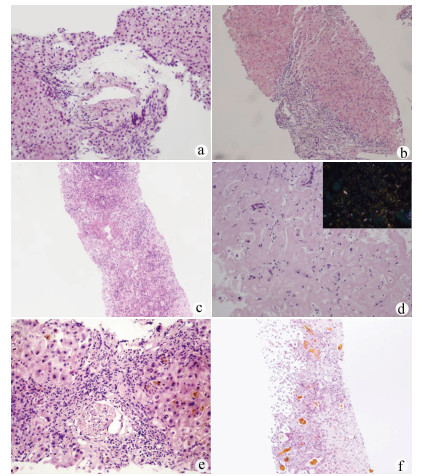
    在肝穿刺病理诊断辅助下,患者出院明确诊断26例(83.87%),其中特发性门静脉高压(idiopathic portal hypertension,IPH)5例,代表病例1:女,39岁,入院诊断“门静脉高压、腹水”(图 1a),结合临床诊断IPH。药物性肝衰竭或肝硬化9例,代表病例2:男,34岁,入院诊断“肝硬化失代偿期”(图 1b),结合临床诊断药物性肝衰竭肝损伤。自身免疫性肝脏疾病包括原发性胆汁性胆管炎(PBC)、自身免疫性肝炎(AIH)、重叠综合征,以及小胆管型原发性硬化性胆管炎(PSC)共5例,代表病例3:女,33岁,入院诊断“乙型肝炎肝衰竭”(图 1c),结合临床诊断AIH慢加急性肝衰竭、乙型肝炎肝硬化。其他原因肝硬化或肝衰竭患者7例,包括肝豆状核变性1例、甲状腺功能减退相关肝硬化1例、肝淀粉样变2例、阻塞性胆管炎1例、酒精性肝衰竭合并溶血性疾病1例,辅助排除肝脏肿瘤1例,代表病例4:男,74岁,入院诊断“肝硬化,布加综合征?”(图 1d),结合临床诊断肝淀粉样变性。尚不能明确病因患者5例(16.13%),代表病例5:男,72岁,入院诊断“肝功能异常,原因待查,腹水”,无明确药物应用史(图 1e)。病理会诊不能明确病因,治疗无效。临床诊断不明原因肝衰竭。此外,为证实诊断及决策是否肝移植患者3例,代表病例6:男,23岁,入院诊断“急性肝衰竭”。结合临床诊断急性药物性肝衰竭,3次人工肝治疗,病情无好转拟行肝移植,肝穿刺后结合病理结果决定保守治疗(图 1f),后经治疗病情好转。

    图  1  部分患者肝活检病理结果
    注: a,汇管区内门静脉壁厚,部分肝窦轻度扩张,未见肝硬化表现(HE染色,×200);b,肝硬化,Laennec分期F4C(HE染色,×100);c,假小叶形成(肝硬化),明显的融合状坏死和桥接坏死,大量的炎性细胞浸润,界面炎明显(HE染色,×10);d,肝窦充满均质粉染物,肝细胞萎缩消失,偏振光下呈苹果绿折光性(右上,刚果红)(HE染色,×400);e,汇管区中度混合性炎细胞浸润,固有胆管尚存,周边细胆管反应增生,有的细胆管扩张含胆栓(HE染色,×200);f,亚急性肝炎,重度肝内淤胆,肝细胞及毛细胆管淤胆易见(HE染色,×10)。

    临床工作中约有20%的肝衰竭或肝硬化患者由于凝血机制障碍或腹水不能实施经PLB获取肝组织行病理检查[6],从而影响疾病的诊治。全球范围内不明原因肝衰竭的占比为10%~50%[7-8]。随着我国人民生活水平的提高,乙型肝炎肝衰竭占比逐年下降,不明原因肝衰竭的占比呈逐年升高趋势,迫切需要病理检查辅助提高疑难重症肝病的确诊率。TJLB因其可克服出血的优势,提供了重症肝病患者病理检测手段。

    本研究纳入的31例患者中5例明确诊断IPH。IPH的发病机制目前未完全清楚,缺乏特异的检测指标,临床诊断较困难,肝活检对于IPH的诊断尤为重要[9-11]。IPH患者一般以肝硬化致门静脉高压合并腹水、消化道出血等起病,失去实施PLB时机,而临床无病理检查诊断较为困难。该组患者16.13%明确诊断为IPH,提示TJLB为隐源性肝硬化患者中IPH的诊断提供了重要手段。药物性肝炎诊断中,肝脏病理检查并不是必要条件,但需要排除其他疾病,特别是需与自身免疫性肝病进行鉴别,而非典型AIH的诊断,肝脏病理检查是必需的[12],因此对于药物性肝损伤的患者病理检查有时格外重要。该组9例患者经病理协助明确诊断药物性肝炎(29.03%),为后续治疗提供帮助。非典型自身免疫性肝脏疾病包括PBC、AIH、重叠综合征、PSC等,这类疾病的诊断依赖病理检查,同时,相关重症患者是否启用激素治疗是救治成功的关键,而激素治疗方案的制订需要病理辅助。该组患者协助明确诊断2例非典型PBC、2例非典型AIH及1例小胆管型PSC,并指导治疗获得较好的临床疗效。肝衰竭的早期精准预后相当重要,特别是指导选择内科保守治疗还是肝移植方面对患者尤为关键,且肝移植的时机也是术后良好恢复的重要参考指标。虽然临床上有较多的模型及指标进行预后判定,但肝脏病理在肝衰竭精准预后方面发挥重要作用,该组患者经过检查有3例患者改变了治疗方式的选择,精准指导肝移植治疗方案的制定。另外,通过肝穿刺病理检查可协助肝硬化晚期或肝衰竭患者确诊罕见病例,包括肝豆状核变性、甲状腺功能减退相关肝硬化、肝淀粉样变性等。

    TJLB在获取肝组织行病理检查协助诊断的同时,可进行HVPG测定,获得血液动力学参数,对于确定是否存在门静脉高压,以及属于窦前、窦性、窦后哪一种类型,有较好的提示意义[13]。本组患者进行HVPG测量对门静脉高压的诊断起到了辅助作用,但HVPG的测量受体位、管路、肝内分流、导管位置等因素影响[14],测量数据可能欠精确,因此测量数值偏差较大,在今后的扩大研究中应注意对同类患者探索对应的分级标准,以便更好的协助诊断和评估预后。

    病理检查在评价肝脏病理严重程度方面较为精准,在医学诊断中俗有“金标准”的地位[15],但是在病因确认方面尚有一定的局限性。一方面缘于病理损害缺乏特征性改变,对于某些肝硬化患者,肝组织病理检查可明确肝纤维化及肝脏炎症程度,但可能无法观察到疾病典型病理特征,不能提出明确的临床诊断。另一方面可能与临床医师及病理医师的知识面不够全面有关。有时肝组织病理检查只是起到排除诊断和提供诊断线索的作用,因此,病理检查只是提供诊断依据的辅助手段,而非临床最终诊断。疑难肝病的诊断需要临床医师具有全面的医学知识和丰富的临床经验,提供病理科医师更全面的信息,并结合其他检查方法,如骨髓穿刺、基因检测、血管成像等才能最终明确。TJLB费用相对较高,且也许不能获得良好预期,临床医师应严格掌握适应证,避免医疗纠纷。

    该组患者肝组织获取成功率为100%,无明显不良反应,且获取肝组织基本符合病理检测要求,提示TJLB手术成功率较高。据文献[16-17]报道,仍有少数患者组织获取质量欠佳。一方面,实施TJLB的患者部分为肝衰竭患者,肝组织炎症水肿明显,获取肝组织过程中容易破碎; 另一方面,部分为肝硬化患者,肝组织纤维化严重,获取肝组织时受纤维组织的牵拉,组织容易断裂,获取的肝组织有时难以达到病理检测的要求,影响病理医师的判定,因此术中应注意观察获取的组织质量,必要时需反复穿刺。

    TJLB为肝硬化合并腹水、肝衰竭及凝血机制障碍的肝病患者提供了肝组织获取的方法,同时可进行HVPG测定,对协助疑难重症肝病诊断及评估预后发挥重要作用,但尚有一定局限性,临床上应注意筛选适宜患者实施。

  • [1]YE SL.Current challenges to primary liver cancer research[J].J Clin Hepatol, 2015, 31 (6) :819-823. (in Chinese) 叶胜龙.原发性肝癌研究当前面临的挑战[J].临床肝胆病杂志, 2015, 31 (6) :819-823.
    [2]QIN N, YANG F, LI A, et al.Alterations of the human gut microbiome in liver cirrhosis[J].Nature, 2014, 513 (7516) :59-64.
    [3]CHEN Y, YANG F, LU H, et al.Characterization of fecal microbial communities in patients with liver cirrhosis[J].Hepatology, 2011, 54 (2) :562-572.
    [4]LU H, WU Z, XU W, et al.Intestinal microbiota was assessed in cirrhotic patients with hepatitis B virus infection.Intestinal microbiota of HBV cirrhotic patients[J].Microb Ecol, 2011, 61 (3) :693-703.
    [5]Ministry of Health of the People's Republic of China.Diagnosis, management, and treatment of hepatocellular carcinoma (V2011) [J].J Clin Hepatol, 2011, 27 (11) :1141-1159. (in Chinese) 中华人民共和国卫生部.原发性肝癌诊疗规范 (2011年版) [J].临床肝胆病杂志, 2011, 27 (11) :1141-1159.
    [6]CAPORASO JG, KUCZYNSKI J, STOMBAUGH J, et al.QIIME allows analysis of high-throughput community sequencing data[J].Nat Methods, 2010, 7 (5) :335-336.
    [7]Greengenes.The greengenes database downloads[DB/OL].http://greengenes.secondgenome.com/downloads/database/13_5.
    [8]TAO X, WANG N, QIN W.Gut Microbiota and Hepatocellular Carcinoma[J].Gastrointest Tumors, 2015, 2 (1) :33-40.
    [9]QIN J, LI R, RAES J, et al.A human gut microbial gene catalogue established by metagenomic sequencing[J].Nature, 2010, 464 (7285) :59-65.
    [10]MADSEN BS, HAVELUND T, KRAG A.Targeting the gut-liver axis in cirrhosis:antibiotics and non-selective beta-blockers[J].Adv Ther, 2013, 30 (7) :659-670.
    [11]SUNG JY, SHAFFER EA, COSTERTON JW.Antibacterial activity of bile salts against common biliary pathogens.Effects of hydrophobicity of the molecule and in the presence of phospholipids[J].Dig Dis Sci, 1993, 38 (11) :2104-2112.
    [12]GUNNARSDOTTIR SA, SADIK R, SHEV S, et al.Small intestinal motility disturbances and bacterial overgrowth in patients with liver cirrhosis and portal hypertension[J].Am J Gastroenterol, 2003, 98 (6) :1362-1370.
    [13]WIEST R, LAWSON M, GEUKING M.Pathological bacterial translocation in liver cirrhosis[J].J Hepatol, 2014, 60 (1) :197-209.
    [14]LI DY, YANG M, EDWARDS S, et al.Nonalcoholic fatty liver disease:for better or worse, blame the gut microbiota?[J].JPEN J Parenter Enteral Nutr, 2013, 37 (6) :787-793.
    [15]SEKI E, de MINICIS S, OSTERREICHER CH, et al.TLR4 enhances TGF-beta signaling and hepatic fibrosis[J].Nat Med, 2007, 13 (11) :1324-1332.
    [16]LOO TM, KAMACHI F, WATANABE Y, et al.Gut microbiota promotes obesity-associated liver cancer through PGE2-mediated suppression of antitumor immunity[J].Cancer Discov, 2017, 7 (5) :522-538
    [17]BRANDI G, de LORENZO S, CANDELA M, et al.Microbiota, NASH, HCC and the potential role of probiotics[J].Carcinogenesis, 2017, 38 (3) :231-240.
    [18]HUANG XF.The role of bile acid signaling in liver carcinogenesis and liver regeneration[D].Fuzhou:Fujian Med Univ, 2007. (in Chinese) 黄雄飞.胆酸信号与肝癌发生和肝脏再生[D].福州:福建医科大学, 2007.
    [19]YOSHIMOTO S, LOO TM, ATARASHI K, et al.Obesity-induced gut microbial metabolite promotes liver cancer through senescence secretome[J].Nature, 2013, 499 (7456) :97-101.
    [20]YU LX, YAN HX, LIU Q, et al.Endotoxin accumulation prevents carcinogen-induced apoptosis and promotes liver tumorigenesis in rodents[J].Hepatology, 2010, 52 (4) :1322-1333.
    [21]DAPITO DH, MENCIN A, GWAK GY, et al.Promotion of hepatocellular carcinoma by the intestinal microbiota and TLR4[J].Cancer Cell, 2012, 21 (4) :504-516.
    [22]ZHANG HL, YU LX, YANG W, et al.Profound impact of gut homeostasis on chemically-induced pro-tumorigenic inflammation and hepatocarcinogenesis in rats[J].J Hepatol, 2012, 57 (4) :803-812.
    [23]MITSUOKA T.Bifidobacteria and their role in human health[J].J Ind Microbiol, 1990, 6 (4) :263-267.
    [24]SHETH AA, GARCIA-TSAO G.Probiotics and liver disease[J].J Clin Gastroenterol, 2008, 42 (Suppl 2) :s80-s84.
    [25]MALAGUARNERA M, GRECO F, BARONE G, et al.Bifidobacterium longum with fructo-oligosaccharide (FOS) treatment in minimal hepatic encephalopathy:a randomized, double-blind, placebo-controlled study[J].Dig Dis Sci, 2007, 52 (11) :3259-3265.
    [26]ZHANG M, ZHENG M, WU Z, et al.Alteration of gut microbial community after N, N-Dimethylformamide exposure[J].J Toxicol Sci, 2017, 42 (2) :241-250.
    [27]HE J, LIU J, KONG Y, et al.Serum activities of liver enzymes in workers exposed to sub-TLV levels of dimethylformamide[J].Int J Occup Med Environ Health, 2015, 28 (2) :395-398.
    [28]KIM KW, CHUNG YH.Hepatotoxicity in rats treated with dimethylformamide or toluene or both[J].Toxicol Res, 2013, 29 (3) :187-193.
    [29]LIU HX, ROCHA CS, DANDEKAR S, et al.Functional analysis of the relationship between intestinal microbiota and the expression of hepatic genes and pathways during the course of liver regeneration[J].J Hepatol, 2016, 64 (3) :641-650.
    [30]ISLAM KB, FUKIYA S, HAGIO M, et al.Bile acid is a host factor that regulates the composition of the cecal microbiota in rats[J].Gastroenterology, 2011, 141 (5) :1773-1781.
    [31]RIDLON JM, ALVES JM, HYLEMON PB, et al.Cirrhosis, bile acids and gut microbiota:unraveling a complex relationship[J].Gut Microbes, 2013, 4 (5) :382-387.
    [32]GRAT M, WRONKA KM, KRASNODEBSKI M, et al.Profile of gut microbiota associated with the presence of hepatocellular cancer in patients with liver cirrhosis[J].Transplant Proc, 2016, 48 (5) :1687-1691.
  • Relative Articles

    [1]Han Fang, Xiao HuiJuan, Zhang Xiang, Han Tao. Association between muscle loss and nutritional risk in patients with liver cirrhosis or hepatocellular carcinoma[J]. Journal of Clinical Hepatology, 2020, 36(8): 1753-1756. doi: 10.3969/j.issn.1001-5256.2020.08.015
    [2]Huang ChunYang, Chen Jie, Liu YanMin, Dan Jing. Role of intestinal flora in the development/progression and treatment of autoimmune liver diseases[J]. Journal of Clinical Hepatology, 2019, 35(1): 205-207. doi: 10.3969/j.issn.1001-5256.2019.01.045
    [3]Chen MuXing, Wang ShaoYang. Role of intestinal dysbacteriosis in the pathogenesis of acute-on-chronic hepatitis B liver failure[J]. Journal of Clinical Hepatology, 2019, 35(3): 665-668. doi: 10.3969/j.issn.1001-5256.2019.03.048
    [4]Li Wei, Xing HuiChun. Intestinal microflora and alcoholic liver disease[J]. Journal of Clinical Hepatology, 2019, 35(3): 481-484. doi: 10.3969/j.issn.1001-5256.2019.03.006
    [5]Cai WenJun, Luan YuTing, Liu ChengHai, Jiang ShiLi, Zhao ZhangQing, Gu HongTu, Yuan JiLi, Xing Feng, Zhou Yang, Lu: Jing, Mu YongPing. Features of intestinal microbiota in hepatitis B cirrhosis patients with amassment or tympanites type of liver-kidney Yin deficiency syndrome: A comparative analysis[J]. Journal of Clinical Hepatology, 2019, 35(4): 785-789. doi: 10.3969/j.issn.1001-5256.2019.04.016
    [6]Li YunPeng, Wang XinHong, Zhang DeKai. Influence of intestinal dysbacteriosis on liver cirrhosis[J]. Journal of Clinical Hepatology, 2019, 35(4): 887-890. doi: 10.3969/j.issn.1001-5256.2019.04.040
    [7]Li Xiang, Cai Jie, Xu YuWen, Yang Fen, Sun ALei, Weng YaLi. A case of hepatolenticular degeneration with hepatocellular carcinoma[J]. Journal of Clinical Hepatology, 2018, 34(9): 1967-1971. doi: 10.3969/j.issn.1001-5256.2018.09.027
    [8]Zhou YiQing, Ceng Min, Zhou Jie, Ruan YongChun, Zhang HaiWang, Su MeiXia, He Wei, Li MingHui. Drug resistance of Enterobacteriaceae bacteria causing liver abscess[J]. Journal of Clinical Hepatology, 2018, 34(8): 1750-1753. doi: 10.3969/j.issn.1001-5256.2018.08.031
    [9]Lu BingJie, Zhao YaHong, An YongTong, Ye Guan, Sun MingYu. Research advances in gut microbiota in liver cirrhosis and related complications[J]. Journal of Clinical Hepatology, 2018, 34(11): 2433-2437. doi: 10.3969/j.issn.1001-5256.2018.11.037
    [10]Huang Rong, Ni JiaJia, Gao Yi. The association between the intestinal microbiota-lipopolysaccharide-Toll-like receptor 4 axis and hepatocellular carcinoma[J]. Journal of Clinical Hepatology, 2018, 34(6): 1325-1328. doi: 10.3969/j.issn.1001-5256.2018.06.041
    [11]Li Peng, Ding HuiGuo, Xu Hui, Zhang ShiBin, Zhang YueNing, Wang ZhenBiao, Wu YanJing, Yu HaiBin. Shortening liver cancer screening interval may improve the prognosis of patients with hepatitis B cirrhosis-related hepatocellular carcinoma[J]. Journal of Clinical Hepatology, 2017, 33(7): 1301-1304. doi: 10.3969/j.issn.1001-5256.2017.07.019
    [12]Han MeiLin, Gong ZhenHua. Mechanism of action of gut microbiota in hepatobiliary diseases[J]. Journal of Clinical Hepatology, 2017, 33(2): 384-388. doi: 10.3969/j.issn.1001-5256.2017.02.040
    [13]Liu JiaQi, Zhou ShaoMing. Research advances in intestinal flora in nonalcoholic fatty liver disease[J]. Journal of Clinical Hepatology, 2017, 33(12): 2453-2456. doi: 10.3969/j.issn.1001-5256.2017.12.045
    [14]Zhang YuanYuan, Li Jing, Chi YuJing, Su Lin, Liu YuLan. Role of intestinal mucosal barrier in the development and progression of nonalcoholic fatty liver disease[J]. Journal of Clinical Hepatology, 2016, 32(12): 2391-2394. doi: 10.3969/j.issn.1001-5256.2016.12.035
    [15]Wei XinPeng, Gao Xiao, Ma YingJie. Relationship between small intestinal bacterial overgrowth and severity of with hepatitis B cirrhosis[J]. Journal of Clinical Hepatology, 2016, 32(4): 716-719. doi: 10.3969/j.issn.1001-5256.2016.04.021
    [16]Wang XiaoTong, Han Tao. A review of relationship between gut microecology and liver diseases[J]. Journal of Clinical Hepatology, 2015, 31(4): 642-645. doi: 10.3969/j.issn.1001-5256.2015.04.041
    [17]Zhang QiuNan, Xiao Ying, Miao RuiXin, Zhang YaQing, Zhu Ying. Changes in intestinal flora in nonalcoholic fatty liver disease: their correlation with insulin resistance index,tumor necrosis factor- α,and interleukin- 6[J]. Journal of Clinical Hepatology, 2015, 31(7): 1078-1081. doi: 10.3969/j.issn.1001-5256.2015.07.019
    [18]Wang AiPing, Wen ChunYang. Recent perspectives in hepatocellular carcinoma (HCC) diagnosis and treatment[J]. Journal of Clinical Hepatology, 2013, 29(1): 32-35.
    [19]Chen Kai, Zhao Ji, Deng XiaoFan, Yang HongJi. A rare case of hepatocellular carcinoma featuring association with the pancreas and duodenum, and giant cells in the portacaval space and the post-superior mesenteric artery region[J]. Journal of Clinical Hepatology, 2012, 28(11): 865-866.
    [20]Li HaiPing, Tao JianShu, Gao Bo, Yu ZongTao, Li WenCui, Zhang JiCai. The expression and clinical significance of resistance gene in hepatocellular carcinoma[J]. Journal of Clinical Hepatology, 2011, 27(9): 972-975.
  • Cited by

    Periodical cited type(2)

    1. 刘继云. 医学防治对HBV携带者子女HBV感染状况的影响研究. 中国实用医药. 2025(04): 107-109 .
    2. 周志强,孙星星,李世勇,万里,罗爱林,韩东吉. 新型冠状病毒肺炎流行期间剖宫产术的麻醉管理. 中华麻醉学杂志. 2020(03): 291-295 .

    Other cited types(0)

  • 加载中

Catalog

    通讯作者: 陈斌, bchen63@163.com
    • 1. 

      沈阳化工大学材料科学与工程学院 沈阳 110142

    1. 本站搜索
    2. 百度学术搜索
    3. 万方数据库搜索
    4. CNKI搜索

    Article Metrics

    Article views (2495) PDF downloads(549) Cited by(2)
    Proportional views
    Related

    /

    DownLoad:  Full-Size Img  PowerPoint
    Return
    Return