中文English
ISSN 1001-5256 (Print)
ISSN 2097-3497 (Online)
CN 22-1108/R

Association of tuberous sclerosis gene 1/2 mutations with the progression of hepatocellular carcinoma and prognosis

DOI: 10.3969/j.issn.1001-5256.2021.01.017
  • Received Date: 2020-07-02
  • Accepted Date: 2020-08-03
  • Published Date: 2021-01-20
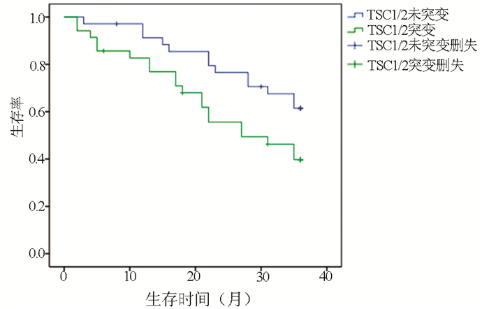
  •   Objective  To investigate the association of tuberous sclerosis gene 1/2 (TSC1/2) mutation with disease severity and prognosis in patients with hepatocellular carcinoma (HCC), and to provide a feasible basis for the diagnosis and treatment of HCC.  Methods  A total of 492 patients with HCC who were admitted to The Affiliated Hospital of Jiangsu University from January 2012 to January 2020 were enrolled, among whom 59 had TSC1/2 mutations (20 with TSC1 mutations, 41 with TSC2 mutations, and 2 had both TSC1 and TSC2 mutations). The clinical features of patients with TSC1/2 mutations were analyzed, and the association of TSC1/2 mutations with the clinical stage of HCC was analyzed. The 35 patients in the mutation group and 35 in the non-mutation group were followed up for 3 years to observe the effect of TSC1/2 mutations on the prognosis of HCC. The chi-square test was used for comparison of categorical data between groups; the Kruskal-Wallis H test was used for comparison of ranked data between groups; a multivariate logistic regression analysis was used to investigate association; the Kaplan-Meier survival analysis was used to analyze follow-up data.  Results  For the 492 patients with HCC, the overall TSC1/2 mutation rate was 11.99%. There were no significant differences in sex, age, Child score, and tumor size between the TSC1/TSC2 mutation group and the non-mutation group (all P > 0.05), while there were significant differences in tumor number, extrahepatic metastasis, and PS score between the two groups (all P < 0.05). The logistic regression analysis showed that TSC1/TSC2 gene mutation was positively correlated with the severity of HCC (odds ratio=1.706, P < 0.05). The follow-up results showed that the TSC1/2 mutation group had a significantly lower survival rate than the non-mutation group, and there was a significant difference in 3-year mortality rate between the TSC1/2 mutation group and the non-mutation group (60.3% vs 38.6%, χ2=3.923, P < 0.05).  Conclusion  TSC1/TSC2 gene mutation may predict the malignant progression of HCC in the early stage, and patients with TSC1/2 mutation tend to have poor prognosis. Targeted drug therapy for gene mutations may have a certain effect in delaying the progression of HCC.


  • HBV慢性感染是肝细胞癌(HCC) 发生的最常见病因,HCC的发病率和病死率在恶性肿瘤中分别占第6位和第4位,严重威胁着人类健康[1]。HCC细胞异常增殖可使血清糖蛋白N-糖链组成和结构发生改变,因此,血清糖蛋白的N-聚糖可作为HCC诊断标志物和HCC治疗的分子靶点[2]。N-聚糖改变的基础在于肝细胞内糖基转移酶活性的变化,其中多数糖基转移酶表达与肝脏功能密切相关。当肝细胞发生癌变时,某些糖基转移酶基因被激活,其表达异常增加,或基因表达受抑制而含量下降,从而导致一些蛋白质异常糖基化修饰。大量研究[3]表明,这种异常糖基化修饰与肿瘤细胞恶性侵袭行为密不可分。笔者前期研究[4]发现,与健康对照相比,HBV相关HCC(HBV-HCC)患者血清中N-聚糖图谱发生一系列特征改变,其中二天线N-聚糖峰1(peak1, NGA2F)丰度升高;三天线N-聚糖峰9(peak9, NA3Fb)丰度特异性升高;其他三天线和四天线N-聚糖峰(peak10 NA3Fc、peak11 NA4、peak12 NA4Fb)丰度也有不同程度的升高。但目前HBV-HCC患者血清N-聚糖变化机制尚未完全阐明。本研究中,通过检测HBV-HCC患者癌组织与癌旁组织中8种重要的糖基转移酶基因(包括岩藻糖基转移酶FUT3、FUT4、FUT6、FUT7、FUT8和N-乙酰氨基葡萄糖转移酶Gn-TⅢ、Gn-TⅣa、Gn-TⅤ)表达水平变化并比较其差异,探索HBV-HCC患者血清N-聚糖变化的可能机制。

    收集解放军总医院2018年9月—2019年11月HBV-HCC行手术患者的肝癌和癌旁组织及正常肝组织标本,同时采集血清标本,置于-80 ℃冰箱保存。患者符合以下入选标准: (1)均为感染HBV的HCC患者;(2)排除HAV、HCV、HDV、HEV等肝炎病毒感染;(3)患者术后病理标本均经医院病理科确诊为HCC,肝癌诊断符合原发性肝癌诊疗规范(2019年版)[5]。同时收集HCC患者临床资料。另外选取20例健康成年人血清作为对照。

    应用SPSS 20.0软件从34例HCC患者中随机选择8例HCC患者血清标本作为HCC试验组,20例健康成年人血清标本作为对照组。采用DSA-FACE法检测和分析血清N-聚糖图谱[6],具体步骤如下:

    (1) 寡糖的释放:取3 μl血清,加入含有2 μl 10 mmol/L NH4HCO3缓冲液和3 μl去离子水的PCR反应板中, 反应板放入PCR仪器,95 ℃加热5 min后冷却至4 ℃, 然后加入3 μl PNGaseF(2.2 U/μl),37 ℃孵育3 h,之后加入100 μl去离子水终止反应,标记为D板。

    (2) 标记寡糖:从D板吸取10 μl溶液加入一新的PCR反应板中,开盖在60 ℃条件下烘干90 min,加入3 μl标记溶液(20 mmol/L APTS∶ 1 mol/L NaCNBH3=1∶ 1),90 ℃反应2 h,加入100 μl去离子水终止反应,标记为L板。

    (3) 去唾液酸:从L板中取2 μl溶液加入一新的PCR反应板,加入0.25 μL 100 mmol/L NH4Ac(pH=5.0)、0.2 μl唾液酸酶(2.5 U/μl)和1.55 μl去离子水, 震荡混匀后42 ℃孵育4 h,加40 μl去离子水终止反应,标记为DE板。

    (4) DNA测序仪上机检测:取DE板10 μl溶液加入ABI测序仪专用96孔板,放入ABI 3500测序仪进行N-聚糖图谱分析,数据经GeneMapper软件分析。

    冻存的肝组织放入超声震荡仪研磨后,用Trizol试剂提取总RNA,用Nano Drop One检测总RNA的浓度和纯度。将总RNA逆转录为cDNA后,用荧光定量PCR仪(ABI 7500 FAST)进行cDNA扩增,反应体系为20 μl。反应条件为:(1)95 ℃ 30 s;(2)95 ℃ 5 s,60 ℃ 34 s共40个循环;(3)溶解曲线:95 ℃ 15 s,60 ℃ 1 min, 95 ℃ 15 s。

    以RPS11作为内参基因,目的基因的相对表达量用2-△△CT表示。癌组织和癌旁组织糖基转移酶mRNA相对表达量检测分析的对照均是正常肝组织。FUT3、FUT4、FUT6、FUT7、FUT8、Gn-TⅢ、Gn-TⅣa和Gn-TⅤ基因的特异引物由生工生物工程股份有限公司合成,引物序列见表 1

    表  1  荧光定量PCR检测糖基转移酶基因引物序列表
    基因 序列号 引物序列(5′→3′)
    FUT3 NM_001374740.1 F: CAA CAG AGA AAG CAG GCA
    R: AAG AAA CAC ACA GCC ACC[7]
    FUT4 XM_032167182.1 F: TCC TAC GGA GAG GCT CAG
    R: TCC TCG TAG TCC AAC ACG[7]
    FUT6 XM_011527879.3 F: CAT TTC TGC TGC CTC AGG
    R: GGG CAA GTC AGG CAA CTC[7]
    FUT7 NM_004479.4 F: CCA CGA TCA CCA TCC TTG
    R: AGG CTT CGG TTG GCA CTC[7]
    FUT8 XM_032180460.1 F: TCT AGC CGA GAA CTG TCC
    R: GCT GCT CTT CTA AAA CGC[7]
    Gn-TⅢ XM_019018480.2 F: CCG CCA CAA GGT GCT CTA T
    R: GAT CTC GTC CGC ATC GTC A[8]
    Gn-TⅣa XM_032178740.1 F: ACC AAG GGC ATA CGC TGG AG
    R: GTT CTT GGT TGC CGC TAT GGA[9]
    Gn-TV XM_032411636.1 F: GCT GCC CAA CTG TAG GAG AC
    R: GAA TCA AGG ACT CGG AGC AT[10]
    RPS11 XM_032159559.1 F: GCC GAG ACT ATC TGC ACT AC
    R: ATG TCC AGC CTC AGA ACT TC[11]
    下载: 导出CSV 
    | 显示表格

    冻存的肝组织放入超声震荡仪研磨后,用含cOmplete蛋白酶抑制剂的RIPA裂解液提取蛋白,用BCA蛋白试剂盒来测定蛋白浓度。电泳分离不同分子量蛋白后,15 V恒压下用半干转电转仪将蛋白转移至PVDF膜,于5%的脱脂奶粉中室温封闭1 h,加入抗-FUT8(1∶ 1000)、抗-Gn-TⅣa(1∶ 1000)、抗-Gn-TⅤ(1∶ 1000)、抗-β-actin(1∶ 4000)一抗,4 ℃过夜,TBST溶液洗膜3次,分别加入抗鼠或抗兔的二抗(1∶ 5000),室温下孵育2 h,TBST溶液洗膜3次,加入增强型ECL化学发光试剂,凝胶成像仪扫描显影的条带,ImageJ软件分析条带灰度。以β-actin作为内参,目的蛋白的相对表达量用目的蛋白灰度值与内参蛋白灰度值的比值来表示。

    应用SPSS 20.0软件对数据进行统计分析。计量资料用x ±s表示,两组间比较采用独立样本t检验。所有统计学分析均采用双侧检验,P<0.05为差异具有统计学意义。

    34例HBV-HCC患者临床特征见表 2

    表  2  34例HBV-HCC患者临床特征
    临床特征 数值
    男性[例(%)] 29 (85.29)
    年龄(岁) 54.85±8.20
    HBV DNA(×106IU/ml) 2.35±7.15
    AFP(ng/ml) 556.82±1995.22
    ALT(U/L) 44.34±60.72
    AST(U/L) 40.71±44.25
    肿瘤直径(例)
    ≤5 cm 21
    5~10 cm 9
    ≥10 cm 4
    下载: 导出CSV 
    | 显示表格

    应用DSA-FACE法分析HCC试验组8例HBV-HCC患者与对照组20例健康成年人血清N-聚糖图谱(图 1),其特征改变与笔者前期研究发现的特征改变相同[4]

    图  1  HCC试验组与对照组血清N-聚糖图谱
    注:a,血清中12种N-聚糖丰度比较;b,12种N-聚糖结构。Peak1-Peak12图引自文献[4],Peak1: 二天线无半乳糖基核心ɑ-l, 6岩藻糖基化N聚糖(NGA2F);Peak2: 二天线无半乳糖基核心ɑ-l, 6岩藻糖基化平分型N聚糖(NGA2FB);Peak3/Peak4: 二天线核心ɑ-l, 6岩藻糖基化单支链半乳糖基N聚糖(NG1A2F);Peak5: 二天线N聚糖(NA2);Peak6: 二天线核心ɑ-l, 6岩藻糖基化N聚糖(NA2F);Peak7: 二天线平分型核心ɑ-l, 6岩藻糖基化N聚糖(NA2FB);Peak8: 三天线N聚糖(NA3);Peak9: 三天线支链ɑ-l, 3岩藻糖基化N聚糖(NA3Fb);Peak9’: 三天线核心ɑ-l, 6岩藻糖基化N聚糖(NA3Fc);Peak10: 三天线支链ɑ-l, 3岩藻糖基化与核心ɑ-l, 6岩藻糖基化N聚糖(NA3Fbc);Peak11: 四天线N聚糖(NA4);Peak12: 四天线支链ɑ-l, 3岩藻糖基化N聚糖(NA4 Fb)。

    与对照组相比,HCC试验组患者三天线N-聚糖峰9(peak9,NA3Fb)丰度明显升高(t=-2.514,P<0.05);血清二天线N-聚糖峰1(peak1,NGA2F)和四天线N-聚糖峰(peak11 NA4、peak12 NA4Fb)的丰度在两组间差异无统计学意义(P值均>0.05)。

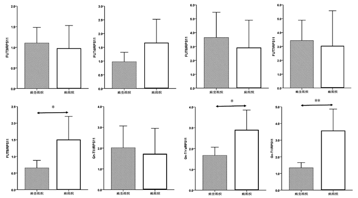
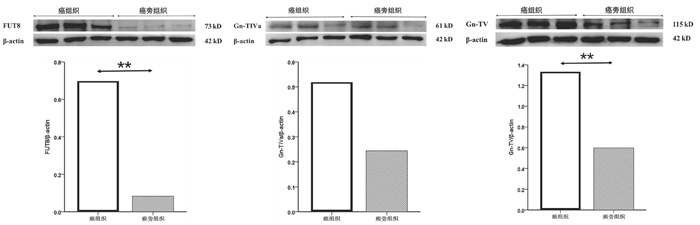
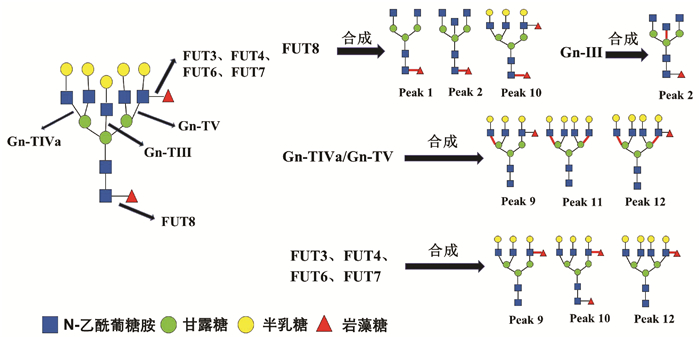
    癌组织中FUT8、Gn-TⅣa和Gn-TⅤ基因mRNA表达水平显著高于癌旁组织(1.50±0.34 vs 0.65± 0.11, t=-2.354,P=0.022; 2.90±0.47 vs 1.68±0.19, t=-2.403,P=0.019; 3.57±0.64 vs 1.33±0.16, t=-3.384,P=0.001),差异均有统计学意义。FUT3、FUT4、FUT6、FUT7和Gn-TⅢ mRNA的表达水平在癌组织与癌旁组织间比较差异均无统计学意义(P值均>0.05)(图 2)。

    图  2  癌组织与癌旁组织中8种糖基转移酶mRNA表达水平比较
    注:*,P<0.05,* *,P<0.01。

    进一步比较了HCC试验组中8例患者癌组织与癌旁组织中8种糖基转移酶mRNA表达水平。与癌旁组织相比,8例HCC试验组患者癌组织中Gn-TⅤ mRNA表达明显升高(Gn-TⅤ: 5.26±1.70 vs 1.49±0.33, t=-2.173, P=0.047);Gn-TⅢ、Gn-TⅣa、FUT4和FUT8 mRNA表达在癌组织与癌旁组织间无显著性差异(Gn-TⅢ: 1.03±0.46 vs 1.55±0.62, t=0.663, P=0.518;Gn-TⅣa: 5.15±1.50 vs 2.39±0.46, t=-1.752, P=0.102; FUT4: 1.56±1.12 vs 0.81±0.27, t=-0.653, P=0.524; FUT8: 2.61±1.26 vs 1.01±0.41, t=-1.213, P=0.245)。

    选取mRNA表达有显著性差异的3个糖基转移酶基因FUT8、Gn-TⅣa和Gn-TⅤ作为蛋白印迹实验的检测因子。结果如图 3所示,癌组织中FUT8与Gn-TⅤ的蛋白表达水平显著高于癌旁组织(0.70±0.11 vs 0.083±0.017, t=9.555, P=0.001; 1.33 ±0.19 vs 0.60±0.15, t=5.097, P=0.007);Gn-TⅣa的蛋白表达水平在癌组织与癌旁组织间比较差异无统计学意义(0.52±0.24 vs 0.24±0.11, t=1.833, P=0.141)。

    图  3  癌组织与癌旁组织糖基转移酶FUT8、Gn-TⅣa和Gn-TⅤ的蛋白表达水平比较
    注:* *,P<0.01

    HCC患者血清中常出现大量异常糖基化N-糖蛋白,分离糖蛋白的N-聚糖链进行表达图谱分析,可发现支链与核心岩藻糖基化N-聚糖和多天线N-聚糖含量增加,这些N-聚糖与HCC发生发展密切相关,可作为筛查和诊断HCC的特异性标志[12]。研究[4, 13]发现在HBV-HCC患者血清中支链岩藻糖基化三天线N-聚糖(peak9, NA3Fb)丰度显著升高,且核心岩藻糖基化二天线N-聚糖(peak1, NGA2F)、核心岩藻糖基化平分型二天线N-聚糖(peak2, NGA2FB)、支链与核心岩藻糖基化三天线N聚糖(peak10, NA3Fc)、四天线N-聚糖(peak11, NA4)和支链岩藻糖基化四天线N-聚糖(peak12, NA4Fb)丰度也有不同程度的升高(N-聚糖结构如图 4)。本研究通过对HBV-HCC患者癌组织与癌旁组织相关糖基转移酶基因表达检测分析,探寻HCC患者特异性血清N-聚糖变化的可能机制。

    图  4  HBV相关HCC患者血清特异变化N-聚糖与8种糖基转移酶之间的关系

    糖基转移酶Gn-TⅤ与Gn-TⅣa催化形成三天线及三天线以上N-聚糖的N-乙酰葡糖胺(GlcNAc)糖链结构[14-15],而Gn-TⅢ催化合成N-聚糖的平分型GlcNAc结构,Gn-TⅢ与Gn-TⅤ、Gn-TⅣa有拮抗作用(图 4)[16]。本研究发现,在癌组织中Gn-TⅤ与Gn-TⅣa mRNA表达水平显著高于癌旁组织(P<0.05),蛋白印迹实验也显示,在癌组织中Gn-TⅤ蛋白表达显著高于癌旁组织(P<0.05)。而Gn-TⅢ mRNA表达水平在癌组织与癌旁组织间无统计学差异(P=0.711)。本研究结果可以解释HCC试验组患者血清中N-聚糖变化:与对照组相比,8例HCC患者血清中三天线N-聚糖(peak9)丰度显著升高(P<0.05),而且这8例HCC患者癌组织Gn-TⅤ表达水平显著高于癌旁组织(P<0.05)。说明HCC患者血清中多天线N-聚糖丰度升高可能与糖基转移酶Gn-TⅤ高水平表达密切相关,促进含GlcNAc多分支(三天线及以上)N-聚糖的合成(图 4)。

    以往研究[4, 13]表明,在HBV-HCC患者血清中核心岩藻糖基化N-聚糖(peak1、peak2、peak10)丰度较高。本研究发现,在癌组织中核心岩藻糖基转移酶FUT8 mRNA与蛋白表达水平显著高于癌旁组织(P<0.05)。FUT8表达上调会导致核心岩藻糖基化修饰结构的N-聚糖增加,可能与癌细胞高转移潜能有关[17]。另外,AFP是临床上最常用的HCC血清学检测指标,有研究[18]发现HCC患者血清中AFP经FUT8催化作用下可形成核心岩藻糖基化AFP(AFP-L3),其与肿瘤的发展速度、肿瘤大小和肿瘤转移密切相关,且诊断HCC效力优于AFP。除了α-1, 6核心岩藻糖基化N-聚糖,α-1, 3分支岩藻糖基化N-聚糖的丰度在HBV-HCC患者血清也特异性升高。α-1, 3分支岩藻糖基化结构是三天线N-聚糖(peak9)中Lewis X结构形成的关键[4],参与合成α-1, 3分支岩藻糖基化结构且与HCC密切相关的糖基转移酶有FUT3、FUT4、FUT6、FUT7[19]。本研究显示,在34例HCC患者癌组织与癌旁组织间FUT3、FUT4、FUT6和FUT7 mRNA表达水平没有显著性差异(P>0.05),不能解释血清N-聚糖水平异常改变。结合以往研究报道分析FUT基因表达的检测结果,可能因为多种岩藻糖基转移酶参与合成α-1, 3分支岩藻糖基化结构,而目前对于这些酶相互作用机制尚未明确,较难判断岩藻糖基转移酶中那一种亚类起主导作用。本研究主要检测8种与HCC密切相关的糖基转移酶基因表达,初步阐述HBV-HCC患者血清中N-聚糖变化的可能机制。但对于不同糖基转移酶之间相互作用的具体机制以及其他血清N-聚糖(如异常唾液酸化修饰的N-聚糖等)变化机制,还有待于进一步研究。

  • [1]
    BUDNY A, KOZŁOWSKI P, KAMIN'SKA M, et al. Epidemiology and risk factors of hepatocellular carcinoma[J]. Pol Merkur Lekarski, 2017, 43(255): 133-139.
    [2]
    ZHU AX, WALLNER KE, FRIVOLD GP, et al. Prostate brachytherapy seed migration to the right coronary artery associated with an acute myocardial infarction[J]. Brachytherapy, 2006, 5(4): 262-265. DOI: 10.1016/j.brachy.2006.08.004
    [3]
    CROCETTI L, BARGELLINI I, CIONI R. Loco-regional treatment of HCC: Current status[J]. Clin Radiol, 2017, 72(8): 626-635. DOI: 10.1016/j.crad.2017.01.013
    [4]
    ALGHAMDI MA, LEE YR, SWIHA M, et al. The effect of sorafenib (S) starting dose and dose intensity on survival in patients with advanced hepatocellular carcinoma (HCC)[J]. J Clin Oncol, 2017, 9(14): 4918-4928.
    [5]
    KAPLAN DE, YU S, TADDEI TH, et al. Up-titration of sorafenib for hepatocellular carcinoma: Impact on duration of exposure and cost[J]. J Clin Oncol, 2017, 35: 385.
    [6]
    CLARK JW, EDER JP, RYAN D, et al. Safety and pharmacokinetics of the dual action Raf kinase and vascular endothelial growth factor receptor inhibitor, BAY 43-9006, in patients with advanced, refractory solid tumors[J]. Clin Cancer Res, 2005, 11(15): 5472-5480. DOI: 10.1158/1078-0432.CCR-04-2658
    [7]
    STRUMBERG D, AWADA A, HIRTE H, et al. Pooled safety analysis of BAY 43-9006 (sorafenib) monotherapy in patients with advanced solid tumours: Is rash associated with treatment outcome?[J]. Eur J Cancer, 2006, 42(4): 548-556. DOI: 10.1016/j.ejca.2005.11.014
    [8]
    ZUCMAN-ROSSI J, VILLANUEVA A, NAULT JC, et al. Genetic landscape and biomarkers of hepatocellular carcinoma[J]. Gastroenterology, 2015, 149(5): 1226-1239.e4. DOI: 10.1053/j.gastro.2015.05.061
    [9]
    JOZWIAK J, JOZWIAK S, WLODARSKI P. Possible mechanisms of disease development in tuberous sclerosis[J]. Lancet Oncol, 2008, 9(1): 73-79. DOI: 10.1016/S1470-2045(07)70411-4
    [10]
    NORTHRUP H, KRUEGER DA, International Tuberous Sclerosis Complex Consensus Group. Tuberous sclerosis complex diagnostic criteria update: Recommendations of the 2012 Iinternational Tuberous Sclerosis Complex Consensus Conference[J]. Pediatr Neurol, 2013, 49(4): 243-254. DOI: 10.1016/j.pediatrneurol.2013.08.001
    [11]
    LI SQ, LI HZ, ZHANG YS. Clinical analysis of spontaneous rupture in tuberous sclerosis-related renal angiomyolipoma[J]. J Clin Urol, 2014, 29(12): 1070-1072. (in Chinese) https://www.cnki.com.cn/Article/CJFDTOTAL-LCMW201412010.htm

    李书强, 李汉忠, 张玉石. 结节性硬化症相关肾错构瘤自发性破裂出血临床诊治分析[J]. 临床泌尿外科杂志, 2014, 29(12): 1070-1072. https://www.cnki.com.cn/Article/CJFDTOTAL-LCMW201412010.htm
    [12]
    XU B, ZHANG Q, JIN J. Laparoscopic aspiration for central renal angiomyolipoma: A novel technique based on single-center initial experience[J]. Urology, 2013, 81(2): 313-318. DOI: 10.1016/j.urology.2012.09.047
    [13]
    HO DWH, CHAN LK, CHIU YT, et al. TSC1/2 mutations define a molecular subset of HCC with aggressive behaviour and treatment implication[J]. Gut, 2017, 66(8): 1496-1506. DOI: 10.1136/gutjnl-2016-312734
    [14]
    Chinese Society of Clinical Oncology. CSCO guidelines for diagnosis and treatment of hepatocellular carcinoma(2020)[M]. Beijing: China People's Health Press, 2020: 28-29. (in Chinese)

    中国临床肿瘤学会. 原发性肝癌诊疗指南2020[M]. 北京:中国人民卫生出版社, 2020: 28-29.
    [15]
    CALDERARO J, COUCHY G, IMBEAUD S, et al. Histological subtypes of hepatocellular carcinoma are related to gene mutations and molecular tumour classification[J]. J Hepatol, 2017, 67(4): 727-738. DOI: 10.1016/j.jhep.2017.05.014
    [16]
    NISHIDA N, KUDO M. Oncogenic signal and tumor microenvironment in hepatocellular carcinoma[J]. Oncology, 2017, 93(Suppl 1): 160-164.
    [17]
    DIBBLE CC, MANNING BD. Signal integration by mTORC1 coordinates nutrient input with biosynthetic output[J]. Nat Cell Biol, 2013, 15(6): 555-564. DOI: 10.1038/ncb2763
    [18]
    HOWELL JJ, RICOULT SJ, BEN-SAHRA I, et al. A growing role for mTOR in promoting anabolic metabolism[J]. Biochem Soc Trans, 2013, 41(4): 906-912. DOI: 10.1042/BST20130041
    [19]
    European Chromosome 16 Tuberous Sclerosis Consortium. Identification and characterization of the tuberous sclerosis gene on chromosome 16[J]. Cell, 1993, 75(7): 1305-1315. DOI: 10.1016/0092-8674(93)90618-Z
    [20]
    CRINO PB, NATHANSON KL, HENSKE EP. The tuberous sclerosis complex[J]. N Engl J Med, 2006, 355(13): 1345-1356. DOI: 10.1056/NEJMra055323
    [21]
    NABBOUT R, BELOUSOVA E, BENEDIK MP, et al. Epilepsy in tuberous sclerosis complex: Findings from the TOSCA Study[J]. Epilepsia Open, 2019, 4(1): 73-84. DOI: 10.1002/epi4.12286
    [22]
    JANSEN AC, BELOUSOVA E, BENEDIK MP, et al. Clinical characteristics of subependymal giant cell astrocytoma in tuberous sclerosis complex[J]. Front Neurol, 2019, 10: 705. DOI: 10.3389/fneur.2019.00705
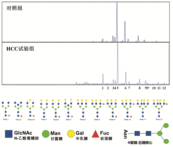
  • Relative Articles

    [1]Tian Bing, Li Fan, Deng BaoCheng. Clinical effect of artificial liver support system in treatment of drug-induced liver failure: A Meta-analysis[J]. Journal of Clinical Hepatology, 2020, 36(4): 823-828. doi: 10.3969/j.issn.1001-5256.2020.04.023
    [2]Lan XiaoQin, Ji YaLi, Chen JinJun, Zhou FuYuan, Wen WeiQun. Effect of artificial liver support therapy on the short-term prognosis of patients with liver failure in the plateau stage: A stratified analysis based on Model for End-Stage Liver Disease score[J]. Journal of Clinical Hepatology, 2020, 36(9): 2005-2009. doi: 10.3969/j.issn.1001-5256.2020.09.019
    [3]Li Shuang, Liu Jing, Chen Yu. Non-bioartificial liver in liver failure: Clinical application and research advances[J]. Journal of Clinical Hepatology, 2019, 35(9): 1909-1915. doi: 10.3969/j.issn.1001-5256.2019.09.004
    [4]Yang XianShan, Zhou Li, Li Lu, Li Shuang, Kong Ming, Li ShanShan, Zou HuaiBin, Xu ManMan. Influence of duration of plasma diafiltration on therapeutic outcome of liver failure[J]. Journal of Clinical Hepatology, 2018, 34(5): 1052-1054. doi: 10.3969/j.issn.1001-5256.2018.05.025
    [5]Guo XiJu, Guo WeiBo, Zhang JiaChang, Lu YaJing, Yang Yan. Clinical effect of double plasma molecular absorption system in treatment of liver failure due to hyperthyroidism: A case report[J]. Journal of Clinical Hepatology, 2018, 34(7): 1526-1528. doi: 10.3969/j.issn.1001-5256.2018.07.033
    [6]Wang XiaoXiao, Huang JianRong. Advances in the application of artificial liver in liver failure[J]. Journal of Clinical Hepatology, 2018, 34(9): 1847-1854. doi: 10.3969/j.issn.1001-5256.2018.09.006
    [7]Li Shuang, Chen Yu. Coping with shortage of plasma-The new therapeutic pattern of non-bioartificial liver[J]. Journal of Clinical Hepatology, 2017, 33(9): 1687-1692. doi: 10.3969/j.issn.1001-5256.2017.09.012
    [8]Ding YiTao. Bioartificial liver and liver transplantation: new modalities for the treatment of liver failure[J]. Journal of Clinical Hepatology, 2017, 33(9): 1693-1698. doi: 10.3969/j.issn.1001-5256.2017.09.013
    [9]Zheng HuanWei, Li Li, Bai GeLan. Clinical efficacy and problems of artificial liver support system in treatment of liver failure[J]. Journal of Clinical Hepatology, 2015, 31(9): 1411-1414. doi: 10.3969/j.issn.1001-5256.2015.09.011
    [10]Yang JianLe, Huang JianRong. Application of artificial liver support system in treatment of liver failure[J]. Journal of Clinical Hepatology, 2015, 31(9): 1405-1410. doi: 10.3969/j.issn.1001-5256.2015.09.010
    [11]Wu ShaoHong, Gan JianHe, Huang XiaoPing, Lin Hua, Lu NianFang, Wu JiangQuan, Zheng RuiQiang. Clinical effect of albumin dialysis combined with plasma perfusion in treatment of liver failure[J]. Journal of Clinical Hepatology, 2014, 30(5): 434-437. doi: 10.3969/j.issn.1001-5256.2014.05.013
    [12]Zhang Lin, Zhao ShouSong. Clinical efficacy of plasma exchange therapy in treatment of liver failure[J]. Journal of Clinical Hepatology, 2014, 30(10): 1015-1019. doi: 10.3969/j.issn.1001-5256.2014.10.010
    [13]Liu XiaoHui, Guo HaiQing, Zhang Jing, Duan ZhongPing. Progress in technology and clinical application of non- bioartificial liver support system[J]. Journal of Clinical Hepatology, 2013, 29(9): 661-665. doi: 10.3969/j.issn.1001-5256.2013.09.007
    [14]You ShaoLi, Liu HongLing, Rong YiHui, Zhu Bing, Zang Hong, Liu WanShu, Wan ZhiHong, Mao PanYong, Xin ShaoJie. Preliminary investigation of hybrid bioartificial liver support system in treatment of HBV- related acute- on- chronic liver failure[J]. Journal of Clinical Hepatology, 2013, 29(9): 685-688. doi: 10.3969/j.issn.1001-5256.2013.09.013
    [15]He HongLiang, Li JianGuo, Gao ZhiLiang. Investigation of artificial liver support system combined with stem cell transplantation in treatment of liver failure[J]. Journal of Clinical Hepatology, 2013, 29(9): 670-673. doi: 10.3969/j.issn.1001-5256.2013.09.009
    [16]Liao JinMao, Li ZhuoRi, Hu XiaoXuan. The therapeutic effect of plasma exchange therapy in patients with severe hepatitis[J]. Journal of Clinical Hepatology, 2012, 28(4): 302-304.
    [17]Zhang AiMin, Xin ShaoJie. Virus infection and liver failure[J]. Journal of Clinical Hepatology, 2012, 28(10): 729-731.
    [18]Duan ZhongPing, Chen Yu. Recent progress  and future perspectives of liver failure diagnosis and treatment strategies [J]. Journal of Clinical Hepatology, 2012, 28(10): 721-725.
    [19]Zhou XinMin, Dong XuYang. Therapeutic strategies of liver failure for clinical cases[J]. Journal of Clinical Hepatology, 2012, 28(2): 93-98.
    [20]Liu ZhiHong, Jiang JianNing, Luo GuangHan, Wu JiZhou, Deng YiMing, Su MingHua, Wu JianLin, Zhang Lu. The value of MELD score for evaluation of plasma exchange in patients with liver failure[J]. Journal of Clinical Hepatology, 2010, 26(6): 615-617.
  • Cited by

    Periodical cited type(3)

    1. 唐飞,梁静,景丽,刘刚,王凤梅. 重组人血小板生成素对D-氨基半乳糖诱导急性肝衰竭大鼠血小板的影响. 中华危重病急救医学. 2024(02): 189-194 .
    2. 闫盼盼,胡兆东,李秀敏,苗明三. 基于中西医临床病症特点的急性肝衰竭动物模型分析. 中华中医药杂志. 2023(10): 5023-5028 .
    3. 黄晓光,林灿峰,林连兴,文之斐,张晓平. 聚乙二醇化重组人粒细胞刺激因子注射液对癌症患者粒细胞的作用. 实用癌症杂志. 2020(07): 1166-1169 .

    Other cited types(1)

  • 加载中

Catalog

    通讯作者: 陈斌, bchen63@163.com
    • 1. 

      沈阳化工大学材料科学与工程学院 沈阳 110142

    1. 本站搜索
    2. 百度学术搜索
    3. 万方数据库搜索
    4. CNKI搜索

    Figures(1)  / Tables(2)

    Article Metrics

    Article views (826) PDF downloads(38) Cited by(4)
    Proportional views
    Related

    /

    DownLoad:  Full-Size Img  PowerPoint
    Return
    Return