中文English
ISSN 1001-5256 (Print)
ISSN 2097-3497 (Online)
CN 22-1108/R

Effect of Cordyceps polysaccharides on hepatocyte apoptosis induced by tumor necrosis factor-α

DOI: 10.3969/j.issn.1001-5256.2021.06.029
  • Received Date: 2021-02-02
  • Accepted Date: 2021-03-10
  • Published Date: 2021-06-20
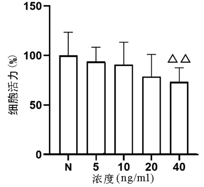
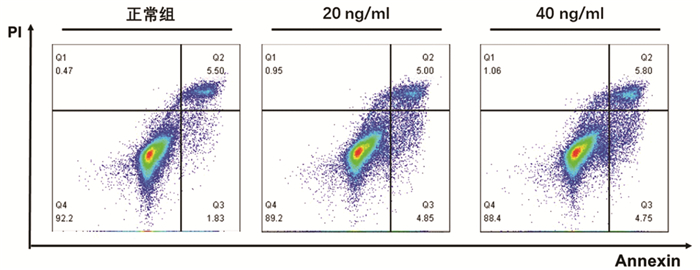
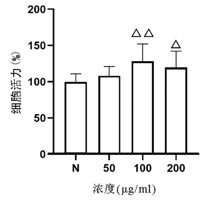
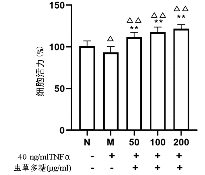
  •   Objective  To investigate the effect of Cordyceps polysaccharides on hepatocyte apoptosis and its mechanism of action.  Methods  Normal human L02 hepatocytes without any drug treatment was established as normal group. The L02 hepatocytes were treated with different concentrations of tumor necrosis factor-α (TNF-α) (5, 10, 20, and 40 ng/ml) for 24 hours to screen out the modeling conditions for the model of hepatocyte apoptosis, which were established as model group. According to the experimental design, L02 hepatocytes pretreated with three different concentrations of Cordyceps polysaccharides (50, 100, and 200 μg/ml) for 12 hours, with or without TNF-α treatment for 24 hours at the selected concentration, were established as experimental group. Related samples were collected for analysis. CCK8 assay was used to measure cell proliferation; Annexin V/PI double staining was used to measure the number of apoptotic hepatocytes; RT-PCR was used to measure the mRNA expression of cell apoptosis genes (Bax, caspase-3, caspase-8, caspase-9, and death receptor Fas); Western blotting was used to measure the protein expression of cleaved-caspase-3 and cleaved-caspase-8. A one-way analysis of variance or the Kruskal-Wallis H test was used for comparison of continuous data between multiple groups, and the least significant difference t-test or the Dunnett-t test was used for further comparison between two groups.  Results  CCK8 assay showed that compared with the normal group, the L02 hepatocytes induced by 40 ng/ml TNF-α for 24 hours had a significant reduction in proliferation (73.54%±14.19% vs 100.00%±23.61%, P < 0.01), and compared with the model group under the premise that Cordyceps polysaccharides had no obvious cytotoxicity to hepatocytes at the three concentrations, the 50, 100, and 200 μg/ml experimental groups had a significant increase in cell proliferation (108.10%±9.05%/114.30%±8.79%/117.70%±9.66% vs 93.02%±7.21%, all P < 0.01). Annexin V/PI double staining showed that compared with the normal group, the model group had a significant increase in the number of apoptotic cells (11.57%±1.41% vs 7.71%±1.20%, P < 0.05), and compared with the model group, the 50, 100, and 200 μg/ml experimental groups had a significant reduction in the number of apoptotic cells (15.16%±0.16%/13.28%±1.57%/16.91%±0.21% vs 18.91%±0.80%, all P < 0.05). RT-PCR and Western blotting showed that compared with the normal group, the L02 hepatocytes induced by 40 ng/ml TNF-α had significant increases in the mRNA expression of the apoptosis-related genes Bax, caspase-9, and death receptor Fas (all P < 0.05), as well as significant increases in the protein expression of the activated forms of cleaved-caspase-3 and cleaved-caspase-8 (P < 0.05). Compared with the model group, the 50 μg/ml experimental group had significant reductions in the mRNA expression of Bax, caspase-3, and caspase-9 and the protein expression of cleaved-caspase-8; the 100 μg/ml experimental group had significant reductions in the mRNA expression of Bax, caspase-3, caspase-8, Fas, and caspase-9 and the protein expression of cleaved-caspase-8; the 200 μg/ml experimental group had significant reductions in the mRNA expression of caspase-3, caspase-8, Fas, and caspase-9 and the protein expression of cleaved-caspase-3 and cleaved-caspase-8. The three concentrations of Cordyceps polysaccharides had a certain dose-effect trend.  Conclusion  Cordyceps polysaccharides can effectively inhibit the apoptosis of normal L02 hepatocytes induced by TNF-α.


  • [1]
    Chinese Society of Hepatology, Chinese Medical Association; Chinese Society of Gastroenterology, Chinese Medical Association; Chinese Society of Infectious Diseases, Chinese Medical Association. Consensus on the diagnosis and therapy of hepatic fibrosis (2019)[J]. J Clin Hepatol, 2019, 35(10): 2163-2172. DOI: 10.3969/j.issn.1001-5256.2019.10.007.

    中华医学会肝病学分会, 中华医学会消化病学分会, 中华医学会感染病学分会. 肝纤维化诊断及治疗共识(2019年)[J]. 临床肝胆病杂志, 2019, 35(10): 2163-2172. DOI: 10.3969/j.issn.1001-5256.2019.10.007.
    [2]
    YAN X, ZHOU T, TAO Y, et al. Salvianolic acid B attenuates hepatocyte apoptosis by regulating mediators in death receptor and mitochondrial pathways[J]. Exp Biol Med (Maywood), 2010, 235(5): 623-632. DOI: 10.1258/ebm.2009.009293.
    [3]
    GUICCIARDI ME, GORES GJ. Apoptosis as a mechanism for liver disease progression[J]. Semin Liver Dis, 2010, 30(4): 402-410. DOI: 10.1055/s-0030-1267540.
    [4]
    PENG JH, LI XM, HU YY, et al. Effect of Cordyceps polysaccharide on lipid peroxidation of rats with dimethylnitrosamine-induced liver fibrosis[J]. China J Chin Mater Med, 2013, 38(3): 391-396. DOI: 10.4268/cjcmm20130320.

    彭景华, 李雪梅, 胡义扬, 等. 虫草多糖对二甲基亚硝胺诱导的肝纤维化大鼠脂质过氧化及肝细胞再生的影响[J]. 中国中药杂志, 2013, 38(3): 391-396. DOI: 10.4268/cjcmm20130320.
    [5]
    WANG XB, LIU P, TANG ZP. Effect of Cordyceps mycelia extract on injury and phenotype shift of liver sinusoidal endothelial cells in rats with dimethylnitrosamine-induced liver cirrhosis[J]. J Beijing Univ Chin Med, 2008, 31(7): 448-451, 505. DOI: 10.3321/j.issn:1006-2157.2008.07.005.

    王宪波, 刘平, 唐志鹏. 虫草菌丝提取物对二甲基亚硝胺大鼠肝窦内皮细胞损伤和表型改变的影响[J]. 北京中医药大学学报, 2008, 31(7): 448-451, 505. DOI: 10.3321/j.issn:1006-2157.2008.07.005.
    [6]
    HU YY. Pay attention to the study on active antiliver fibrosis components of Chinese herbal medicine[J]. Chin J Integr Med, 2012, 18(8): 563-564. DOI: 10.1007/s11655-012-1029-7.
    [7]
    PENG J, LI X, FENG Q, et al. Anti-fibrotic effect of Cordyceps sinensis polysaccharide: Inhibiting HSC activation, TGF-β1/Smad signalling, MMPs and TIMPs[J]. Exp Biol Med (Maywood), 2013, 238(6): 668-677. DOI: 10.1177/1535370213480741.
    [8]
    TIAN H, LIU L, LI Z, et al. Chinese medicine CGA formula ameliorates liver fibrosis induced by carbon tetrachloride involving inhibition of hepatic apoptosis in rats[J]. J Ethnopharmacol, 2019, 232: 227-235. DOI: 10.1016/j.jep.2018.11.027.
    [9]
    CROW MT, MANI K, NAM YJ, et al. The mitochondrial death pathway and cardiac myocyte apoptosis[J]. Circ Res, 2004, 95(10): 957-970. DOI: 10.1161/01.RES.0000148632.35500.d9.
    [10]
    PATEL T. Apoptosis in hepatic pathophysiology[J]. Clin Liver Dis, 2000, 4(2): 295-317. DOI: 10.1016/s1089-3261(05)70112-4.
  • Relative Articles

    [1]Gong Jing, Jie XinKe. Effect of endoplasmic reticulum stress and autophagy on hepatocyte apoptosis[J]. Journal of Clinical Hepatology, 2019, 35(12): 2828-2832. doi: 10.3969/j.issn.1001-5256.2019.12.041
    [2]Gao Si, Zhao XiangXuan, Lu ZaiMing. Role of cell apoptosis regulated by P53 in treatment of hepatocellular carcinoma[J]. Journal of Clinical Hepatology, 2017, 33(7): 1373-1376. doi: 10.3969/j.issn.1001-5256.2017.07.038
    [3]Zhang ChaoYa, Zhao XiangXuan, Lu ZaiMing, Guo QiYong. Research advances in sorafenib- induced apoptotic signaling pathways in liver cancer cells[J]. Journal of Clinical Hepatology, 2016, 32(4): 816-820. doi: 10.3969/j.issn.1001-5256.2016.04.048
    [4]Pan LiuLan, Jia ShengNan, Ma JingTing, Tai JingHua, Jin ZhenJing. Effect of decoy receptor 3 gene on hepatocyte apoptosis[J]. Journal of Clinical Hepatology, 2016, 32(7): 1330-1333. doi: 10.3969/j.issn.1001-5256.2016.07.023
    [5]Zhang Xu, Wang Yu, Ma Juan, Wang ZhenZhen, Yuan Juan, Ma Yan, Liu Xin. Role of endoplasmic reticulum stress-mediated apoptosis in liver fibrosis[J]. Journal of Clinical Hepatology, 2015, 31(9): 1537-1539. doi: 10.3969/j.issn.1001-5256.2015.09.044
    [6]Wang YingChun, Kong WeiZong, Jin QingMei, Chen Juan. Effect of salvianolic acid B on hepatocyte apoptosis in rats with nonalcoholic steatohepatitis[J]. Journal of Clinical Hepatology, 2014, 30(12): 1325-1329. doi: 10.3969/j.issn.1001-5256.2014.12.022
    [7]Wang XiXun, Jiang LiXin, Wang JingLin, Liang Jing, Zhai HuiYuan, Xia XianMing. Experimental study on hepatocyte apoptosis induced by acute biliary tract infection[J]. Journal of Clinical Hepatology, 2014, 30(11): 1164-1168. doi: 10.3969/j.issn.1001-5256.2014.11.018
    [8]Pan YuanWei, Ping Jian, Xu LieMing. Research advances in main signaling pathways for inducing apoptosis of activated hepatic stellate cells[J]. Journal of Clinical Hepatology, 2014, 30(2): 182-185. doi: 10.3969/j.issn.1001-5256.2014.02.021
    [9]Xu Li, Ma KeXin, Dong Bing, Liu HaiWang, Liu QinLong, Gao ZhenMing, Sun DeGuang, Zhang RiXin, Wang HaiBo, Wang LiMing, Liang Rui. Regulatory effect of miRNAs on apoptosis, migration, and cell cycle of hepatocellular carcinoma cells[J]. Journal of Clinical Hepatology, 2013, 29(7): 554-557.
    [10]Tai BoJun, Yao Min, Gu Xing, Shi Yun, Yu DanDan, Chen Jie, Zheng WenJie, Yao DengFu, Lu ShaoLin. Effects of silencing glypican- 3 gene transcription on proliferation and apoptosis of hepatoma cells[J]. Journal of Clinical Hepatology, 2013, 29(11): 863-866. doi: 10.3969/j.issn.1001-5256.2013.10.015
    [11]Weng ShanGeng, Xu ChangGuo, Sun Ying, Shi Zheng, Lin LiJuan. Study on the apoptosis of rat hepatic stellate cells induced by kapamycin[J]. Journal of Clinical Hepatology, 2012, 28(3): 219-222.
    [12]Qiao ChunPing, Chen YueXiang. Advances in study of the relationship between apoptosis and alcoholic liver disease[J]. Journal of Clinical Hepatology, 2012, 28(5): 386-390.
    [13]Sun YunTao, Ban Bo, Wang YiBo, Liu Yang. Effect of fenofibrate on hepatocyte apoptosis and hepatic glycogen in rats with obstructive jaundice[J]. Journal of Clinical Hepatology, 2011, 27(9): 947-950.
    [14]Gao Jie, Xu ChunHai, Liang Ming, Yu JianWu, Li ShuChen. Effect of fibronectin on protection of rat hepatic stellate cells HSC-T6 from TRAIL-induced apoptosis[J]. Journal of Clinical Hepatology, 2011, 27(11): 1199-1202.
    [15]Zhang LiLi, Guo XiaoHong, Liu LiXin, Zhang QianQian. IGFBPrP1 siRNA induced apoptosis in rat hepatic stellate cells and its mechanism[J]. Journal of Clinical Hepatology, 2011, 27(9): 965-969+979.
    [17]Zhang Biao, Sui Tao, Yu XiuYing, Cao YongXian, Yao Yuan, He Hong. Significance of detecting apoptosis of PBMC and cytokine of supernatant induced by PHA in chronic hepatitis B[J]. Journal of Clinical Hepatology, 2004, 20(3): 138-140.
    [20]Wang JunXue, Wang GuoJun, Cai Xiong. The effects of oxymatrine on apoptosis induced by TNF-α in human liver cell line L02[J]. Journal of Clinical Hepatology, 2001, 17(4): 210-211+217.
  • Cited by

    Periodical cited type(5)

    1. 王雪莹,侯惠宁,安永升,卢佳,李日顺,张才. 甘草多糖对H_2O_2诱导肉鸡肝细胞氧化应激损伤的保护作用. 中国兽医学报. 2024(08): 1773-1781 .
    2. 马传贵,张志秀,贺宗毅. 虫草属真菌多糖的分离提取、理化性质及生物活性研究进展. 食用菌. 2023(02): 1-3+7 .
    3. 李爽,韩淑贞,戴瑜婷,修明慧,杜籼芹,和建政,蔺兴遥. 基于多种化疗肠损伤发生机制的中医药防治进展. 中国临床药理学与治疗学. 2023(05): 583-593 .
    4. 韩思婕,潘翔,张梦美,魏琼,叶晓川. 白蔹乙酸乙酯部位体内抗肿瘤的药效作用机制及其成分分析研究. 中药新药与临床药理. 2022(12): 1623-1631 .
    5. 常久,张琪,郑宏,季巍巍,向宇雁. 药用真菌抗肿瘤作用机制研究进展. 中国中医药信息杂志. 2021(12): 140-144 .

    Other cited types(5)

  • 加载中

Catalog

    通讯作者: 陈斌, bchen63@163.com
    • 1. 

      沈阳化工大学材料科学与工程学院 沈阳 110142

    1. 本站搜索
    2. 百度学术搜索
    3. 万方数据库搜索
    4. CNKI搜索

    Figures(6)  / Tables(2)

    Article Metrics

    Article views (618) PDF downloads(37) Cited by(10)
    Proportional views
    Related

    /

    DownLoad:  Full-Size Img  PowerPoint
    Return
    Return