中文English
ISSN 1001-5256 (Print)
ISSN 2097-3497 (Online)
CN 22-1108/R

Value of alpha-fetoprotein combined with gamma-glutamyl transpeptidase/aspartate aminotransferase ratio in diagnosis of HBV-associated hepatocellular carcinoma

DOI: 10.3969/j.issn.1001-5256.2021.09.021
Research funding:

National Natural Science Foundation of China (81773444);

Hubei Provincial Natural Science Foundation of China (2019CFC846)

  • Received Date: 2021-01-09
  • Accepted Date: 2021-04-02
  • Published Date: 2021-09-20
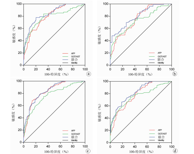
  •   Objective  To investigate the clinical value of alpha-fetoprotein (AFP) combined with gamma-glutamyl transpeptidase (GGT)/aspartate aminotransferase (AST) ratio in the diagnosis of hepatitis B virus (HBV)-associated hepatocellular carcinoma (HCC).  Methods  A total of 352 subjects who received treatment or underwent physical examination in Renmin Hospital of Wuhan University from January 15 to June 15, 2020, were enrolled, among whom there were 86 healthy controls (HC group), 68 patients with chronic hepatitis B (CHB group), 69 patients with liver cirrhosis (LC group), and 129 patients with HCC (HCC group), and a retrospective analysis was performed for the serological test results of all subjects. An analysis of variance was used for comparison of normally distributed continuous data between multiple groups, and the least significant difference t-test was used for further comparison between two groups; the Kruskal-Wallis H test was used for comparison of continuous data with skewed distribution between multiple groups, and the Nemenyi method was used for further comparison between two groups. A binary logistic regression analysis was used to calculate predictor variables; a receiver operating characteristic (ROC) curve was plotted for AFP, GGT/AST, and the predictor variables used alone or in combination, and the area under the ROC curve (AUC), sensitivity, and specificity were calculated; the Z test was used for comparison of AUC.  Results  The HCC group had significantly higher GGT/AST ratio and AFP than the other groups (all P < 0.05). The ROC curve analysis showed that AFP combined with GGT/AST ratio had a significantly higher AUC than AFP alone in the HCC group vs the LC group, the HCC group vs the HC+CHB+LC groups, and the HCC group vs the CHB+LC groups (Z=2.684, 2.241, and 2.415, P=0.007, 0.025, and 0.016).  Conclusion  AFP combined with GGT/AST ratio can improve the clinical diagnostic performance of HBV-related HCC and thus has a certain diagnostic value.


  • 目前我国慢性乙型肝炎(CHB)患者主要使用核苷(酸)类似物(NUC)抗病毒治疗[1],作为目前最重要的抗HBV治疗手段,NUC主要以抑制HBV的逆转录来降低循环血中的HBV DNA水平。目前的NUC药物并不能完全清除肝细胞内的HBV共价闭合环状DNA(cccDNA)[2-3],cccDNA的存在是CHB患者HBV持续感染的关键因素,因此肝内HBV cccDNA的消失是目前国内公认的CHB患者最可能达到病毒学治愈的状态。HBV cccDNA存在于肝细胞内,其检测具有创伤性,在临床难以广泛开展[4]。目前HBV相关的血清学标志物包括HBV DNA、HBeAg、HBsAg,经NUC治疗后HBV DNA阴转并不代表HBV cccDNA的清除,即使HBV cccDNA转录沉默,整合HBV S基因区也可转录翻译成HBsAg[5],在前C区以及基本cp区变异后可导致HBeAg载量下调甚至消失,但检测HBV cccDNA仍存在一定的转录活性。因此,目前常用的血清学标志物反映肝内HBV cccDNA的转录水平存在一定的局限性。

    新型生物学标志物HBV前基因组RNA(pgRNA)、乙型肝炎核心相关抗原(HBcrAg)已引起广泛关注,HBV pgRNA作为cccDNA的直接转录产物,HBcAg作为HBV衣壳的主要结构蛋白,表达于完整的HBV Dane颗粒中[6],其水平与cccDNA的转录活性和水平具有良好的相关性[7-8]。本团队在研究了经NUC治疗CHB患者各时间段的HBV pgRNA、HBcrAg水平之后,收集到治疗至少5年到达完全应答并满足2017年版欧洲肝病学会(EASL)指南[9]停药标准的CHB患者,研究其停药后各时间段HBV pgRNA、HBcrAg水平,分析pgRNA、HBcrAg水平变化与复发的相关性,探索其在临床的应用价值。

    研究对象来自2019年12月—2022年7月川北医学院附属医院门诊中抗HBV治疗至少5年到达完全应答并满足2017年版EASL指南停药标准的随访队列。所有研究对象年龄均满18周岁且具有民事行为能力。排除标准:合并其他嗜肝/非嗜肝病毒感染及其他原因所致肝组织炎症;合并肝硬化、肝癌等其他疾病。完全应答标准:满足生化学、血清学以及病毒学联合应答,生化指标ALT、AST正常,e抗原阴转,HBV DNA检测不到。复发标准:病毒学复发指的是获得病毒学应答的患者停药后,间隔1个月2次检测HBV DNA均>2×10³ IU/mL;临床复发指的是病毒学复发且ALT>2倍正常值上限,但排除其他因素引起的ALT增高。

    收集CHB患者停药时、停药4周、12周以及24周时的空腹静脉血样本3 mL于肝素抗凝管内,在6 h内以1000 r/min离心15 min并获取上清液,置于-80 ℃冰箱保存。

    1.3.1   HBV pgRNA检测

    采用湖南圣湘公司HBV pgRNA定量测定试剂盒检测血清中HBV RNA含量。

    1.3.2   HBcrAg检测

    采用ELISA酶联免疫分析试剂盒,运用CObase601全自动免疫分析系统检测。

    1.3.3   HBV DNA水平检测

    运用实时荧光定量法高精度检测样品中HBV DNA载量,标准值>20 IU/mL,如低于检测值下限按20 IU/mL进行统计学计算,严格按照说明书专人操作。

    数据运用SPSS 21.0统计软件进行分析。符合正态分布的计量资料以x±s表示,两组间比较采用t检验;多组间比较采用单因素方差分析,进一步两两比较采用LSD-t检验。采用Pearson相关检验分析循环血中各指标间的相关性。P<0.05表示差异有统计学意义。

    本研究收集CHB患者108例,其中失访者26例,82例随访者在4周、12周、24周的复发情况见表 1,累积复发率达到29.3%(24/82)。复发者均为病毒学复发,无临床复发者,病毒学复发的患者从复发时结束随访观察。

    表  1  CHB患者停药后随访24周内病毒学复发人数及累积复发率
    Table  1.  The number of viral recurrences and cumulative recurrence rate of CHB patients within 24 weeks of follow-up after drug withdrawal
    组别 复发率(%)
    停药4周 0
    停药12周 17.1(14/82)
    HBV DNA复阳 64.3(9/14)
    HBeAg复阳 28.6(4/14)
    HBV DNA、HBeAg同时复阳 7.1(1/14)
    停药24周 14.7(10/68)
    HBV DNA复阳 60.0(6/10)
    HBeAg复阳 20.0(2/10)
    HBV DNA、HBeAg同时复阳 20.0(2/10)
    下载: 导出CSV 
    | 显示表格

    停药24周时,CHB患者循环血清中HBV pgRNA、HBcrAg、HBV DNA表达水平较停药时及停药4周表达明显升高(P值均<0.05);停药后各时段组间CHB患者循环血清中HBV pgRNA、HBcrAg、HBV DNA表达水平差异均有统计学意义(P值均<0.05)(表 2)。

    表  2  CHB患者停药后各时段循环血清中HBV pgRNA、HBcrAg、HBV DNA表达水平比较
    Table  2.  Comparison of HBV pgRNA, HBcrAg and HBV DNA expression levels in circulating serum of CHB patients at different periods after drug withdrawal
    组别 例数 HBV pgRNA (log10拷贝/mL) HBcrAg (log10 U/L) HBV DNA (log10 IU/mL)
    停药时 82 1.36±0.12 1.31±0.16 1.30±0.18
    停药4周 82 1.37±0.10 1.31±0.17 1.61±0.20
    停药12周 82 1.39±0.12 1.40±0.13 2.38±0.26
    停药24周 68 1.42±0.141)2) 1.51±0.161)2) 2.50±0.351)2)
    F 10.276 14.359 21.473
    P 0.022 0.013 < 0.001
    注:与停药时相比,1)P<0.05;与停药4周相比,2)P<0.05。
    下载: 导出CSV 
    | 显示表格

    对CHB患者停药后复发组选择复发当时循环血清中各指标水平状态,未复发组选择随访24周后循环血清中各指标的水平状态,复发组循环血清中HBV pgRNA、HBcrAg、HBV DNA水平较未复发组显著高表达(P值均<0.05)(表 3)。对复发组进一步分析发现,在12~24周时复发患者循环血清中HBV pgRNA[(1.59±0.16)log10拷贝/mL vs (1.42±0.14)log10拷贝/mL,t=2.903,P<0.05]、HBcrAg[(2.43±0.12)log10 U/L vs (1.73±0.06)log10 U/L,t=18.163,P<0.05]以及HBV DNA[(3.97±0.44)log10 IU/mL vs (3.57±0.31)log10 IU/mL,t=2.640,P<0.05]水平较4~12周时高表达。

    表  3  CHB患者停药后复发组与未复发组循环血清中HBV pgRNA、HBcrAg、HBV DNA、HBsAg水平比较
    Table  3.  Comparison of HBV pgRNA, HBcrAg, HBV DNA and HBsAg levels in circulating serum between relapse group and non relapse group of CHB patients after drug withdrawal
    组别 例数 HBV pgRNA (log10拷贝/mL) HBcrAg (log10 U/L) HBV DNA (log10 IU/mL)
    复发组 24 1.49±0.15 2.02±0.11 3.74±0.39
    未复发组 58 1.39±0.13 1.35±0.17 2.24±0.15
    t 2.549 8.654 25.680
    P 0.015 < 0.001 < 0.001
    下载: 导出CSV 
    | 显示表格

    复发组时循环血清中HBV pgRNA、HBcrAg的表达水平较未复发组显著升高(P值均<0.05)(表 4)。

    表  4  停药时复发组与未复发组CHB患者循环血清中HBV pgRNA、HBcrAg表达水平比较
    Table  4.  Comparison of HBV pgRNA, HBcrAg expression levels in circulating serum of CHB patients in relapse and non relapse groups at the time of drug withdrawal
    组别 例数 HBV pgRNA (log10拷贝/mL) HBcrAg (log10 U/L)
    复发组 24 1.42±0.11 1.55±0.20
    未复发组 58 1.34±0.07 1.21±0.11
    t 18.561 6.152
    P < 0.001 < 0.001
    下载: 导出CSV 
    | 显示表格

    CHB患者停药后复发组循环血清中HBV pgRNA、HBcrAg与HBV DNA均呈正相关(r值分别为0.81、0.66,P值均<0.001);未复发组循环血清中HBV pgRNA、HBcrAg与HBV DNA均无相关性(r值分别为0.14、0.04,P值均>0.05)(图 1~4)。

    图  1  复发组HBV pgRNA与HBV DNA的关系
    Figure  1.  Relationship between HBV pgRNA and HBV DNA in relapse group
    图  2  复发组HBcrAg与HBV DNA的关系
    Figure  2.  Relationship between HBcrAg and HBV DNA in relapse group
    图  3  未复发组HBV pgRNA与HBV DNA的关系
    Figure  3.  Relationship between HBV pgRNA and HBV DNA in non relapse group
    图  4  未复发组HBcrAg与HBV DNA的关系
    Figure  4.  Relationship between HBcrAg and HBV DNA in non relapse group

    CHB患者经NUC药物治疗后可以达到HBV DNA阴转,但CHB患者体内仍存在一定量的HBV cccDNA,HBV cccDNA的持续低表达是HBV不断复制的根源,目前只有HBV cccDNA彻底清除才是CHB等肝病患者根治的核心指标[5]。已有研究[10]表明,CHB患者循环血清中HBV pgRNA水平检测可以反映肝细胞内cccDNA水平;也有研究[11-12]发现,HBcrAg表达水平与HBV cccDNA水平呈正相关。因此,本研究通过检测CHB患者停药后各时段循环血清中HBV pgRNA、HBcrAg的表达水平以及复发组与未复发组循环血清中HBV pgRNA、HBcrAg的表达差异,分析HBV pgRNA、HBcrAg停药时水平与复发的相关性以及其水平能否作为停药的参考临界值指标,HBV pgRNA、HBcrAg的检测可能为后续选择停药时机、评估药物疗效等提供临床价值基础。

    本研究显示,CHB患者停药后随访4~12周时复发率17.1%,随访24周时累积复发率达到29.3%。其中单独HBV DNA复阳者各占64.3%、60.0%,所有复发者均为病毒学复发,未发现临床复发患者。目前CHB患者抗病毒治疗主要依赖于NUC药物。NUC药物主要是抑制HBV逆转录酶来抑制HBV DNA的转录,降低CHB患者循环血清中HBV DNA的表达水平,对肝细胞内的HBV cccDNA清除能力较低,HBV pgRNA作为HBV cccDNA的直接转录产物即可长期以低水平存在于CHB患者体内。本研究的CHB患者经过至少5年的NUC抗病毒治疗,满足2017年版EASL指南停药标准后停药,由结果可见,尽管CHB患者经治疗后HBV DNA阴转,e抗原发生转换,在停药后随访中发现,仍有一部分患者发生HBV DNA和/或HBeAg复阳,其中HBV DNA单独复阳者占多数,这可能是由于CHB患者循环血清中的HBV DNA来源于HBV pgRNA,pgRNA再经过逆转录形成HBV rcDNA,被病毒蛋白包裹后释放到肝细胞外成为新的HBV DNA,HBV DNA形成与HBV pgRNA关系密切,在后续研究结果中发现低水平的HBV pgRNA是HBV DNA复阳的关键,既往也有研究[13]证实,停止NUC治疗后,血清pgRNA水平与病毒复发风险有密切关系。

    本研究在CHB患者停药24周内的随访中发现,随着停药时间的延长,CHB患者循环血清中HBV pgRNA、HBcrAg、HBV DNA表达水平成上升趋势,差异具有统计学意义;进一步对复发组进行分析,复发组CHB患者循环血清中HBV pgRNA、HBcrAg、HBV DNA表达水平较未复发组高表达,且12~14周复发组患者循环血清中HBV pgRNA、HBcrAg、HBV DNA表达水平较4~12周复发组高表达;停药24周后未复发组患者循环血中HBV pgRNA、HBcrAg、HBV DNA的表达水平相比较于最初停药时有所上升,但未达到复发组患者水平。由于本研究观察时间较短,在后续的停药过程中,未复发组患者循环血中各指标水平是否会进一步上升甚至达到复发组患者水平还需要进一步观察;根据未复发组患者24周时循环血中各指标的表达水平能否预测其未来复发的可能性还需要进一步研究证明。本研究的CHB患者停药时HBV DNA阴转,但检测结果提示其循环血清中HBV pgRNA、HBcrAg仍微量存在,在停药后CHB患者循环血清中各指标水平逐渐升高,以HBV DNA水平上升为著,这可能是由于CHB患者体内HBV pgRNA、HBcrAg的持续存在,在失去NUC药物的抑制后,HBV pgRNA经过逆转录酶重新生成HBV DNA,导致循环血清中HBV DNA表达水平上升明显。也有研究[14]显示,在CHB患者达到2017年版EASL指南停药标准后,仍有约半数的患者循环血清中pgRNA未阴转,这表明肝细胞内HBV cccDNA仍有一定的转录活性,在停药后其复制活跃程度增高,导致患者发生病毒学复发。

    进一步分析复发组和未复发组在停药时其循环血清中HBV pgRNA、HBcrAg的水平差异,研究结果提示复发组在停药时其循环血清中HBV pgRNA、HBcrAg表达水平较未复发组高表达。这表明,相比较于未复发患者,复发患者在停药时,其循环血清中存在较高水平的HBV pgRNA、HBcrAg,这可能是导致复发组患者体内HBV再活跃的关键指标,因本研究停药后观察时间较短,未复发组循环血清中HBV pgRNA、HBcrAg停药时水平是否能作为停药的临界指标尚需进一步观察研究。免疫系统的激活与应答对乙型肝炎的进展和临床结局起着关键作用,清除CHB患者体内HBV需要固有免疫以及获得性免疫的协同作用。在大多数CHB患者体内免疫细胞处于“无应答”状态,经NUC治疗后,部分免疫细胞激活,免疫系统重新构建。近期研究[15-16]表明,经长期NUC治疗后停药复发与患者体内各种细胞因子激活等也有一定关系。

    复发组以及未复发组循环血清中HBV pgRNA、HBcrAg与HBV DNA之间的相关性分析发现,复发组循环血清中HBV pgRNA、HBcrAg与HBV DNA呈正相关,未复发组循环血清中HBV pgRNA、HBcrAg与HBV DNA无相关性,这与部分学者[17-18]研究结果一致。作者认为在CHB患者经NUC药物治疗至HBV DNA阴转后,其HBV pgRNA、HBcrAg水平下降滞后于HBV DNA,此时CHB患者体内HBV pgRNA、HBcrAg水平不能反映HBV DNA活性,而更能准确反映HBV cccDNA的转录活性与药物治疗疗效;在复发组患者循环血清中存在较高水平的HBV pgRNA、HBcrAg,HBV cccDNA的转录活性更强,经逆转录酶生成HBV DNA能力提高,HBV DNA载量越高,HBV cccDNA活性越强,因此HBV DNA与HBV pgRNA、HBcrAg呈正相关。

    综上所述,目前CHB患者治疗主要依赖NUC药物,根据2017年版EASL指南停药标准,即使HBV DNA阴转,循环血清中仍存在一定量的HBV pgRNA、HBcrAg,本研究发现复发组与未复发组停药时循环血清中HBV pgRNA、HBcrAg水平存在差异,但由于随访时间较短,未复发组患者在停药时HBV pgRNA、HBcrAg水平是否能代表安全停药的临界值指标尚需进一步观察研究。本研究仅纳入了HBV DNA阴转检测HBV pgRNA仍呈阳性的患者,未检测出HBV pgRNA阴性的患者,两者未予以比较,对于停药时HBV pgRNA阴性的患者停药后其水平是否会进一步升高、是否发生病毒学复发、其水平是否更能代表安全停药的临界值指标等诸多问题需要进一步扩大样本,延长随访时间进行研究观察。

  • [1]
    CHEN Y, TIAN Z. HBV-induced immune imbalance in the development of HCC[J]. Front Immunol, 2019, 10: 2048. DOI: 10.3389/fimmu.2019.02048.
    [2]
    ORTEGA-PRIETO AM, DORNER M. Immune evasion strategies during chronic hepatitis B and C virus infection[J]. Vaccines (Basel), 2017, 5(3): 24. DOI: 10.3390/vaccines5030024.
    [3]
    TAROCCHI M, POLVANI S, MARRONCINI G, et al. Molecular mechanism of hepatitis B virus-induced hepatocarcinogenesis[J]. World J Gastroenterol, 2014, 20(33): 11630-11640. DOI: 10.3748/wjg.v20.i33.11630.
    [4]
    AFFO S, YU LX, SCHWABE RF. The role of cancer-associated fibroblasts and fibrosis in liver cancer[J]. Annu Rev Pathol, 2017, 12: 153-186. DOI: 10.1146/annurev-pathol-052016-100322.
    [5]
    DIMITROULIS D, DAMASKOS C, VALSAMI S, et al. From diagnosis to treatment of hepatocellular carcinoma: An epidemic problem for both developed and developing world[J]. World J Gastroenterol, 2017, 23(29): 5282-5294. DOI: 10.3748/wjg.v23.i29.5282.
    [6]
    National Health and Family Planning Commission of the People' s Republic of China. Diagnosis, management, and treatment of hepatocellular carcinoma (V2017)[J]. J Clin Hepatol, 2017, 33(8): 1419-1431. DOI: 10.3969/j.issn.1001-5256.2017.08.003.

    中华人民共和国国家卫生和计划生育委员会. 原发性肝癌诊疗规范(2017年版)[J]. 临床肝胆病杂志, 2017, 33(8): 1419-1431. DOI: 10.3969/j.issn.1001-5256.2017.08.003.
    [7]
    Chinese Journal of Hepatology, Liver Cancer Study Group, Chinese Society of Hepatology, Chinese Medical Association. Expert consensus on multidisciplinary diagnosis and treatment of precancerous lesions of hepatocellular carcinoma(2020 edition)[J]. J Clin Hepatol, 2020, 36(3): 514-518. DOI: 10.3969/j.issn.1001-5256.2020.03.007.

    《中华肝脏病杂志》编辑委员会, 中华医学会肝病学分会肝癌学组. 肝细胞癌癌前病变的诊断和治疗多学科专家共识(2020版)[J]. 临床肝胆病杂志, 2020, 36(3): 514-518. DOI: 10.3969/j.issn.1001-5256.2020.03.007.
    [8]
    ZHENG Y, ZHU M, LI M. Effects of alpha-fetoprotein on the occurrence and progression of hepatocellular carcinoma[J]. J Cancer Res Clin Oncol, 2020, 146(10): 2439-2446. DOI: 10.1007/s00432-020-03331-6.
    [9]
    LUO P, WU S, YU Y, et al. Current status and perspective biomarkers in AFP negative HCC: Towards screening for and diagnosing hepatocellular carcinoma at an earlier stage[J]. Pathol Oncol Res, 2020, 26(2): 599-603. DOI: 10.1007/s12253-019-00585-5.
    [10]
    SONG P, TOBE RG, INAGAKI Y, et al. The management of hepatocellular carcinoma around the world: A comparison of guidelines from 2001 to 2011[J]. Liver Int, 2012, 32(7): 1053-1063. DOI: 10.1111/j.1478-3231.2012.02792.x.
    [11]
    GALLE PR, FOERSTER F, KUDO M, et al. Biology and significance of alpha-fetoprotein in hepatocellular carcinoma[J]. Liver Int, 2019, 39(12): 2214-2229. DOI: 10.1111/liv.14223.
    [12]
    WANG T, ZHANG KH. New blood biomarkers for the diagnosis of AFP-negative hepatocellular carcinoma[J]. Front Oncol, 2020, 10: 1316. DOI: 10.3389/fonc.2020.01316.
    [13]
    GAO C, FANG L, YAO SK. Correlation of GGT with AFP and diagnostic value of GGT for hepatocellular carcinoma[J]. J Clin Hepatol, 2014, 30(9): 921-925. DOI: 10.3969/j.issn.1001-5256.2014.09.021.

    高春, 房龙, 姚树坤. γ-谷氨酰转肽酶和甲胎蛋白表达的相关性及其在肝细胞癌诊断中的价值[J]. 临床肝胆病杂志, 2014, 30(9): 921-925. DOI: 10.3969/j.issn.1001-5256.2014.09.021.
    [14]
    XIA J, SONG P, SUN Z, et al. Advances of diagnostic and mechanistic studies of γ-glutamyl transpeptidase in hepatocellular carcinoma[J]. Drug Discov Ther, 2016, 10(4): 181-187. DOI: 10.5582/ddt.2016.01052.
    [15]
    ZHOU L, WANG SB, CHEN SG, et al. Prognostic value of ALT, AST, and AAR in hepatocellular carcinoma with B-type hepatitis-associated cirrhosis after radical hepatectomy[J]. Clin Lab, 2018, 64(10): 1739-1747. DOI: 10.7754/Clin.Lab.2018.180532.
    [16]
    YANG JG, HE XF, HUANG B, et al. Rule of changes in serum GGT levels and GGT/ALT and AST/ALT ratios in primary hepatic carcinoma patients with different AFP levels[J]. Cancer Biomark, 2018, 21(4): 743-746. DOI: 10.3233/CBM-170088.
  • Relative Articles

    [1]Meng LU, Mingjie WANG, Li CHEN. Advances in the application of elastography in noninvasive diagnosis of liver fibrosis in nonalcoholic fatty liver disease[J]. Journal of Clinical Hepatology, 2023, 39(2): 408-412. doi: 10.3969/j.issn.1001-5256.2023.02.025
    [2]Zhiran YANG, Linheng WANG, Yuan LI, Fusheng LIU, Yu WANG, Jianfang WANG, Runhua CHEN. Diagnostic value of transient elastography in the staging of hepatic fibrosis in patients with autoimmune liver disease: A Meta-analysis[J]. Journal of Clinical Hepatology, 2022, 38(1): 97-103. doi: 10.3969/j.issn.1001-5256.2022.01.015
    [3]Shuang FENG, Huifen FENG, Jing XU, Gongchao YU, Shan ZHENG. Value of FibroScan and platelet count-to-spleen thickness ratio in predicting the degree of esophageal and gastric varices in liver cirrhosis[J]. Journal of Clinical Hepatology, 2021, 37(12): 2819-2823. doi: 10.3969/j.issn.1001-5256.2021.12.018
    [4]Kaimin SONG, Jun LIU. Advances in the application of transient elastography in chronic hepatitis B[J]. Journal of Clinical Hepatology, 2021, 37(2): 419-424. doi: 10.3969/j.issn.1001-5256.2021.02.036
    [5]Xuemei WANG, Yao ZHANG, Xueping YANG, Yue WANG, Mixue WANG. Clinical value of two-dimensional shear wave elastography in predicting esophageal varices in patients with hepatitis B cirrhosis by evaluating liver and spleen stiffness[J]. Journal of Clinical Hepatology, 2021, 37(5): 1081-1084. doi: 10.3969/j.issn.1001-5256.2021.05.021
    [6]Minrui YU, Jie YANG, Jinyong WANG, Bo ZHOU, Bin JIANG, Baocheng DENG. Value of spleen stiffness measured by two-dimensional shear wave elastography combined with platelet count/spleen diameter ratio in evaluating moderate-to-severe gastroesophageal varices in patients with hepatitis B cirrhosis[J]. Journal of Clinical Hepatology, 2021, 37(7): 1572-1577. doi: 10.3969/j.issn.1001-5256.2021.07.019
    [7]Lin JingQi, Tian Bing, Deng BaoCheng. Application of ultrasound elastography in evaluating portal hypertension[J]. Journal of Clinical Hepatology, 2020, 36(10): 2310-2314. doi: 10.3969/j.issn.1001-5256.2020.10.033
    [8]Liu LinXiang, Nie Yuan, Zhu Xuan. Clinical application of transient elastography in liver cirrhosis and its complications[J]. Journal of Clinical Hepatology, 2020, 36(6): 1362-1365. doi: 10.3969/j.issn.1001-5256.2020.06.037
    [9]Zhang DaKun, Chen Min, Zhang WenHui, Wang RuiFang, Shi XiaoJuan. Clinical value of acoustic radiation force impulse elastography in predicting esophageal variceal bleeding in liver cirrhosis[J]. Journal of Clinical Hepatology, 2020, 36(3): 561-564. doi: 10.3969/j.issn.1001-5256.2020.03.018
    [10]SUN YiFei, HUANG LiPing. Noninvasive evaluation of esophageal and gastric varices in liver cirrhosis by shear-wave ultrasound elastography[J]. Journal of Clinical Hepatology, 2020, 36(12): 2815-2818. doi: 10.3969/j.issn.1001-5256.2020.12.036
    [11]Li HongYan, Tang TongYu. Application of reverse ligation in treatment of gastroesophageal varices[J]. Journal of Clinical Hepatology, 2019, 35(3): 648-650. doi: 10.3969/j.issn.1001-5256.2019.03.044
    [12]Xu FengMing, Sheng QingShou. Research advances in serum markers and transient elastography in the evaluation of liver fibrosis[J]. Journal of Clinical Hepatology, 2018, 34(3): 618-622. doi: 10.3969/j.issn.1001-5256.2018.03.040
    [13]Chen Min, Zhang DaKun, Gao YueJuan, Li Meng, Wang RuiFang, Zhang WenHui. Clinical value of acoustic radiation force impulse in quantitative prediction of the degree of esophageal varices in patients with liver cirrhosis[J]. Journal of Clinical Hepatology, 2018, 34(1): 80-83. doi: 10.3969/j.issn.1001-5256.2018.01.016
    [14]Dong Xue, Huang LiPing. Research advances in the value of ultrasound elastography in the diagnosis of gastroesophageal varices in patients with liver cirrhosis[J]. Journal of Clinical Hepatology, 2018, 34(11): 2424-2427. doi: 10.3969/j.issn.1001-5256.2018.11.035
    [15]Zhuang XiaoFang, Sun Jie, Wang XiaoBo, Wang Yan, Wu QiQi, Wang XiaoZhong. Performance of transient elastography in diagnosis of nonalcoholic fatty liver disease[J]. Journal of Clinical Hepatology, 2017, 33(12): 2366-2371. doi: 10.3969/j.issn.1001-5256.2017.12.022
    [16]Ge Na, He Qian. Ultrasound elastography for evaluation of classification of liver fibrosis[J]. Journal of Clinical Hepatology, 2016, 32(12): 2379-2382. doi: 10.3969/j.issn.1001-5256.2016.12.032
    [17]Zhang GuoSheng, Wang TianYi, Xu YouQing, Zhou Li, Geng Nan, Chen Yu. Diagnostic value of real-time tissue elastography for liver fibrosis in patients with chronic hepatitis B[J]. Journal of Clinical Hepatology, 2014, 30(7): 616-619. doi: 10.3969/j.issn.1001-5256.2014.07.010
    [18]Liu LingFeng, Lu HaiYing, Yu YanYan, Wu ChiHong, Tian XiuLan. A comparative study of FibroScan and B ultrasound in diagnosis of liver fibrosis[J]. Journal of Clinical Hepatology, 2014, 30(10): 1045-1049. doi: 10.3969/j.issn.1001-5256.2014.10.018
    [19]Mai Jing, Gao YueQiu. The application of magnetic resonace elastography in the non-invasive diagnosis of hepatic fibrosis[J]. Journal of Clinical Hepatology, 2011, 27(3): 318-320+324.
    [20]Zhang XiaoHong, Zheng RongQin, Lu CuiRong, Chen Qing, Yang ShaoJi. Ultrasonography prediction of the portal hypertensive gastropathy in patients with liver cirrhosis[J]. Journal of Clinical Hepatology, 2004, 20(1): 30-31.
  • Cited by

    Periodical cited type(14)

    1. 刘波,陈昌兰. 研究ELISA联合荧光定量PCR检测丙型肝炎病毒的效果. 系统医学. 2024(02): 79-82 .
    2. 王学英. HBV pgRNA联合cccDNA对慢性乙型肝炎患者抗病毒疗效的预测价值. 罕少疾病杂志. 2024(04): 54-56 .
    3. 陈春燕,樊子勉. 慢性乙型肝炎者外周血SAA/CRP、NLR水平与HBV-DNA载量、病情程度的相关性分析. 昆明医科大学学报. 2024(05): 144-150 .
    4. 隋娟,钟芳芳,郑秀霞. 乙肝患者HBV-DNA载量与血清标志物水平的相关性及其对抗病毒疗效的预测价值. 中国民康医学. 2024(10): 148-150+154 .
    5. 周芳,王永平,欧阳宇. HBV pgRNA联合HBcrAg对慢性乙型肝炎患者停药后复发的预测价值. 中国肝脏病杂志(电子版). 2024(02): 42-47 .
    6. 朱启泰,黄帆,宋华峰,陈慧,周芬,胥萍. 苏州地区HBV感染患者血清中乙型肝炎核心相关抗原表达特点分析. 标记免疫分析与临床. 2024(09): 1648-1652+1764 .
    7. 蒋贤静,潘小平,周颖,潘强,池林峰,饶芳. 慢性乙型肝炎中西医双重诊疗体系的实践与应用进展. 中西医结合肝病杂志. 2024(11): 1041-1044 .
    8. 王丽莉. 不同抗病毒方案对慢性乙型肝炎患者的影响. 实用中西医结合临床. 2024(22): 42-44 .
    9. 巫翟,祝达. TDF与TAF交替使用治疗慢性乙型肝炎的临床效果观察. 现代养生. 2024(24): 1849-1853 .
    10. 田晓晓,张娟,李琳彬,赵小芬,刘力瑜,余焰. Th1/Th2细胞因子表达水平对慢性乙型肝炎患者停用核苷(酸)类似物后复发的预测价值. 中国药物经济学. 2024(12): 59-63+67 .
    11. 陈柏伶. 动态护理模式对门诊慢性乙型肝炎患者疾病认知及遵医行为的影响. 现代养生. 2023(18): 1410-1413 .
    12. 刘虹,王鹏雁,刘友德. 监测血清HBV RNA水平变化对于NAs治疗CHB停药复发的预测价值. 国际医药卫生导报. 2023(19): 2762-2766 .
    13. 陈长红. 肝爽颗粒联合恩替卡韦对乙型肝炎肝硬化患者HBV-DNA水平及肝功能的影响. 黑龙江医药. 2023(06): 1349-1351 .
    14. 戎云清,徐敬轩. 加味补肾生髓成肝方联合聚乙二醇干扰素α对慢性乙型肝炎患者肝纤维化的影响. 世界复合医学. 2023(12): 98-102 .

    Other cited types(1)

  • 加载中

Catalog

    通讯作者: 陈斌, bchen63@163.com
    • 1. 

      沈阳化工大学材料科学与工程学院 沈阳 110142

    1. 本站搜索
    2. 百度学术搜索
    3. 万方数据库搜索
    4. CNKI搜索

    Figures(1)  / Tables(2)

    Article Metrics

    Article views (633) PDF downloads(74) Cited by(15)
    Proportional views
    Related

    /

    DownLoad:  Full-Size Img  PowerPoint
    Return
    Return