中文English
ISSN 1001-5256 (Print)
ISSN 2097-3497 (Online)
CN 22-1108/R

Research advances in the risk factors for recurrence of common bile duct stone after choledocholithotomy

DOI: 10.3969/j.issn.1001-5256.2023.01.036
Research funding:

National Natural Science Foundation of China (82174136);

Capacity building Project of Key Clinical Specialty of Traditional Chinese Medicine of Liaoning Provincial Health Commission(Liaoning Health Office 2019(169)) ( )

More Information
  • Corresponding author: ZHANG Guixin, zgx0109@126.com (ORCID: 0000-0002-6171-394X)
  • Received Date: 2022-06-23
  • Accepted Date: 2022-09-01
  • Published Date: 2023-01-20
  • Cholelithiasis is a common and frequent disease of the digestive system, and its incidence rate tends to increase with the improvement of living standards. Patients suffering from both gallbladder stones and common bile duct stones account for 5%-15%. Choledocholithiasis can cause a series of serious complications such as acute cholangitis and biliary pancreatitis. Choledocholithotomy is the main method for the treatment of choledocholithiasis, but there is still a high recurrence rate after surgery. The recurrence of choledocholithiasis seriously affects the life of patients and increases their economic burden. With reference to the latest published clinical studies, this article summarizes the influencing factors for the recurrence of choledocholithiasis from the aspects of anatomical factors, stone-related factors, biliary factors, and surgical factors, so as to provide a reference for the treatment of choledocholithiasis and the prevention of its recurrence.


  • HBV慢性感染是肝细胞癌(HCC) 发生的最常见病因,HCC的发病率和病死率在恶性肿瘤中分别占第6位和第4位,严重威胁着人类健康[1]。HCC细胞异常增殖可使血清糖蛋白N-糖链组成和结构发生改变,因此,血清糖蛋白的N-聚糖可作为HCC诊断标志物和HCC治疗的分子靶点[2]。N-聚糖改变的基础在于肝细胞内糖基转移酶活性的变化,其中多数糖基转移酶表达与肝脏功能密切相关。当肝细胞发生癌变时,某些糖基转移酶基因被激活,其表达异常增加,或基因表达受抑制而含量下降,从而导致一些蛋白质异常糖基化修饰。大量研究[3]表明,这种异常糖基化修饰与肿瘤细胞恶性侵袭行为密不可分。笔者前期研究[4]发现,与健康对照相比,HBV相关HCC(HBV-HCC)患者血清中N-聚糖图谱发生一系列特征改变,其中二天线N-聚糖峰1(peak1, NGA2F)丰度升高;三天线N-聚糖峰9(peak9, NA3Fb)丰度特异性升高;其他三天线和四天线N-聚糖峰(peak10 NA3Fc、peak11 NA4、peak12 NA4Fb)丰度也有不同程度的升高。但目前HBV-HCC患者血清N-聚糖变化机制尚未完全阐明。本研究中,通过检测HBV-HCC患者癌组织与癌旁组织中8种重要的糖基转移酶基因(包括岩藻糖基转移酶FUT3、FUT4、FUT6、FUT7、FUT8和N-乙酰氨基葡萄糖转移酶Gn-TⅢ、Gn-TⅣa、Gn-TⅤ)表达水平变化并比较其差异,探索HBV-HCC患者血清N-聚糖变化的可能机制。

    收集解放军总医院2018年9月—2019年11月HBV-HCC行手术患者的肝癌和癌旁组织及正常肝组织标本,同时采集血清标本,置于-80 ℃冰箱保存。患者符合以下入选标准: (1)均为感染HBV的HCC患者;(2)排除HAV、HCV、HDV、HEV等肝炎病毒感染;(3)患者术后病理标本均经医院病理科确诊为HCC,肝癌诊断符合原发性肝癌诊疗规范(2019年版)[5]。同时收集HCC患者临床资料。另外选取20例健康成年人血清作为对照。

    应用SPSS 20.0软件从34例HCC患者中随机选择8例HCC患者血清标本作为HCC试验组,20例健康成年人血清标本作为对照组。采用DSA-FACE法检测和分析血清N-聚糖图谱[6],具体步骤如下:

    (1) 寡糖的释放:取3 μl血清,加入含有2 μl 10 mmol/L NH4HCO3缓冲液和3 μl去离子水的PCR反应板中, 反应板放入PCR仪器,95 ℃加热5 min后冷却至4 ℃, 然后加入3 μl PNGaseF(2.2 U/μl),37 ℃孵育3 h,之后加入100 μl去离子水终止反应,标记为D板。

    (2) 标记寡糖:从D板吸取10 μl溶液加入一新的PCR反应板中,开盖在60 ℃条件下烘干90 min,加入3 μl标记溶液(20 mmol/L APTS∶ 1 mol/L NaCNBH3=1∶ 1),90 ℃反应2 h,加入100 μl去离子水终止反应,标记为L板。

    (3) 去唾液酸:从L板中取2 μl溶液加入一新的PCR反应板,加入0.25 μL 100 mmol/L NH4Ac(pH=5.0)、0.2 μl唾液酸酶(2.5 U/μl)和1.55 μl去离子水, 震荡混匀后42 ℃孵育4 h,加40 μl去离子水终止反应,标记为DE板。

    (4) DNA测序仪上机检测:取DE板10 μl溶液加入ABI测序仪专用96孔板,放入ABI 3500测序仪进行N-聚糖图谱分析,数据经GeneMapper软件分析。

    冻存的肝组织放入超声震荡仪研磨后,用Trizol试剂提取总RNA,用Nano Drop One检测总RNA的浓度和纯度。将总RNA逆转录为cDNA后,用荧光定量PCR仪(ABI 7500 FAST)进行cDNA扩增,反应体系为20 μl。反应条件为:(1)95 ℃ 30 s;(2)95 ℃ 5 s,60 ℃ 34 s共40个循环;(3)溶解曲线:95 ℃ 15 s,60 ℃ 1 min, 95 ℃ 15 s。

    以RPS11作为内参基因,目的基因的相对表达量用2-△△CT表示。癌组织和癌旁组织糖基转移酶mRNA相对表达量检测分析的对照均是正常肝组织。FUT3、FUT4、FUT6、FUT7、FUT8、Gn-TⅢ、Gn-TⅣa和Gn-TⅤ基因的特异引物由生工生物工程股份有限公司合成,引物序列见表 1

    表  1  荧光定量PCR检测糖基转移酶基因引物序列表
    基因 序列号 引物序列(5′→3′)
    FUT3 NM_001374740.1 F: CAA CAG AGA AAG CAG GCA
    R: AAG AAA CAC ACA GCC ACC[7]
    FUT4 XM_032167182.1 F: TCC TAC GGA GAG GCT CAG
    R: TCC TCG TAG TCC AAC ACG[7]
    FUT6 XM_011527879.3 F: CAT TTC TGC TGC CTC AGG
    R: GGG CAA GTC AGG CAA CTC[7]
    FUT7 NM_004479.4 F: CCA CGA TCA CCA TCC TTG
    R: AGG CTT CGG TTG GCA CTC[7]
    FUT8 XM_032180460.1 F: TCT AGC CGA GAA CTG TCC
    R: GCT GCT CTT CTA AAA CGC[7]
    Gn-TⅢ XM_019018480.2 F: CCG CCA CAA GGT GCT CTA T
    R: GAT CTC GTC CGC ATC GTC A[8]
    Gn-TⅣa XM_032178740.1 F: ACC AAG GGC ATA CGC TGG AG
    R: GTT CTT GGT TGC CGC TAT GGA[9]
    Gn-TV XM_032411636.1 F: GCT GCC CAA CTG TAG GAG AC
    R: GAA TCA AGG ACT CGG AGC AT[10]
    RPS11 XM_032159559.1 F: GCC GAG ACT ATC TGC ACT AC
    R: ATG TCC AGC CTC AGA ACT TC[11]
    下载: 导出CSV 
    | 显示表格

    冻存的肝组织放入超声震荡仪研磨后,用含cOmplete蛋白酶抑制剂的RIPA裂解液提取蛋白,用BCA蛋白试剂盒来测定蛋白浓度。电泳分离不同分子量蛋白后,15 V恒压下用半干转电转仪将蛋白转移至PVDF膜,于5%的脱脂奶粉中室温封闭1 h,加入抗-FUT8(1∶ 1000)、抗-Gn-TⅣa(1∶ 1000)、抗-Gn-TⅤ(1∶ 1000)、抗-β-actin(1∶ 4000)一抗,4 ℃过夜,TBST溶液洗膜3次,分别加入抗鼠或抗兔的二抗(1∶ 5000),室温下孵育2 h,TBST溶液洗膜3次,加入增强型ECL化学发光试剂,凝胶成像仪扫描显影的条带,ImageJ软件分析条带灰度。以β-actin作为内参,目的蛋白的相对表达量用目的蛋白灰度值与内参蛋白灰度值的比值来表示。

    应用SPSS 20.0软件对数据进行统计分析。计量资料用x ±s表示,两组间比较采用独立样本t检验。所有统计学分析均采用双侧检验,P<0.05为差异具有统计学意义。

    34例HBV-HCC患者临床特征见表 2

    表  2  34例HBV-HCC患者临床特征
    临床特征 数值
    男性[例(%)] 29 (85.29)
    年龄(岁) 54.85±8.20
    HBV DNA(×106IU/ml) 2.35±7.15
    AFP(ng/ml) 556.82±1995.22
    ALT(U/L) 44.34±60.72
    AST(U/L) 40.71±44.25
    肿瘤直径(例)
    ≤5 cm 21
    5~10 cm 9
    ≥10 cm 4
    下载: 导出CSV 
    | 显示表格

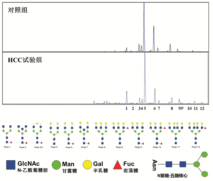
    应用DSA-FACE法分析HCC试验组8例HBV-HCC患者与对照组20例健康成年人血清N-聚糖图谱(图 1),其特征改变与笔者前期研究发现的特征改变相同[4]

    图  1  HCC试验组与对照组血清N-聚糖图谱
    注:a,血清中12种N-聚糖丰度比较;b,12种N-聚糖结构。Peak1-Peak12图引自文献[4],Peak1: 二天线无半乳糖基核心ɑ-l, 6岩藻糖基化N聚糖(NGA2F);Peak2: 二天线无半乳糖基核心ɑ-l, 6岩藻糖基化平分型N聚糖(NGA2FB);Peak3/Peak4: 二天线核心ɑ-l, 6岩藻糖基化单支链半乳糖基N聚糖(NG1A2F);Peak5: 二天线N聚糖(NA2);Peak6: 二天线核心ɑ-l, 6岩藻糖基化N聚糖(NA2F);Peak7: 二天线平分型核心ɑ-l, 6岩藻糖基化N聚糖(NA2FB);Peak8: 三天线N聚糖(NA3);Peak9: 三天线支链ɑ-l, 3岩藻糖基化N聚糖(NA3Fb);Peak9’: 三天线核心ɑ-l, 6岩藻糖基化N聚糖(NA3Fc);Peak10: 三天线支链ɑ-l, 3岩藻糖基化与核心ɑ-l, 6岩藻糖基化N聚糖(NA3Fbc);Peak11: 四天线N聚糖(NA4);Peak12: 四天线支链ɑ-l, 3岩藻糖基化N聚糖(NA4 Fb)。

    与对照组相比,HCC试验组患者三天线N-聚糖峰9(peak9,NA3Fb)丰度明显升高(t=-2.514,P<0.05);血清二天线N-聚糖峰1(peak1,NGA2F)和四天线N-聚糖峰(peak11 NA4、peak12 NA4Fb)的丰度在两组间差异无统计学意义(P值均>0.05)。

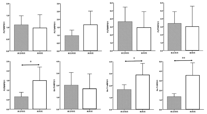
    癌组织中FUT8、Gn-TⅣa和Gn-TⅤ基因mRNA表达水平显著高于癌旁组织(1.50±0.34 vs 0.65± 0.11, t=-2.354,P=0.022; 2.90±0.47 vs 1.68±0.19, t=-2.403,P=0.019; 3.57±0.64 vs 1.33±0.16, t=-3.384,P=0.001),差异均有统计学意义。FUT3、FUT4、FUT6、FUT7和Gn-TⅢ mRNA的表达水平在癌组织与癌旁组织间比较差异均无统计学意义(P值均>0.05)(图 2)。

    图  2  癌组织与癌旁组织中8种糖基转移酶mRNA表达水平比较
    注:*,P<0.05,* *,P<0.01。

    进一步比较了HCC试验组中8例患者癌组织与癌旁组织中8种糖基转移酶mRNA表达水平。与癌旁组织相比,8例HCC试验组患者癌组织中Gn-TⅤ mRNA表达明显升高(Gn-TⅤ: 5.26±1.70 vs 1.49±0.33, t=-2.173, P=0.047);Gn-TⅢ、Gn-TⅣa、FUT4和FUT8 mRNA表达在癌组织与癌旁组织间无显著性差异(Gn-TⅢ: 1.03±0.46 vs 1.55±0.62, t=0.663, P=0.518;Gn-TⅣa: 5.15±1.50 vs 2.39±0.46, t=-1.752, P=0.102; FUT4: 1.56±1.12 vs 0.81±0.27, t=-0.653, P=0.524; FUT8: 2.61±1.26 vs 1.01±0.41, t=-1.213, P=0.245)。

    选取mRNA表达有显著性差异的3个糖基转移酶基因FUT8、Gn-TⅣa和Gn-TⅤ作为蛋白印迹实验的检测因子。结果如图 3所示,癌组织中FUT8与Gn-TⅤ的蛋白表达水平显著高于癌旁组织(0.70±0.11 vs 0.083±0.017, t=9.555, P=0.001; 1.33 ±0.19 vs 0.60±0.15, t=5.097, P=0.007);Gn-TⅣa的蛋白表达水平在癌组织与癌旁组织间比较差异无统计学意义(0.52±0.24 vs 0.24±0.11, t=1.833, P=0.141)。

    图  3  癌组织与癌旁组织糖基转移酶FUT8、Gn-TⅣa和Gn-TⅤ的蛋白表达水平比较
    注:* *,P<0.01

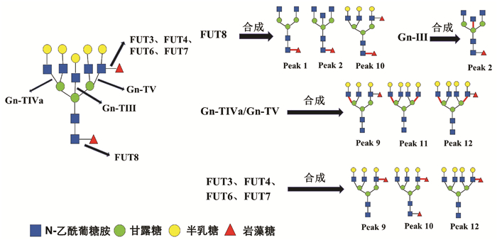
    HCC患者血清中常出现大量异常糖基化N-糖蛋白,分离糖蛋白的N-聚糖链进行表达图谱分析,可发现支链与核心岩藻糖基化N-聚糖和多天线N-聚糖含量增加,这些N-聚糖与HCC发生发展密切相关,可作为筛查和诊断HCC的特异性标志[12]。研究[4, 13]发现在HBV-HCC患者血清中支链岩藻糖基化三天线N-聚糖(peak9, NA3Fb)丰度显著升高,且核心岩藻糖基化二天线N-聚糖(peak1, NGA2F)、核心岩藻糖基化平分型二天线N-聚糖(peak2, NGA2FB)、支链与核心岩藻糖基化三天线N聚糖(peak10, NA3Fc)、四天线N-聚糖(peak11, NA4)和支链岩藻糖基化四天线N-聚糖(peak12, NA4Fb)丰度也有不同程度的升高(N-聚糖结构如图 4)。本研究通过对HBV-HCC患者癌组织与癌旁组织相关糖基转移酶基因表达检测分析,探寻HCC患者特异性血清N-聚糖变化的可能机制。

    图  4  HBV相关HCC患者血清特异变化N-聚糖与8种糖基转移酶之间的关系

    糖基转移酶Gn-TⅤ与Gn-TⅣa催化形成三天线及三天线以上N-聚糖的N-乙酰葡糖胺(GlcNAc)糖链结构[14-15],而Gn-TⅢ催化合成N-聚糖的平分型GlcNAc结构,Gn-TⅢ与Gn-TⅤ、Gn-TⅣa有拮抗作用(图 4)[16]。本研究发现,在癌组织中Gn-TⅤ与Gn-TⅣa mRNA表达水平显著高于癌旁组织(P<0.05),蛋白印迹实验也显示,在癌组织中Gn-TⅤ蛋白表达显著高于癌旁组织(P<0.05)。而Gn-TⅢ mRNA表达水平在癌组织与癌旁组织间无统计学差异(P=0.711)。本研究结果可以解释HCC试验组患者血清中N-聚糖变化:与对照组相比,8例HCC患者血清中三天线N-聚糖(peak9)丰度显著升高(P<0.05),而且这8例HCC患者癌组织Gn-TⅤ表达水平显著高于癌旁组织(P<0.05)。说明HCC患者血清中多天线N-聚糖丰度升高可能与糖基转移酶Gn-TⅤ高水平表达密切相关,促进含GlcNAc多分支(三天线及以上)N-聚糖的合成(图 4)。

    以往研究[4, 13]表明,在HBV-HCC患者血清中核心岩藻糖基化N-聚糖(peak1、peak2、peak10)丰度较高。本研究发现,在癌组织中核心岩藻糖基转移酶FUT8 mRNA与蛋白表达水平显著高于癌旁组织(P<0.05)。FUT8表达上调会导致核心岩藻糖基化修饰结构的N-聚糖增加,可能与癌细胞高转移潜能有关[17]。另外,AFP是临床上最常用的HCC血清学检测指标,有研究[18]发现HCC患者血清中AFP经FUT8催化作用下可形成核心岩藻糖基化AFP(AFP-L3),其与肿瘤的发展速度、肿瘤大小和肿瘤转移密切相关,且诊断HCC效力优于AFP。除了α-1, 6核心岩藻糖基化N-聚糖,α-1, 3分支岩藻糖基化N-聚糖的丰度在HBV-HCC患者血清也特异性升高。α-1, 3分支岩藻糖基化结构是三天线N-聚糖(peak9)中Lewis X结构形成的关键[4],参与合成α-1, 3分支岩藻糖基化结构且与HCC密切相关的糖基转移酶有FUT3、FUT4、FUT6、FUT7[19]。本研究显示,在34例HCC患者癌组织与癌旁组织间FUT3、FUT4、FUT6和FUT7 mRNA表达水平没有显著性差异(P>0.05),不能解释血清N-聚糖水平异常改变。结合以往研究报道分析FUT基因表达的检测结果,可能因为多种岩藻糖基转移酶参与合成α-1, 3分支岩藻糖基化结构,而目前对于这些酶相互作用机制尚未明确,较难判断岩藻糖基转移酶中那一种亚类起主导作用。本研究主要检测8种与HCC密切相关的糖基转移酶基因表达,初步阐述HBV-HCC患者血清中N-聚糖变化的可能机制。但对于不同糖基转移酶之间相互作用的具体机制以及其他血清N-聚糖(如异常唾液酸化修饰的N-聚糖等)变化机制,还有待于进一步研究。

  • [1]
    CIANCI P, RESTINI E. Management of cholelithiasis with choledocholithiasis: Endoscopic and surgical approaches[J]. World J Gastroenterol, 2021, 27(28): 4536-4554. DOI: 10.3748/wjg.v27.i28.4536.
    [2]
    LI ZQ, SUN JX, LI B, et al. Meta-analysis of single-stage versus two-staged management for concomitant gallstones and common bile duct stones[J]. J Minim Access Surg, 2020, 16(3): 206-214. DOI: 10.4103/jmas.JMAS_146_18.
    [3]
    WU Y, XU CJ, XU SF. Advances in risk factors for recurrence of common bile duct stones[J]. Int J Med Sci, 2021, 18(4): 1067-1074. DOI: 10.7150/ijms.52974.
    [4]
    WANG LM, CHEN C, DING H, et al. Analysis of risk factors for recurrence of cholecystolithiasis after retrograde cholecystectomy with laparoscopic cholecystectomy[J/CD]. Chin Arch Gen Surg(Electronic Edition), 2019, 13(3): 224-228. DOI: 10.3877/cma.j.issn.1674-0793.2019.03.012.

    王雷鸣, 陈晨, 丁辉, 等. 经内镜逆行胰胆管造影联合腹腔镜胆囊切除术后胆囊结石合并胆总管结石复发的危险因素分析[J/CD]. 中华普通外科学文献(电子版), 2019, 13(3): 224-228. DOI: 10.3877/cma.j.issn.1674-0793.2019.03.012.
    [5]
    RYU S, JO IH, KIM S, et al. Clinical impact of common bile duct angulation on the recurrence of common bile duct stone: a meta-analysis and review[J]. Korean J Gastroenterol, 2020, 76(4): 199-205. DOI: 10.4166/kjg.2020.76.4.199.
    [6]
    KEIZMAN D, SHALOM MI, KONIKOFF FM. An angulated common bile duct predisposes to recurrent symptomatic bile duct stones after endoscopic stone extraction[J]. Surg Endosc, 2006, 20(10): 1594-1599. DOI: 10.1007/s00464-005-0656-x.
    [7]
    CHONG CC, CHIU PW, TAN T, et al. Correlation of CBD/CHD angulation with recurrent cholangitis in patients treated with ERCP[J]. Endosc Int Open, 2016, 4(1): E62-E67. DOI: 10.1055/s-0035-1569689.
    [8]
    JANSSEN BV, van LAARHOVEN S, ELSHAER M, et al. Comprehensive classification of anatomical variants of the main biliary ducts[J]. Br J Surg, 2021, 108(5): 458-462. DOI: 10.1093/bjs/znaa147.
    [9]
    CHOI SJ, YOON JH, KOH DH, et al. Low insertion of cystic duct increases risk for common bile duct stone recurrence[J]. Surg Endosc, 2022, 36(5): 2786-2792. DOI: 10.1007/s00464-021-08563-2.
    [10]
    TSITOURIDIS I, LAZARAKI G, PAPASTERGIOU C, et al. Low conjunction of the cystic duct with the common bile duct: does it correlate with the formation of common bile duct stones?[J]. Surg Endosc, 2007, 21(1): 48-52. DOI: 10.1007/s00464-005-0498-6.
    [11]
    LU ZH, NIU J, XU PP, et al. Analysis of related risk factors for recurrence of choledocholithiasis after the operation[J]. Chin J Curr Adv Gen Surg, 2016, 19(5): 372-375. DOI: 10.3969/j.issn.1009-9905.2016.05.011.

    鲁志华, 牛军, 许平平, 等. 胆总管取石并胆囊切除术后胆总管结石复发的相关危险因素分析[J]. 中国现代普通外科进展, 2016, 19(5): 372-375. DOI: 10.3969/j.issn.1009-9905.2016.05.011.
    [12]
    CAO J, LI TY, ZHOU J, et al. The effect of periampullary diverticulum on the stone composition and biliary flora in patients with choledocholithiasis[J]. Chin J Microecol, 2022, 34(3): 300-305. DOI: 10.13381/j.cnki.cjm.202203010.

    曹洁, 李天亚, 周静, 等. 壶腹周围憩室对胆总管结石患者结石成分及胆道菌群的影响[J]. 中国微生态学杂志, 2022, 34(3): 300-305. DOI: 10.13381/j.cnki.cjm.202203010.
    [13]
    SANDSTAD O, OSNES T, SKAR V, et al. Structure and composition of common bile duct stones in relation to duodenal diverticula, gastric resection, cholecystectomy and infection[J]. Digestion, 2000, 61(3): 181-188. DOI: 10.1159/000007755.
    [14]
    WANG Y, JIE J, QIAN B, et al. Analysis of the relationship between periampullary diverticulum and recurrent bile duct stones after endoscopy on magnetic resonance imaging of magnetic nanoparticles[J]. J Biomed Nanotechnol, 2022, 18(2): 607-615. DOI: 10.1166/jbn.2022.3270.
    [15]
    YOO ES, YOO BM, KIM JH, et al. Evaluation of risk factors for recurrent primary common bile duct stone in patients with cholecystectomy[J]. Scand J Gastroenterol, 2018, 53(4): 466-470. DOI: 10.1080/00365521.2018.1438507.
    [16]
    WU RZ. Analysis of related factors of recurrent choledocholithiasis after cholecystectomy and choledocholithiasis[J]. Chin Foreign Med Res, 2017, 15(5): 45-47. DOI: 10.14033 / j.carol carroll nki CFMR. 2017.5.024.

    吴润芝. 胆总管取石并胆囊切除术后胆总管结石复发的相关因素分析[J]. 中外医学研究, 2017, 15(5): 45-47. DOI: 10.14033/j.cnki.cfmr.2017.5.024.
    [17]
    MENG H. Risk factors for recurrent choledocholithiasis after ERCP lithotomy[J]. Hebei Med J, 2016, 38(5): 716-718. DOI: 10.3969/j.issn.1002-7386.2016.05.025.

    孟环. ERCP取石后胆总管结石复发的相关危险因素研究[J]. 河北医药, 2016, 38(5): 716-718. DOI: 10.3969/j.issn.1002-7386.2016.05.025.
    [18]
    LUJIAN P, XIANNENG C, LEI Z. Risk factors of stone recurrence after endoscopic retrograde cholangiopancreatography for common bile duct stones[J]. Medicine (Baltimore), 2020, 99(27): e20412. DOI: 10.1097/MD.0000000000020412.
    [19]
    PARK SY, HONG TH, LEE SK, et al. Recurrence of common bile duct stones following laparoscopic common bile duct exploration: a multicenter study[J]. J Hepatobiliary Pancreat Sci, 2019, 26(12): 578-582. DOI: 10.1002/jhbp.675.
    [20]
    QUARESIM S, BAlla A, GUERRIERI M et al. Results of medium seventeen years' follow-up after laparoscopic choledochotomy for ductal stones. [J]. Gastroenterol Res Pract, 2016, 2016: 9506406. DOI: 10.1155/2016/9506406
    [21]
    CUI ML, CHO JH, KIM TN. Long-term follow-up study of gallbladder in situ after endoscopic common duct stone removal in Korean patients. [J]. Surg Endosc, 2013, 27: 1711-6. DOI: 10.1007/s00464-012-2662-0
    [22]
    THISTLE JL. Pathophysiology of bile duct stones[J]. World J Surg, 1998, 22(11): 1114-1118. DOI: 10.1007/s002689900529.
    [23]
    KUNDUMADAM S, FOGEL EL, GROMSKI MA. Gallstone pancreatitis: general clinical approach and the role of endoscopic retrograde cholangiopancreatography[J]. Korean J Intern Med, 2021, 36(1): 25-31. DOI: 10.3904/kjim.2020.537.
    [24]
    CAI Y, HE YA. Risk factors for recurrence of stones after LC combined LCHTD in patients with gallbladder and common bile duct stones[J]. J Hepatobiliary Surg, 2016, 24 (3): 203-205. https://www.cnki.com.cn/Article/CJFDTOTAL-GDWZ201603015.htm

    蔡宇, 何彦安. 胆囊结石合并胆总管结石患者行腹腔镜胆囊切除联合胆总管切开取石T管引流术后结石复发危险因素分析[J]. 肝胆外科杂志, 2016, 24(3): 203-205. https://www.cnki.com.cn/Article/CJFDTOTAL-GDWZ201603015.htm
    [25]
    NIJIATIJIANG A, ALIMUJIANG A, ALIDAN A, et al. Efficacy of LC combined with LCHTD in treatment of choledocholithiasis complicated with cholecystolithiasis and the related factors of postoperative recurrence of choledocholithiasis[J]. J Hepatobiliary Surg, 2021, 33(7): 419-422. DOI: 10.11952/j.issn.1007-1954.2021.07.007.

    尼加提江·艾比不拉, 阿力木江·阿布力米提, 阿里旦·艾尔肯, 等. LC联合LCHTD治疗胆总管结石合并胆囊结石的效果及术后胆总管结石复发的相关因素分析[J]. 肝胆胰外科杂志, 2021, 33(7): 419-422. DOI: 10.11952/j.issn.1007-1954.2021.07.007.
    [26]
    LYU YH, ZHANG HC, SONG CY, et al. Logistic regression analysis of related factors of stone recurrence after laparoscopic cholecystectomy combined with choledocholithotomy and T-tube drainage[J]. Chongqing Med, 2016, 45(9): 1262-1264. DOI: 10.3969/j.issn.1671-8348.2016.09.033.

    吕运海, 张焕常, 宋朝阳, 等. 腹腔镜胆囊切除联合胆总管切开取石T管引流术后结石复发相关因素的Logistic回归分析[J]. 重庆医学, 2016, 45(9): 1262-1264. DOI: 10.3969/j.issn.1671-8348.2016.09.033.
    [27]
    QIU WG, XU J. Analysis of risk factors for postoperative recurrence of gallbladder stones with common bile duct stones[J]. Chin J Gen Surg, 2014, 23 (2): 170-173. DOI: 10.7659/j.issn.1005-6947.2014.02.006.

    裘文刚, 徐江. 胆囊结石合并胆总管结石术后复发的危险因素分析[J]. 中国普通外科杂志, 2014, 23(2): 170-173. DOI: 10.7659/j.issn.1005-6947.2014.02.006.
    [28]
    XU L, SUN WR, LI Y. Laparoscopic cholecystectomy combined with choledocholithot- omy and T-tube drainage for treatment of choledocholithiasis combined with cho-ledocholithiasis and its influencing factors[J]. J Clin Psychosom Dis, 2021, 27(2): 156-159. DOI: 10.3969/j.issn.1672-187X.2021.02.036.

    徐璐, 孙万日, 黎洋. 腹腔镜胆囊切除联合胆总管切开取石T管引流术治疗胆囊结石合并胆总管结石术后复发率及影响因素[J]. 临床心身疾病杂志, 2021, 27(2): 156-159. DOI: 10.3969/j.issn.1672-187X.2021.02.036.
    [29]
    AL-HABBAL Y, REID I, TIANG T, et al. Retrospective comparative analysis of choledochoscopic bile duct exploration versus ERCP for bile duct stones[J]. Sci Rep, 2020, 10(1): 14736. DOI: 10.1038/s41598-020-71731-2.
    [30]
    ZHANG Q, YE M, SU W, et al. Sphincter of Oddi laxity alters bile duct microbiota and contributes to the recurrence of choledocholithiasis[J]. Ann Transl Med, 2020, 8(21): 1383. DOI: 10.21037/atm-20-3295.
    [31]
    GUPTA N. Role of laparoscopic common bile duct exploration in the management of choledocholithiasis[J]. World J Gastrointest Surg, 2016, 8(5): 376-381. DOI: 10.4240/wjgs.v8.i5.376.
    [32]
    TAKIMOTO Y, IRISAWA A, HOSHI K, et al. The impact of endoscopic sphincterotomy incision size on common bile duct stone recurrence: A propensity score matching analysis[J]. J Hepatobiliary Pancreat Sci, 2021. DOI: 10.1002/jhbp.1083.[Online ahead of print]
    [33]
    HUANG B, CHEN YF, ZHAI M, et al. Comparison of the efficacy of primary suture and T-tube drainage in patients with choledocholithiasis and analysis of influencing factors for postoperative recurrence[J]. Chin J Med, 2021, 56(9): 980-984. DOI: 10.3969/j.issn.1008-1070.2021.09.016.

    黄博, 陈一帆, 翟敏, 等. 胆总管结石术后一期缝合与T管引流的疗效对比及复发因素分析[J]. 中国医刊, 2021, 56(9): 980-984. DOI: 10.3969/j.issn.1008-1070.2021.09.016.
    [34]
    WANG X, WANG X, SUN H, et al. Endoscopic papillary large balloon dilation reduces further recurrence in patients with recurrent common bile duct stones: a randomized controlled trial[J]. Am J Gastroenterol, 2022, 117(5): 740-747. DOI: 10.14309/ajg.0000000000001690.
    [35]
    PARK BK, SEO JH, JEON HH, et al. A nationwide population-based study of common bile duct stone recurrence after endoscopic stone removal in Korea[J]. J Gastroenterol, 2018, 53(5): 670-678. DOI: 10.1007/s00535-017-1419-x.
    [36]
    LEE SJ, CHOI IS, MOON JI, et al. Optimal treatment for concomitant gallbladder stones with common bile duct stones and predictors for recurrence of common bile duct stones[J]. Surg Endosc, 2022, 36(7): 4748-4756. DOI: 10.1007/s00464-021-08815-1.
    [37]
    LAN T, CUI NQ. Analysis of risk factors for postoperative recurrence of common bile duct stones[J]. Chin J Surg Integr Tradit West Med, 2016, 22 (6): 538-541. DOI: 10.3969/j.issn.1007-6948.2016.06.005.

    兰涛, 崔乃强. 胆总管结石术后复发危险因素的分析[J]. 中国中西医结合外科杂志, 2016, 22(6): 538-541. DOI: 10.3969/j.issn.1007-6948.2016.06.005.
    [38]
    ZHU WG, ZHENG XP. Analysis of risk factors related to postoperative recurrence of common bile duct stones[J]. J Qiqihar Med Coll, 2016, 37 (16): 2050-2052. https://www.cnki.com.cn/Article/CJFDTOTAL-QQHB201616018.htm

    朱卫国, 郑小平. 胆总管结石术后复发的相关危险因素分析[J]. 齐齐哈尔医学院学报, 2016, 37(16): 2050-2052. https://www.cnki.com.cn/Article/CJFDTOTAL-QQHB201616018.htm
    [39]
    FEI J, HAN TQ, JIANG ZY, et al. Primary study on inheritant characteristics in cholelithiasis pedigrees[J]. J Hepatobiliary Surg, 2002, 14(1): 4-6. DOI: 10.3969/j.issn.1007-1954.2002.01.003.

    费健, 韩天权, 蒋兆彦, 等. 胆囊结石病家系遗传特征的初步研究[J]. 肝胆胰外科杂志, 2002, 14(1): 4-6. DOI: 10.3969/j.issn.1007-1954.2002.01.003.
    [40]
    von SCHÖNFELS W, BUCH S, WÖLK M, et al. Recurrence of gallstones after cholecystectomy is associated with ABCG5/8 genotype[J]. J Gastroenterol, 2013, 48(3): 391-396. DOI: 10.1007/s00535-012-0639-3.
    [41]
    ZHAO HD, GAO P, ZHAN L. The mechanism of intestinal flora and its metabolites in the formation of cholesterol gallstones[J]. J Clin Hepatol, 2022, 38(4): 947-950. DOI: 10.3969/j.issn.1001-5256.2022.04.042.

    赵瀚东, 高鹏, 詹丽. 肠道菌群及其代谢物在胆囊胆固醇结石形成中的作用机制[J]. 临床肝胆病杂志, 2022, 38(4): 947-950. DOI: 10.3969/j.issn.1001-5256.2022.04.042.
    [42]
    ZHANG Z, XIAO HL, ZHAO YH. Gallstone composition and lipid metabolism can influence the recurrence of common bile duct calculi after ERCP[J]. Chin J Health Care Med, 2021, 23(6): 615-618. DOI: 10.3969/j.issn.1674-3245.2021.06.016.

    张芝, 肖红利, 赵云华. 胆石成分和脂质代谢指标对胆总管结石ERCP术后结石复发的影响[J]. 中华保健医学杂志, 2021, 23(6): 615-618. DOI: 10.3969/j.issn.1674-3245.2021.06.016.
    [43]
    LIU QX, HAN W, LI T, et al. Clinical analysis of laparoscopic cholecystectomy combined with choledochoscopy in the treatment of recurrent choledocholithiasis[J]. Mod Med, 2017, 45 (9): 1333-1337. DOI: 10.3969/j.issn.1671-7562.2017.09.026.

    刘全新, 韩威, 李涛, 等. 腹腔镜联合胆道镜治疗胆总管结石复发预后的临床分析[J]. 现代医学, 2017, 45(9): 1333-1337. DOI: 10.3969/j.issn.1671-7562.2017.09.026.
    [44]
    FUKUBA N, ISHIHARA S, SONOYAMA H, et al. Proton pump inhibitor is a risk factor for recurrence of common bile duct stones after endoscopic sphincterotomy - propensity score matching analysis[J]. Endosc Int Open, 2017, 5(4): E291-E296. DOI: 10.1055/s-0043-102936.
    [45]
    SBEIT W, ABUKAES H, SAID AHMAD H, et al. The possible association of proton pump inhibitor use with acute cholangitis in patients with choledocholithiasis: a multi-center study[J]. Scand J Gastroenterol, 2022 : 1-5. DOI: 10.1080/00365521.2022.2106150.
    [46]
    RYUICHI Y, TAZUMA S, KANNO K et al. Ursodeoxycholic acid after bile duct stone removal and risk factors for recurrence: a randomized trial. [J]. J Hepatobiliary Pancreat Sci, 2016, 23(2): 132-136. DOI: 10.1002/jhbp.316.
    [47]
    CHEN X, YAN XR, ZHANG LP. Ursodeoxycholic acid after common bile duct stones removal for prevention of recurrence: A systematic review and meta-analysis of randomized controlled trials[J]. Medicine (Baltimore), 2018, 97(45): e13086. DOI: 10.1097/MD.0000000000013086.
    [48]
    ENDO R, SATOH A, TANAKA Y, et al. Saline solution irrigation of the bile duct after stone removal reduces the recurrence of common bile duct stones[J]. Tohoku J Exp Med, 2020, 250(3): 173-179. DOI: 10.1620/tjem.250.173.
  • Relative Articles

    [1]Tian Bing, Li Fan, Deng BaoCheng. Clinical effect of artificial liver support system in treatment of drug-induced liver failure: A Meta-analysis[J]. Journal of Clinical Hepatology, 2020, 36(4): 823-828. doi: 10.3969/j.issn.1001-5256.2020.04.023
    [2]Lan XiaoQin, Ji YaLi, Chen JinJun, Zhou FuYuan, Wen WeiQun. Effect of artificial liver support therapy on the short-term prognosis of patients with liver failure in the plateau stage: A stratified analysis based on Model for End-Stage Liver Disease score[J]. Journal of Clinical Hepatology, 2020, 36(9): 2005-2009. doi: 10.3969/j.issn.1001-5256.2020.09.019
    [3]Li Shuang, Liu Jing, Chen Yu. Non-bioartificial liver in liver failure: Clinical application and research advances[J]. Journal of Clinical Hepatology, 2019, 35(9): 1909-1915. doi: 10.3969/j.issn.1001-5256.2019.09.004
    [4]Yang XianShan, Zhou Li, Li Lu, Li Shuang, Kong Ming, Li ShanShan, Zou HuaiBin, Xu ManMan. Influence of duration of plasma diafiltration on therapeutic outcome of liver failure[J]. Journal of Clinical Hepatology, 2018, 34(5): 1052-1054. doi: 10.3969/j.issn.1001-5256.2018.05.025
    [5]Guo XiJu, Guo WeiBo, Zhang JiaChang, Lu YaJing, Yang Yan. Clinical effect of double plasma molecular absorption system in treatment of liver failure due to hyperthyroidism: A case report[J]. Journal of Clinical Hepatology, 2018, 34(7): 1526-1528. doi: 10.3969/j.issn.1001-5256.2018.07.033
    [6]Wang XiaoXiao, Huang JianRong. Advances in the application of artificial liver in liver failure[J]. Journal of Clinical Hepatology, 2018, 34(9): 1847-1854. doi: 10.3969/j.issn.1001-5256.2018.09.006
    [7]Li Shuang, Chen Yu. Coping with shortage of plasma-The new therapeutic pattern of non-bioartificial liver[J]. Journal of Clinical Hepatology, 2017, 33(9): 1687-1692. doi: 10.3969/j.issn.1001-5256.2017.09.012
    [8]Ding YiTao. Bioartificial liver and liver transplantation: new modalities for the treatment of liver failure[J]. Journal of Clinical Hepatology, 2017, 33(9): 1693-1698. doi: 10.3969/j.issn.1001-5256.2017.09.013
    [9]Zheng HuanWei, Li Li, Bai GeLan. Clinical efficacy and problems of artificial liver support system in treatment of liver failure[J]. Journal of Clinical Hepatology, 2015, 31(9): 1411-1414. doi: 10.3969/j.issn.1001-5256.2015.09.011
    [10]Yang JianLe, Huang JianRong. Application of artificial liver support system in treatment of liver failure[J]. Journal of Clinical Hepatology, 2015, 31(9): 1405-1410. doi: 10.3969/j.issn.1001-5256.2015.09.010
    [11]Wu ShaoHong, Gan JianHe, Huang XiaoPing, Lin Hua, Lu NianFang, Wu JiangQuan, Zheng RuiQiang. Clinical effect of albumin dialysis combined with plasma perfusion in treatment of liver failure[J]. Journal of Clinical Hepatology, 2014, 30(5): 434-437. doi: 10.3969/j.issn.1001-5256.2014.05.013
    [12]Zhang Lin, Zhao ShouSong. Clinical efficacy of plasma exchange therapy in treatment of liver failure[J]. Journal of Clinical Hepatology, 2014, 30(10): 1015-1019. doi: 10.3969/j.issn.1001-5256.2014.10.010
    [13]Liu XiaoHui, Guo HaiQing, Zhang Jing, Duan ZhongPing. Progress in technology and clinical application of non- bioartificial liver support system[J]. Journal of Clinical Hepatology, 2013, 29(9): 661-665. doi: 10.3969/j.issn.1001-5256.2013.09.007
    [14]You ShaoLi, Liu HongLing, Rong YiHui, Zhu Bing, Zang Hong, Liu WanShu, Wan ZhiHong, Mao PanYong, Xin ShaoJie. Preliminary investigation of hybrid bioartificial liver support system in treatment of HBV- related acute- on- chronic liver failure[J]. Journal of Clinical Hepatology, 2013, 29(9): 685-688. doi: 10.3969/j.issn.1001-5256.2013.09.013
    [15]He HongLiang, Li JianGuo, Gao ZhiLiang. Investigation of artificial liver support system combined with stem cell transplantation in treatment of liver failure[J]. Journal of Clinical Hepatology, 2013, 29(9): 670-673. doi: 10.3969/j.issn.1001-5256.2013.09.009
    [16]Liao JinMao, Li ZhuoRi, Hu XiaoXuan. The therapeutic effect of plasma exchange therapy in patients with severe hepatitis[J]. Journal of Clinical Hepatology, 2012, 28(4): 302-304.
    [17]Zhang AiMin, Xin ShaoJie. Virus infection and liver failure[J]. Journal of Clinical Hepatology, 2012, 28(10): 729-731.
    [18]Duan ZhongPing, Chen Yu. Recent progress  and future perspectives of liver failure diagnosis and treatment strategies [J]. Journal of Clinical Hepatology, 2012, 28(10): 721-725.
    [19]Zhou XinMin, Dong XuYang. Therapeutic strategies of liver failure for clinical cases[J]. Journal of Clinical Hepatology, 2012, 28(2): 93-98.
    [20]Liu ZhiHong, Jiang JianNing, Luo GuangHan, Wu JiZhou, Deng YiMing, Su MingHua, Wu JianLin, Zhang Lu. The value of MELD score for evaluation of plasma exchange in patients with liver failure[J]. Journal of Clinical Hepatology, 2010, 26(6): 615-617.
  • Cited by

    Periodical cited type(3)

    1. 唐飞,梁静,景丽,刘刚,王凤梅. 重组人血小板生成素对D-氨基半乳糖诱导急性肝衰竭大鼠血小板的影响. 中华危重病急救医学. 2024(02): 189-194 .
    2. 闫盼盼,胡兆东,李秀敏,苗明三. 基于中西医临床病症特点的急性肝衰竭动物模型分析. 中华中医药杂志. 2023(10): 5023-5028 .
    3. 黄晓光,林灿峰,林连兴,文之斐,张晓平. 聚乙二醇化重组人粒细胞刺激因子注射液对癌症患者粒细胞的作用. 实用癌症杂志. 2020(07): 1166-1169 .

    Other cited types(1)

  • 加载中
    Created with Highcharts 5.0.7Chart context menuAccess Class DistributionFULLTEXT: 3.7 %FULLTEXT: 3.7 %META: 92.1 %META: 92.1 %PDF: 4.2 %PDF: 4.2 %FULLTEXTMETAPDF
    Created with Highcharts 5.0.7Chart context menuAccess Area Distribution其他: 7.4 %其他: 7.4 %India: 0.2 %India: 0.2 %上海: 2.2 %上海: 2.2 %丽水: 0.5 %丽水: 0.5 %北京: 5.9 %北京: 5.9 %南宁: 0.2 %南宁: 0.2 %卡罗尔顿: 0.2 %卡罗尔顿: 0.2 %台州: 1.5 %台州: 1.5 %吉林: 1.0 %吉林: 1.0 %哥伦布: 0.5 %哥伦布: 0.5 %墨尔本: 0.2 %墨尔本: 0.2 %张家口: 8.1 %张家口: 8.1 %昆明: 0.5 %昆明: 0.5 %杭州: 2.7 %杭州: 2.7 %沈阳: 0.7 %沈阳: 0.7 %深圳: 1.0 %深圳: 1.0 %渭南: 0.2 %渭南: 0.2 %湖州: 1.5 %湖州: 1.5 %芒廷维尤: 15.8 %芒廷维尤: 15.8 %芜湖: 0.2 %芜湖: 0.2 %莫斯科: 2.5 %莫斯科: 2.5 %衢州: 1.2 %衢州: 1.2 %西宁: 43.3 %西宁: 43.3 %都伯林: 0.2 %都伯林: 0.2 %金华: 0.2 %金华: 0.2 %长春: 1.5 %长春: 1.5 %长沙: 0.2 %长沙: 0.2 %其他India上海丽水北京南宁卡罗尔顿台州吉林哥伦布墨尔本张家口昆明杭州沈阳深圳渭南湖州芒廷维尤芜湖莫斯科衢州西宁都伯林金华长春长沙

Catalog

    通讯作者: 陈斌, bchen63@163.com
    • 1. 

      沈阳化工大学材料科学与工程学院 沈阳 110142

    1. 本站搜索
    2. 百度学术搜索
    3. 万方数据库搜索
    4. CNKI搜索

    Article Metrics

    Article views (2288) PDF downloads(140) Cited by(4)
    Proportional views
    Related

    /

    DownLoad:  Full-Size Img  PowerPoint
    Return
    Return