中文English
ISSN 1001-5256 (Print)
ISSN 2097-3497 (Online)
CN 22-1108/R

Association of 25-hydro xyvitamin D and ferritin with metabolic associated fatty liver disease and fibrosis-4 index

DOI: 10.3969/j.issn.1001-5256.2023.08.015
Research funding:

Key Project of Wuhan Municipal Health Commission (WX20A08)

More Information
  • Corresponding author: LIU Zhiping, xiao6599@sina.com (ORCID: 0000-0002-3447-0356)
  • Received Date: 2022-11-12
  • Accepted Date: 2022-12-12
  • Published Date: 2023-08-20
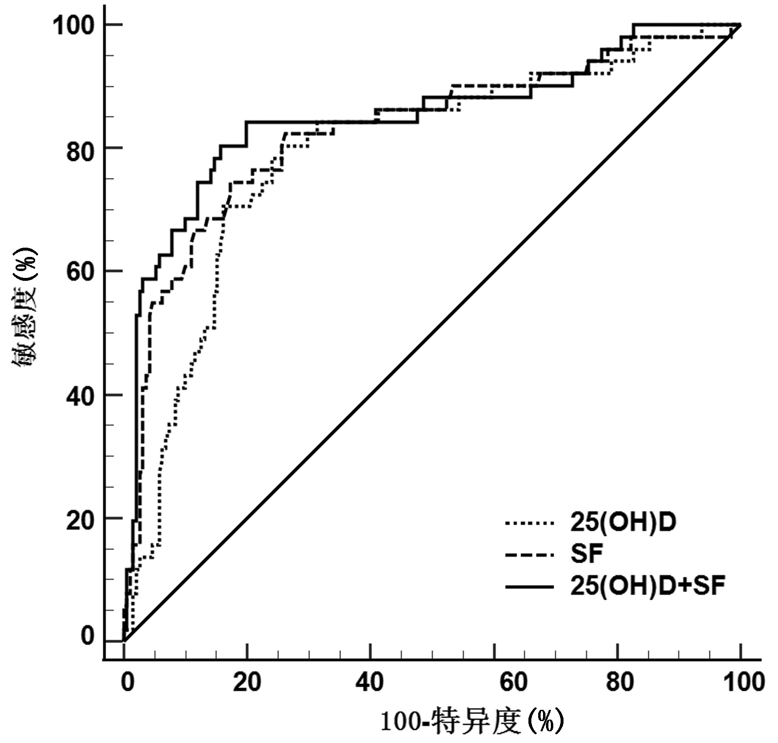
  •   Objective  To investigate the association of serum 25-hydroxyvitamin D [25(OH)D] and serum ferritin (SF) with metabolic associated fatty liver disease (MAFLD) and fibrosis-4 (FIB-4) index.  Methods  A retrospective analysis was performed for the clinical data of 595 patients who were hospitalized in Department of Gastroenterology, Wuhan Hankou Hospital, from August 2020 to December 2021. Clinical features were compared between 242 patients with MAFLD and 353 patients without MAFLD, and the prevalence rate of MAFLD and SF level were compared between the groups with different 25(OH)D levels. The non-normally distributed continuous data were expressed as M(P25-P75), and the Mann-Whitney U test was used for comparison between two groups; the chi-square test was used for comparison of categorical data between groups. A Spearman correlation analysis was used to investigate the correlation between serum 25(OH)D and SF in different populations; a binary logistic regression analysis was used to investigate the association of 25(OH)D and SF with the risk of MAFLD and FIB-4 index; the receiver operating characteristic (ROC) curves were used to assess the value of 25(OH)D and SF in the diagnosis of liver fibrosis in patients with MAFLD.  Results  Compared with the non-MAFLD patients, the MAFLD patients had a significant reduction in serum 25(OH)D level [15.35(11.26-20.02) vs 21.71(15.39-27.84), Z=-9.761, P < 0.05] and a significant increase in SF level [365.50(251.75-525.00) vs 205.00(112.50-275.00), Z=-13.317, P < 0.05]. The prevalence rate of MAFLD and SF level tended to increase with the reduction in serum 25(OH)D level (Z=75.512, P < 0.05). Serum 25(OH)D level was significantly negatively correlated with SF in MAFLD patients (r=-0.460, P < 0.05). The logistic regression analysis showed that the reduction in serum 25(OH)D level (odds ratio [OR]=0.934, 95% confidence interval [CI]: 0.879-0.992, P=0.028) and the increase in SF level (OR=1.009, 95%CI: 1.006-1.013, P < 0.001) were independent risk factors for MAFLD, and the reduction in serum 25(OH)D level (OR=0.852, 95%CI: 0.752-0.965, P=0.012) was also an independent risk factor for elevated FIB-4 index (> 2.67) in MAFLD patients. The ROC curve analysis showed that serum 25(OH)D, SF, and their combination had an area under the ROC curve of 0.793, 0.829, and 0.851, respectively, in predicting elevated FIB-4 index (> 2.67) in MAFLD patients (all P < 0.05).  Conclusion  Serum 25(OH)D is negatively correlated with SF, and the reduction in serum 25(OH)D and the increase in SF are associated with the risk of MAFLD and elevated FIB-4 index. Serum 25(OH)D and SF levels have a certain value in predicting liver fibrosis in patients with MAFLD.


  • 全世界约有一半新诊断的肝细胞癌(hepatocellular cell carcinoma,HCC)病例发生在我国,其中HBV感染是主要危险因素,占全球HCC病例的50%~80%[1]。HBV通过多种机制在宿主细胞中持续存在,从而导致慢性HBV感染[2],并且诱导HCC发生[3]。超过80%的HCC在肝硬化基础上发展,这表明肝硬化在肝癌前环境中起着重要作用[4]。因此慢性HBV感染和肝硬化是肝癌发生的高风险因素。由于缺乏明显的症状和有效的筛查策略,80%的HCC患者被诊断时已为中晚期,其中仅有30%~40%患者符合当前有效治疗方案的条件[5],故在早期将慢性HBV感染、肝硬化与HCC区分诊断是延长患者生存期甚至根治HCC的关键。现急需一种检测方式能够在患有良性肝病的肝癌高危人群中早期筛查出HCC,从而降低病死率。影像学检查作为有效筛查方法被应用于临床,但是受限于设备以及检测人员。实验室检测则更能筛查大量人群,目前主要的早期实验室筛查方法为检测患者血清AFP,但单一指标检测的敏感度与特异度存在限制。考虑联合指标检测能够提高诊断效率,本文将探究AFP与GGT/AST联合检测在良性肝病和HCC区别诊断中发挥的作用,以期为临床诊断提供新的参考依据。

    选取2019年1月15日—6月15日于本院诊治的慢性乙型肝炎患者(CHB组)、乙型肝炎肝硬化患者(LC组)、HBV相关HCC患者(HCC组),另选取同期健康体检者作为对照(HC组)。纳入标准:(1)HCC组患者HBsAg为阳性且诊断完全符合《原发性肝癌诊疗规范(2017年版)》[6]; (2)HCC组、CHB组和LC组患者均为首次诊断,在接受治疗前收集生化指标; (3)临床资料完整。排除标准:(1)排除HCC之外的恶性肿瘤; (2)排除HBV以外的其他肝炎病毒感染; (3)排除患有严重糖尿病、甲状腺亢进以及心血管疾病等患者; (4)排除妊娠患者。研究对象均自愿参与本项研究。

    采集所有研究对象空腹6~8 h后的静脉血5 ml,以转速3500 r/min(离心半径=16 cm)离心5 min,分离血清后-80 ℃冷冻备用。采用Siemens公司ADVIACENTUAR XP全自动化学发光免疫分析仪及配套试剂盒检测血清AFP水平,参考区间为0~8.0 ng/ml。使用Siemens公司ADVIA2400全自动生化分析仪检测AST、ALT、GGT水平,参考区间分别为15~40、5~40、10~60 U/L。以上操作严格依据说明书进行,所有检测均在试剂盒说明书规定时间内完成,且严格遵守试验相关操作规程。

    本研究方案经由武汉大学人民医院临床研究伦理委员会审批,批号:WDRY2018-K047。

    采用SPSS 23.0软件、GraphPad Prism 6.0和MedCalc 18.2.1对数据进行统计分析。正态分布计量资料以x±s表示,多组间比较采用方差分析,进一步两两比较采用LSD-t检验。偏态分布计量资料以M(P25~P75)表示,多组间比较采用Kruskal-Wallis H检验,进一步两两比较采用Nemenyi检验。计数资料组间比较采用χ2检验。通过二元logistic回归分析,计算出预测变量,并绘制AFP、GGT/AST以及预测变量单独或联合检测的受试者工作特征曲线(ROC曲线),计算曲线下面积(AUC)及敏感度、特异度,采用Z检验对AUC进行比较。P<0.05为差异有统计学意义。

    共纳入研究对象352例,其中HC组86例,男42例,女44例,年龄23~82岁,平均(55.90±15.19)岁; CHB组68例,男38例,女30例,年龄22~77岁,平均(51.40±10.59)岁,HBV DNA阳性50例; LC组69例,男34例,女35例,年龄28~79岁,平均(53.49±11.35)岁,HBV DNA阳性36例,Child-Pugh A级25例,B级32例,C级12例; HCC组129例,男70例,女59例,年龄24~78岁,平均(54.60±11.17)岁,HBV DNA阳性50例,Child-Pugh A级79例,B级41例,C级9例,BCLC分期A期21例,B期31例,C期77例。4组研究对象年龄(F=1.455,P=0.227)与性别分布(χ2=1.346,P=0.718)差异均无统计学意义。

    HCC组与HC组、CHB组、LC组之间患者的AFP、GGT/AST、GGT比较差异均有统计学意义(P值均<0.05)(表 1)。

    表  1  各组AFP、GGT、AST及GGT/AST血清学水平比较
    组别 例数 AFP(ng/ml) GGT/AST GGT(U/L) AST(U/L)
    HC组 86 3.35(2.20~4.70) 0.98(0.71~1.36) 20.00(15.00~27.00) 21.00(18.00~24.00)
    CHB组 68 4.35(2.35~15.75) 0.82(0.46~1.25) 51.00(23.75~140.75)1) 61.00(34.25~125.00)1)
    LC组 69 8.60(2.70~54.20)1) 0.97(0.55~1.81) 69.00(27.00~113.00)1) 47.00(31.00~88.00)1)
    HCC组 129 157.10(9.90~6126.40)1)2)3) 2.00(1.19~3.11)1)2)3) 98.00(45.00~207.00)1)2)3) 46.00(29.00~88.00)1)2)
    H 124.018 70.202 126.282 135.987
    P <0.001 <0.001 <0.001 <0.001
    注:与HC组比较,1)P<0.05;与CHB组比较,2)P<0.05;与LC组比较,3)P<0.05。
    下载: 导出CSV 
    | 显示表格

    采用二元logistic回归分析得到不同组别之间两种指标的联合回归模型,然后进一步绘制其ROC曲线(图 1),计算AUC(表 2),结果显示,在HCC组与LC组、HCC组与HC组+CHB组+LC组、HCC组与CHB组+LC组中,AFP与GGT/AST联合诊断的AUC均显著高于AFP单独诊断的AUC(Z值分别为2.684、2.241、2.415,P值分别为0.007、0.025、0.016)。

    图  1  AFP与GGT/AST单独或联合检测诊断HCC的ROC曲线
    注:a,HCC组与CHB组; b,HCC组与LC组; c,HCC组与HC组+CHB组+LC组; d,HCC组与CHB组+LC组。
    表  2  AFP与GGT/AST单独或联合检测在辅助HCC诊断中的价值
    分组 标志物 AUC 95%CI 敏感度(%) 特异度(%) P
    HCC组vs CHB组 AFP 0.803 0.742~0.864 72.9 72.1 <0.05
    GGT/AST 0.789 0.717~0.861 67.4 83.8 <0.05
    联合 0.846 0.790~0.910 79.8 77.9 <0.05
    HCC组vs LC组 AFP 0.760 0.693~0.826 55.8 84.1 <0.05
    GGT/AST 0.727 0.650~0.803 90.7 46.4 <0.05
    联合 0.802 0.741~0.862 48.1 97.1 <0.05
    HCC组vs HC组+CHB组+LC组 AFP 0.835 0.791~0.879 86.0 68.2 <0.05
    GGT/AST 0.768 0.718~0.817 67.4 74.9 <0.05
    联合 0.843 0.802~0.885 73.6 78.5 <0.05
    HCC组vs CHB组+LC组 AFP 0.781 0.727~0.835 55.8 85.4 <0.05
    GGT/AST 0.758 0.700~0.815 67.4 77.5 <0.05
    联合 0.823 0.775~0.871 81.4 66.4 <0.05
    下载: 导出CSV 
    | 显示表格

    HCC的发生通常伴有前期的慢性HBV感染和肝硬化,在慢性HBV感染以及患有肝硬化的高危人群中,利用有效的筛查方式早期发现HCC至关重要,是患者可能获得根治的关键[7]。本文纳入352例研究对象,基于相关纳排标准将其分为4组,统计分析结果发现HCC组与HC组、CHB组、LC组AFP和GGT/AST比较,差异均有统计学意义(P值均<0.05),此结果与以往研究结果[8]相符。

    AFP作为最早被发现的蛋白肿瘤标志物之一[9],其可反映肝功能情况,因此广泛用于实验室检查。综合全球多种指南,AFP在我国可作为良好的血清学指标[10],但其特异度低,易引起误判[11]。在CHB、LC患者中AFP水平也会升高,因此鉴别诊断HBV相关HCC患者时,AFP具有一定局限性,且部分HBV相关HCC患者的AFP水平并未达到筛查标准,因此容易漏查[12]

    GGT在哺乳动物组织中广泛分布,是一种质膜结合蛋白,血清中的GGT主要来源于肝胆系统[13]。已有研究[14]表明GGT通过诱导DNA损伤来促进肿瘤进展和不良预后,释放活性氧以激活与入侵相关的信号通路,与HCC发展关联密切。AST主要分布于肝细胞线粒体内,当肝脏严重病变坏死时,血清中AST水平会显著升高[15],作为肝功能检查常用生化指标,GGT与AST在临床中应用广泛[16]。本研究计算GGT/AST比值并发现其单独或联合AFP时在HBV相关HCC的诊断中具有一定价值,且该指标计算简单,不用另增检验项目,便于在HCC诊断过程中应用。

    本研究ROC曲线分析结果显示,在HCC组与LC组中,GGT/AST联合AFP后AUC明显提高,表明GGT/AST与AFP联合在鉴别HCC与LC时有良好的诊断效果; 在HCC组与HC组+CHB组+LC组以及CHB组+LC组分别区别诊断时,二者联合的AUC均明显高于AFP单独诊断的AUC,进一步说明GGT/AST联合AFP在鉴别HCC与良性肝病患者中有较好的诊断价值。

    综上所述,本研究表明GGT/AST联合AFP在HBV相关HCC的临床诊断中具有一定价值,提高了AFP的单独诊断效率。由于本研究样本量较小,因此相关研究结果仍需未来增加样本量予以验证。

  • [1]
    LAZARUS JV, MARK HE, ANSTEE QM, et al. Advancing the global public health agenda for NAFLD: A consensus statement[J]. Nat Rev Gastroenterol Hepatol, 2022, 19(1): 60-78. DOI: 10.1038/s41575-021-00523-4.
    [2]
    DU SX, LU LL, GENG N, et al. Association of serum ferritin with non-alcoholic fatty liver disease: A meta-analysis[J]. Lipids Health Dis, 2017, 16(1): 228. DOI: 10.1186/s12944-017-0613-4.
    [3]
    ALI SANGOUNI A, GHAVAMZADEH S, JAMALZEHI A. A narrative review on effects of vitamin D on main risk factors and severity of non-alcoholic fatty liver disease[J]. Diabetes MetabSyndr, 2019, 13(3): 2260-2265. DOI: 10.1016/j.dsx.2019.05.013.
    [4]
    ESLAM M, NEWSOME PN, SARIN SK, et al. A new definition for metabolic dysfunction-associated fatty liver disease: An international expert consensus statement[J]. J Hepatol, 2020, 73(1): 202-209. DOI: 10.1016/j.jhep.2020.03.039.
    [5]
    LIN S, HUANG JF, WANG MF, et al. Comparison of MAFLD and NAFLD diagnostic criteria in real world[J]. Liver Int, 2020, 40(9): 2082-2089. DOI: 10.1111/liv.14548.
    [6]
    CIARDULLO S, PERSEGHIN G. Prevalence of NAFLD, MAFLD and associated advanced fibrosis in the contemporary United States population[J]. Liver Int, 2021, 41(6): 1290-1293. DOI: 10.1111/liv.14828.
    [7]
    SHAH AG, LYDECKER A, MURRAY K, et al. Comparison of noninvasive markers of fibrosis in patients with nonalcoholic fatty liver disease[J]. Clin Gastroenterol Hepatol, 2009, 7(10): 1104-1112. DOI: 10.1016/j.cgh.2009.05.033.
    [8]
    LIAO XP, ZHANG ZL, ZHANG HH, et al. Application guideline for vitamin D and bone health in adult Chinese(2014 standard edition) vitamin D working group of osteoporosis committee of China gerontological society[J]. Chin J Osteoporos, 2014, 20(9): 1011-1030. DOI: 10.3969/j.issn.1006-7108.2014.09.002.

    廖祥鹏, 张增利, 张红红, 等. 维生素D与成年人骨骼健康应用指南(2014年标准版)[J]. 中国骨质疏松杂志, 2014, 20(9): 1011-1030. DOI: 10.3969/j.issn.1006-7108.2014.09.002.
    [9]
    ZHOU Q, LI JQ, LI XW. Influence of vitamin D deficiency on fibrosis-4 index and disease severity in patients with nonalcoholic steatohepatitis[J]. J Clin Hepatol, 2022, 38(6): 1293-1298. DOI: 10.3969/j.issn.1001-5256.2022.06.015.

    周荃, 李金强, 黎晓武. 维生素D缺乏对非酒精性脂肪性肝炎患者FIB-4指数及病情严重程度的影响[J]. 临床肝胆病杂志, 2022, 38(6): 1293-1298. DOI: 10.3969/j.issn.1001-5256.2022.06.015.
    [10]
    SUN CJ, ZUO XQ, YAO N, et al. Study on the correlation between serum ferritin and non-alcoholic fatty liver disease[J]. Zhejiang J Integr Tradit Chin West Med, 2019, 29(5): 371-375. DOI: 10.3969/j.issn.1005-4561.2019.05.008.

    孙彩娟, 左昔清, 姚娜, 等. 血清铁蛋白与非酒精性脂肪性肝病相关性研究[J]. 浙江中西医结合杂志, 2019, 29(5): 371-375. DOI: 10.3969/j.issn.1005-4561.2019.05.008.
    [11]
    BACCHETTA J, ZARITSKY JJ, SEA JL, et al. Suppression of iron-regulatory hepcidin by vitamin D[J]. J Am Soc Nephrol, 2014, 25(3): 564-572. DOI: 10.1681/ASN.2013040355.
    [12]
    LIU ZP, ZHANG JH, WANG XN, et al. Effect of vitamin D on serum markers of iron metabolism in patients with non-alcoholic fatty liver disease[J]. Tianjin Med J, 2018, 46(12): 1316-1318. DOI: 10.11958/20180847.

    刘志平, 张金华, 王湘宁, 等. 维生素D对非酒精性脂肪性肝病患者铁代谢的影响[J]. 天津医药, 2018, 46(12): 1316-1318. DOI: 10.11958/20180847.
    [13]
    PATEL YA, GIFFORD EJ, GLASS LM, et al. Identifying nonalcoholic fatty liver disease advanced fibrosis in the veterans health administration[J]. Dig Dis Sci, 2018, 63(9): 2259-2266. DOI: 10.1007/s10620-018-5123-3.
    [14]
    MA XH, ZHANG X, YOU Y, et al. Diagnostic value of APRI combined with FIB-4 for significant liver fibrosis in patients with chronic hepatitis B[J]. Chin J Gastroenterol, 2017, 22(9): 544-547. DOI: 10.3969/j.issn.1008-7125.2017.09.007.

    马晓辉, 张新, 游云, 等. APRI、FIB-4联合对慢性乙型肝炎患者显著肝纤维化的诊断价值[J]. 胃肠病学, 2017, 22(9): 544-547. DOI: 10.3969/j.issn.1008-7125.2017.09.007.
    [15]
    YANG BB, CHEN YH, ZHANG C, et al. Low vitamin D status is associated with advanced liver fibrosis in patients with nonalcoholic fatty liver disease[J]. Endocrine, 2017, 55(2): 582-590. DOI: 10.1007/s12020-016-1152-x.
    [16]
    UDOMSINPRASERT W, JITTIKOON J. Vitamin D and liver fibrosis: Molecular mechanisms and clinical studies[J]. Biomed Pharmacother, 2019, 109: 1351-1360. DOI: 10.1016/j.biopha.2018.10.140.
    [17]
    KOWDLEY KV, BELT P, WILSON LA, et al. Serum ferritin is an independent predictor of histologic severity and advanced fibrosis in patients with nonalcoholic fatty liver disease[J]. Hepatology, 2012, 55(1): 77-85. DOI: 10.1002/hep.24706.
    [18]
    BUZZETTI E, PETTA S, MANUGUERRA R, et al. Evaluating the association of serum ferritin and hepatic iron with disease severity in non-alcoholic fatty liver disease[J]. Liver Int, 2019, 39(7): 1325-1334. DOI: 10.1111/liv.14096.
    [19]
    YONEDA M, THOMAS E, SUMIDA Y, et al. Clinical usage of serum ferritin to assess liver fibrosis in patients with non-alcoholic fatty liver disease: Proceed with caution[J]. Hepatol Res, 2014, 44(14): E499-E502. DOI: 10.1111/hepr.12327.
    [20]
    ZENG J, FAN JG. Clinical significance of renaming nonalcoholic fatty liver disease[J]. J Clin Hepatol, 2020, 36(6): 1205-1207. DOI: 10.3969/j.issn.1001-5256.2020.06.002.

    曾静, 范建高. 非酒精性脂肪性肝病更名的临床意义[J]. 临床肝胆病杂志, 2020, 36(6): 1205-1207. DOI: 10.3969/j.issn.1001-5256.2020.06.002.
  • Relative Articles

    [1]Yuan WEN, Juanjuan ZHU. Clinical efficacy of double plasma molecular absorption system and sequential plasma exchange combined with continuous renal replacement therapy in treatment of acute-on-chronic liver failure with acute kidney injury[J]. Journal of Clinical Hepatology, 2024, 40(3): 556-561. doi: 10.12449/JCH240319
    [2]Jie LU, Dingchun LI, Ye LIU, Linna YUAN, Zhiwen DUAN, Wu LI. Clinical efficacy of low-dose plasma exchange combined with double plasma molecular absorption system/hemoperfusion in treatment of acute-on-chronic liver failure[J]. Journal of Clinical Hepatology, 2022, 38(11): 2526-2531. doi: 10.3969/j.issn.1001-5256.2022.11.017
    [3]Lan XiaoQin, Ji YaLi, Chen JinJun, Zhou FuYuan, Wen WeiQun. Effect of artificial liver support therapy on the short-term prognosis of patients with liver failure in the plateau stage: A stratified analysis based on Model for End-Stage Liver Disease score[J]. Journal of Clinical Hepatology, 2020, 36(9): 2005-2009. doi: 10.3969/j.issn.1001-5256.2020.09.019
    [4]Yao YunHai, Gan JianHe, Zhao WeiFeng. Effect of plasma exchange combined with continuous renal replacement therapy on the prognosis of patients with HBV-related acute-on-chronic liver failure and acute kidney injury[J]. Journal of Clinical Hepatology, 2019, 35(5): 1065-1069. doi: 10.3969/j.issn.1001-5256.2019.05.026
    [5]Guo XiJu, Guo WeiBo, Zhang JiaChang, Lu YaJing, Yang Yan. Clinical effect of double plasma molecular absorption system in treatment of liver failure due to hyperthyroidism: A case report[J]. Journal of Clinical Hepatology, 2018, 34(7): 1526-1528. doi: 10.3969/j.issn.1001-5256.2018.07.033
    [6]Wu YiChen, Yao HongYu, Wang KaiLi, Jiang QiYu, Liu Chang, Xing HanQian, Liu SuXia, You ShaoLi, Zhao Jun. Effect of plasma exchange on bioactivity of peripheral blood CD34+ cells in patients with acute-on-chronic (subacute) liver failure[J]. Journal of Clinical Hepatology, 2018, 34(11): 2383-2387. doi: 10.3969/j.issn.1001-5256.2018.11.023
    [7]Li Shuang, Chen Yu. Coping with shortage of plasma-The new therapeutic pattern of non-bioartificial liver[J]. Journal of Clinical Hepatology, 2017, 33(9): 1687-1692. doi: 10.3969/j.issn.1001-5256.2017.09.012
    [8]Zhou Jian, Wan Hong, Yang ZhengMao, Wang ZhaoXun. Clinical effect of plasma perfusion combined with plasma exchange in treatment of patients with acute-on-chronic liver failure[J]. Journal of Clinical Hepatology, 2017, 33(4): 715-718. doi: 10.3969/j.issn.1001-5256.2017.04.023
    [9]Hou HuanRong, Shang Jia, Kang Yi, Li YuKui, Ceng YanLi, Ding GangQiang, Mao ZhongShan, Xiao ErHui. Clinical effect of plasma exchange combined with hemofiltration in patients with subacute liver failure induced by antitubercular agents[J]. Journal of Clinical Hepatology, 2016, 32(2): 342-346. doi: 10.3969/j.issn.1001-5256.2016.02.030
    [10]Zhang LiangJie, Chen HuiJuan, Zhao ShouSong. Effects of artificial liver plasma exchange on cytokines in patients with liver failure[J]. Journal of Clinical Hepatology, 2015, 31(4): 578-581. doi: 10.3969/j.issn.1001-5256.2015.04.024
    [11]Wu ShaoHong, Gan JianHe, Huang XiaoPing, Lin Hua, Lu NianFang, Wu JiangQuan, Zheng RuiQiang. Clinical effect of albumin dialysis combined with plasma perfusion in treatment of liver failure[J]. Journal of Clinical Hepatology, 2014, 30(5): 434-437. doi: 10.3969/j.issn.1001-5256.2014.05.013
    [12]Liu XiaoHui, Guo HaiQing, Zhang Jing, Duan ZhongPing. Progress in technology and clinical application of non- bioartificial liver support system[J]. Journal of Clinical Hepatology, 2013, 29(9): 661-665. doi: 10.3969/j.issn.1001-5256.2013.09.007
    [13]You ShaoLi, Liu HongLing, Rong YiHui, Zhu Bing, Zang Hong, Liu WanShu, Wan ZhiHong, Mao PanYong, Xin ShaoJie. Preliminary investigation of hybrid bioartificial liver support system in treatment of HBV- related acute- on- chronic liver failure[J]. Journal of Clinical Hepatology, 2013, 29(9): 685-688. doi: 10.3969/j.issn.1001-5256.2013.09.013
    [14]Hu QiJiang, Jiang YingAn. Evaluation of the therapeutic efficacy of lamivudine combined with plasma exchange for treating acute-on-chronic hepatitis B liver failure [J]. Journal of Clinical Hepatology, 2013, 29(2): 107-109.
    [15]Zhou Jian, Wan Hong. Relationship between changes in serum cytokine levels after artificial liver therapy and clinical prognosis in patients with liver failure[J]. Journal of Clinical Hepatology, 2013, 29(7): 535-537. doi: 1001-5256 (2013) 07-0535-03
    [16]Zhang AiMin, You ShaoLi, Wan ZhiHong, Rong YiHui, Zhu Bing, Zang Hong, Xin ShaoJie. Combination therapy of plasma perfusion, plasma exchange, and entecavir in patients with hepatitis B acute-on-chronic liver failure[J]. Journal of Clinical Hepatology, 2012, 28(10): 744-747.
    [17]Zhang WenHua, Jia WenLing, Zhou ZiYing, Tang HaiTao, Wang FengMei. Investigation on the method and safety of the non-heparinized plasma exchange therapy in treating patients with hepatic failure[J]. Journal of Clinical Hepatology, 2012, 28(1): 50-52.
    [18]Liao JinMao, Li ZhuoRi, Hu XiaoXuan. The therapeutic effect of plasma exchange therapy in patients with severe hepatitis[J]. Journal of Clinical Hepatology, 2012, 28(4): 302-304.
    [19]Liu ZhiHong, Jiang JianNing, Luo GuangHan, Wu JiZhou, Deng YiMing, Su MingHua, Wu JianLin, Zhang Lu. The value of MELD score for evaluation of plasma exchange in patients with liver failure[J]. Journal of Clinical Hepatology, 2010, 26(6): 615-617.
  • Cited by

    Periodical cited type(3)

    1. 姚爱武,廖和壁,张璟. 血清AFP、AFP-L3与肝细胞癌经肝动脉化疗栓塞术后疗效的关系分析. 分子诊断与治疗杂志. 2023(04): 690-693+698 .
    2. 朱富平,刘红强,常清,冷伟业. miR-222-3p在乙型肝炎病毒相关肝细胞癌血清外泌体中的表达及临床意义. 局解手术学杂志. 2022(08): 702-708 .
    3. 高武林,韦超,郭晓烨. 术前血清MMP-9水平对HBV相关肝细胞癌患者肝切除术后生存的预测作用. 东南大学学报(医学版). 2022(05): 652-659 .

    Other cited types(2)

  • 加载中

Catalog

    通讯作者: 陈斌, bchen63@163.com
    • 1. 

      沈阳化工大学材料科学与工程学院 沈阳 110142

    1. 本站搜索
    2. 百度学术搜索
    3. 万方数据库搜索
    4. CNKI搜索

    Figures(2)  / Tables(4)

    Article Metrics

    Article views (497) PDF downloads(54) Cited by(5)
    Proportional views
    Related

    /

    DownLoad:  Full-Size Img  PowerPoint
    Return
    Return