中文English
ISSN 1001-5256 (Print)
ISSN 2097-3497 (Online)
CN 22-1108/R

Role of neuroimmune communication via the gut-brain axis in the pathogenesis of hepatic encephalopathy

DOI: 10.12449/JCH241224
Research funding:

National Natural Science Foundation of China (82260907);

National Natural Science Foundation of China (82260899);

National Natural Science Foundation of China (82274434);

National Youth Qihuang Scholar Program (Guozhong Pharmaceutical Education Letter [2022] No.256);

Guangxi Youth Qihuang Scholar Project (Guizhong Medical Science and Education Development [2023] No.7)

More Information
  • Corresponding author: MAO Dewen, mdwboshi2005@163.com (ORCID: 0000-0001-9438-9325)
  • Received Date: 2024-03-23
  • Accepted Date: 2024-04-24
  • Published Date: 2024-12-25
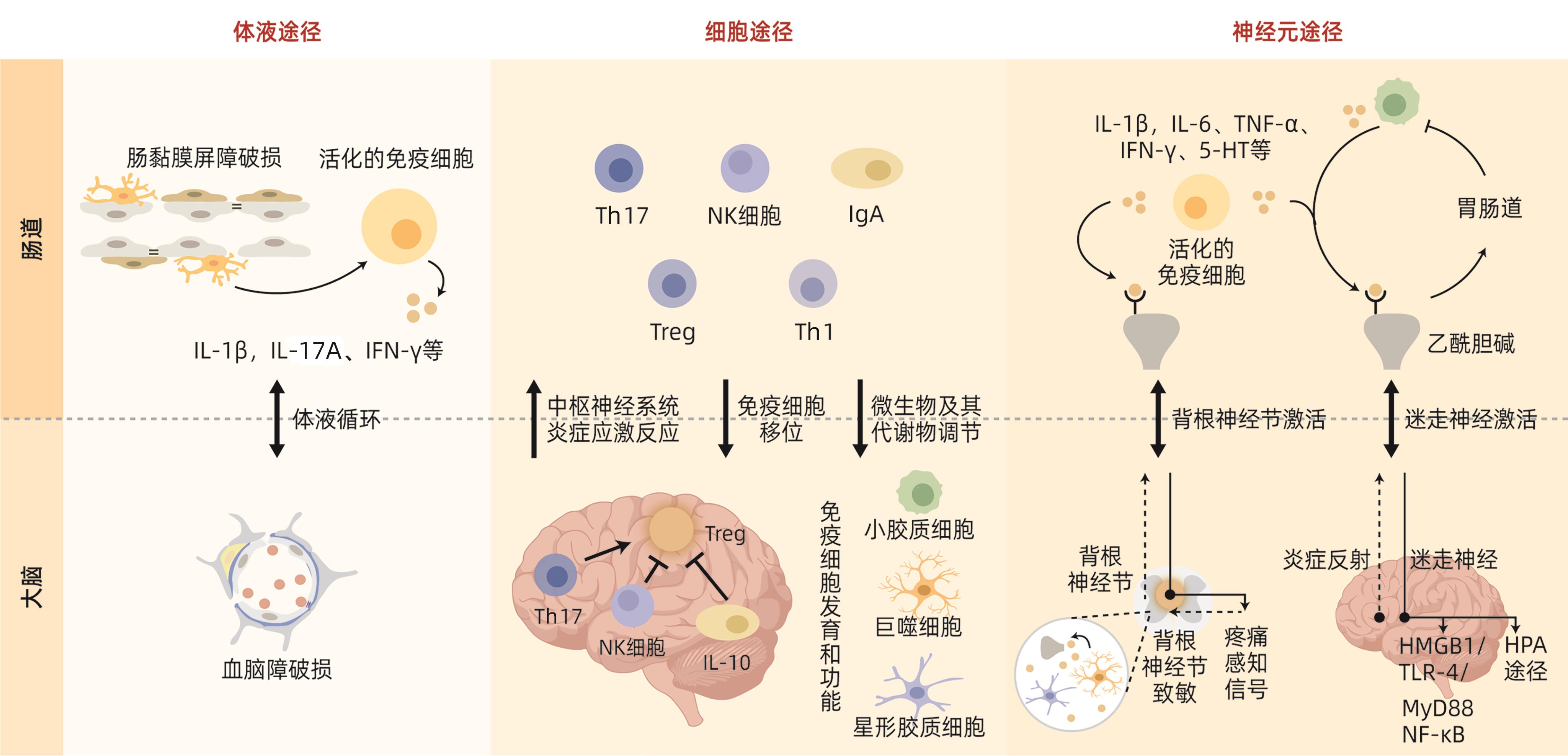
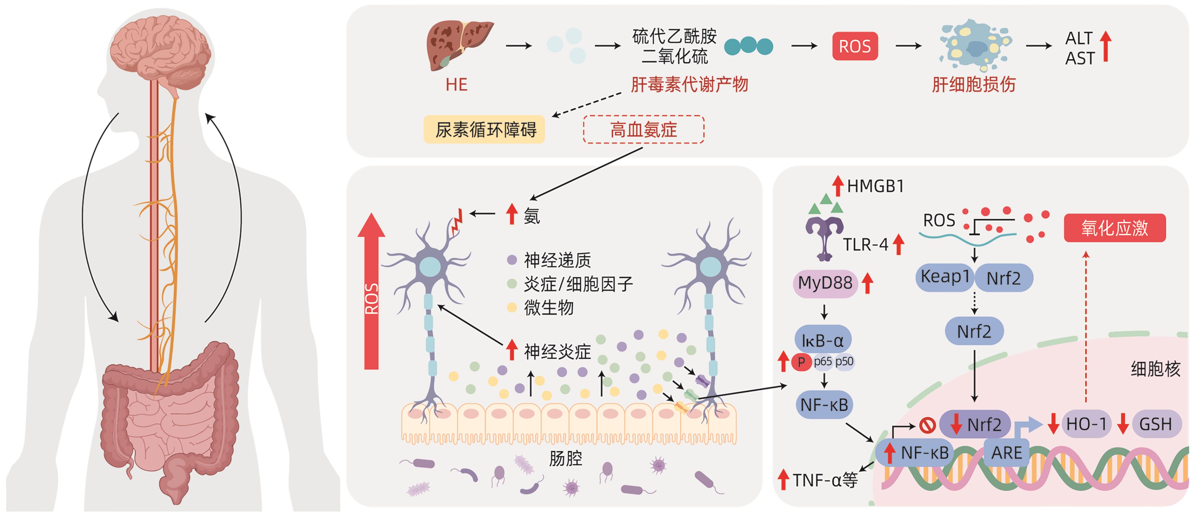
  • Hepatic encephalopathy (HE) is a common severe liver disease syndrome in clinical practice and is one of the critical and severe diseases in internal medicine, and more than half of liver failure patients diagnosed with overt HE have a survival time of less than 1 year. A comprehensive analysis of the complex pathogenesis of HE and the development of diagnosis and treatment regimens based on evidence-based medicine are of great importance for alleviating high medical resource consumption, high medical expenses, and high incidence and mortality rates in clinical practice. The latest studies have shown that the intestinal tract and the central nervous system can perform bidirectional continuous interaction and signal transmission and regulate the function of inflammation signals, molecules, cells, and organs, which is known as neuroimmune communication and is highly consistent with the main pathological features of HE. With a focus on the mechanism of neuroimmune communication in HE, this article reviews the association between inflammation signal transduction via the gut-brain axis and neurotransmitter regulation and its role in neuroimmune communication in HE, which provides new ideas for the clinical diagnosis and treatment of HE and the research and development of related drugs.


  • [1]
    RUDLER M, WEISS N, BOUZBIB C, et al. Diagnosis and management of hepatic encephalopathy[J]. Clin Liver Dis, 2021, 25( 2): 393- 417. DOI: 10.1016/j.cld.2021.01.008.
    [2]
    Liver Failure and Artificial Liver Group, Chinese Society of Infectious Diseases, Chinese Medical Association; Severe Liver Disease and Artificial Liver Group, Chinese Society of Hepatology, Chinese Medical Association. Guideline for diagnosis and treatment of liver failure(2018)[J]. J Clin Hepatol, 2019, 35( 1): 38- 44. DOI: 10.3969/j.issn.1001-5256.2019.01.007.

    中华医学会感染病学分会肝衰竭与人工肝学组, 中华医学会肝病学分会重型肝病与人工肝学组. 肝衰竭诊治指南(2018年版)[J]. 临床肝胆病杂志, 2019, 35( 1): 38- 44. DOI: 10.3969/j.issn.1001-5256.2019.01.007.
    [3]
    ELSAID MI, RUSTGI VK. Epidemiology of hepatic encephalopathy[J]. Clin Liver Dis, 2020, 24( 2): 157- 174. DOI: 10.1016/j.cld.2020.01.001.
    [4]
    LIN Y, YAN GJ, FENG F, et al. Association between cholesterol and liver regeneration and its significance and potential value in clinical treatment of liver failure[J]. J Clin Hepatol, 2022, 38( 3): 708- 713. DOI: 10.3969/j.issn.1001-5256.2022.03.044.

    林镛, 颜耿杰, 冯逢, 等. 胆固醇与肝再生关系及其在肝衰竭治疗中的意义和潜在价值[J]. 临床肝胆病杂志, 2022, 38( 3): 708- 713. DOI: 10.3969/j.issn.1001-5256.2022.03.044.
    [5]
    DU YQ, WANG M, HUANG GC, et al. Therapeutic effect of retention enema with compound rhubarb decoction on a rat model of minimal hepatic encephalopathy based on bile acid metabolomics[J]. J Clin Hepatol, 2023, 39( 10): 2348- 2357. DOI: 10.3969/j.issn.1001-5256.2023.10.012.

    杜沅沁, 王萌, 黄国初, 等. 基于胆汁酸代谢组学探讨复方大黄煎剂保留灌肠对轻微型肝性脑病大鼠模型的治疗作用[J]. 临床肝胆病杂志, 2023, 39( 10): 2348- 2357. DOI: 10.3969/j.issn.1001-5256.2023.10.012.
    [6]
    European Association for the Study of the Liver. EASL clinical practice guidelines on the management of hepatic encephalopathy[J]. J Hepatol, 2022, 77( 3): 807- 824. DOI: 10.1016/j.jhep.2022.06.001.
    [7]
    PUN CK, HUANG HC, CHANG CC, et al. Hepatic encephalopathy: From novel pathogenesis mechanism to emerging treatments[J]. J Chin Med Assoc, 2023, 87( 3): 245- 251. DOI: 10.1097/JCMA.0000000000001041.
    [8]
    RONALDSON PT, DAVIS TP. Regulation of blood-brain barrier integrity by microglia in health and disease: A therapeutic opportunity[J]. J Cereb Blood Flow Metab, 2020, 40( 1_suppl): S6- S24. DOI: 10.1177/0271678X20951995.
    [9]
    OCHOA-SANCHEZ R, TAMNANLOO F, ROSE CF. Hepatic encephalopathy: From metabolic to neurodegenerative[J]. Neurochem Res, 2021, 46( 10): 2612- 2625. DOI: 10.1007/s11064-021-03372-4.
    [10]
    COOPER AJ, PLUM F. Biochemistry and physiology of brain ammonia[J]. Physiol Rev, 1987, 67( 2): 440- 519. DOI: 10.1152/physrev.1987.67.2.440.
    [11]
    AGIRMAN G, YU KB, HSIAO EY. Signaling inflammation across the gut-brain axis[J]. Science, 2021, 374( 6571): 1087- 1092. DOI: 10.1126/science.abi6087.
    [12]
    VIDAL-CEVALLOS P, CHÁVEZ-TAPIA NC, URIBE M. Current approaches to hepatic encephalopathy[J]. Ann Hepatol, 2022, 27( 6): 100757. DOI: 10.1016/j.aohep.2022.100757.
    [13]
    ORZEŁ‍-GAJOWIK K, MILEWSKI K, ZIELIŃSKA M. miRNA-ome plasma analysis unveils changes in blood-brain barrier integrity associated with acute liver failure in rats[J]. Fluids Barriers CNS, 2023, 20( 1): 92. DOI: 10.1186/s12987-023-00484-7.
    [14]
    SCOTT SA, FU JJ, CHANG PV. Microbial tryptophan metabolites regulate gut barrier function via the aryl hydrocarbon receptor[J]. Proc Natl Acad Sci U S A, 2020, 117( 32): 19376- 19387. DOI: 10.1073/pnas.2000047117.
    [15]
    KHALIL HMA, ELIWA HA, EL-SHIEKH RA, et al. Ashwagandha(Withania somnifera) root extract attenuates hepatic and cognitive deficits in thioacetamide-induced rat model of hepatic encephalopathy via induction of Nrf2/HO-1 and mitigation of NF-κB/MAPK signaling pathways[J]. J Ethnopharmacol, 2021, 277: 114141. DOI: 10.1016/j.jep.2021.114141.
    [16]
    ALI SA, DATUSALIA AK. Protective effects of Tinospora cordifolia miers extract against hepatic and neurobehavioral deficits in thioacetamide-induced hepatic encephalopathy in rats via modulating hyperammonemia and glial cell activation[J]. J Ethnopharmacol, 2024, 323: 117700. DOI: 10.1016/j.jep.2023.117700.
    [17]
    CRYAN JF, O’RIORDAN KJ, COWAN CSM, et al. The microbiota-gut-brain axis[J]. Physiol Rev, 2019, 99( 4): 1877- 2013. DOI: 10.1152/physrev.00018.2018.
    [18]
    XU CL, LEE SK, ZHANG DC, et al. The gut microbiome regulates psychological-stress-induced inflammation[J]. Immunity, 2020, 53( 2): 417- 428. DOI: 10.1016/j.immuni.2020.06.025.
    [19]
    LOUVEAU A, HERZ J, ALME MN, et al. CNS lymphatic drainage and neuroinflammation are regulated by meningeal lymphatic vasculature[J]. Nat Neurosci, 2018, 21( 10): 1380- 1391. DOI: 10.1038/s41593-018-0227-9.
    [20]
    JAMESON KG, OLSON CA, KAZMI SA, et al. Toward understanding microbiome-neuronal signaling[J]. Mol Cell, 2020, 78( 4): 577- 583. DOI: 10.1016/j.molcel.2020.03.006.
    [21]
    JIA W, RAJANI C, KADDURAH-DAOUK R, et al. Expert insights: The potential role of the gut microbiome-bile acid-brain axis in the development and progression of Alzheimer’s disease and hepatic encephalopathy[J]. Med Res Rev, 2020, 40( 4): 1496- 1507. DOI: 10.1002/med.21653.
    [22]
    LIU T, ZHANG LY, JOO D, et al. NF-κB signaling in inflammation[J]. Signal Transduct Target Ther, 2017, 2: 17023. DOI: 10.1038/sigtrans.2017.23.
    [23]
    ESSAM RM, SAADAWY MA, GAMAL M, et al. Lactoferrin averts neurological and behavioral impairments of thioacetamide-induced hepatic encephalopathy in rats via modulating HGMB1/TLR-4/MyD88/Nrf2 pathway[J]. Neuropharmacology, 2023, 236: 109575. DOI: 10.1016/j.neuropharm.2023.109575.
    [24]
    KIGERL KA, LAI WM, WALLACE LM, et al. High mobility group box-1(HMGB1) is increased in injured mouse spinal cord and can elicit neurotoxic inflammation[J]. Brain Behav Immun, 2018, 72: 22- 33. DOI: 10.1016/j.bbi.2017.11.018.
    [25]
    YOU XH, WANG LX, FENG DC. Relationship of the intestinal flora and irritable bowel syndrome sub-typing with the functional changes in the neuroendocrine axis[J]. J Clin Exp Med, 2022, 21( 15): 1667- 1670. DOI: 10.3969/j.issn.1671-4695.2022.15.027.

    游先辉, 王利霞, 冯大超. 肠道菌群和肠易激综合征分型与神经内分泌轴功能变化的关系研究[J]. 临床和实验医学杂志, 2022, 21( 15): 1667- 1670. DOI: 10.3969/j.issn.1671-4695.2022.15.027.
    [26]
    PENNY HA, DOMINGUES RG, KRAUSS MZ, et al. Rhythmicity of intestinal IgA responses confers oscillatory commensal microbiota mutualism[J]. Sci Immunol, 2022, 7( 75): eabk2541. DOI: 10.1126/sciimmunol.abk2541.
    [27]
    ROJAS OL, PRÖBSTEL AK, PORFILIO EA, et al. Recirculating intestinal IgA-producing cells regulate neuroinflammation via IL-10[J]. Cell, 2019, 176( 3): 610- 624. DOI: 10.1016/j.cell.2018.11.035.
    [28]
    BONAZ B, BAZIN T, PELLISSIER S. The vagus nerve at the interface of the microbiota-gut-brain axis[J]. Front Neurosci, 2018, 12: 49. DOI: 10.3389/fnins.2018.00049.
    [29]
    BLUTHÉ RM, MICHAUD B, KELLEY KW, et al. Vagotomy blocks behavioural effects of interleukin-1 injected via the intraperitoneal route but not via other systemic routes[J]. Neuroreport, 1996, 7( 15-17): 2823- 2827. DOI: 10.1097/00001756-199611040-00083.
    [30]
    MCVEY NEUFELD KA, BIENENSTOCK J, BHARWANI A, et al. Oral selective serotonin reuptake inhibitors activate vagus nerve dependent gut-brain signalling[J]. Sci Rep, 2019, 9( 1): 14290. DOI: 10.1038/s41598-019-50807-8.
    [31]
    MORAIS LH, SCHREIBER HL 4th. MAZMANIAN SK The gut microbiotabrain axis in behaviour and brain disorders[J]. Nat Rev Microbiol, 2021, 19( 4): 241- 255. DOI: 10.1038/s41579-020-00460-0.
    [32]
    MARTÍNEZ-LOZADA Z, ORTEGA A. Glutamatergic transmission: A matter of three[J]. Neural Plast, 2015, 2015: 787396. DOI: 10.1155/2015/787396.
    [33]
    CABRERA-PASTOR A, LLANSOLA M, MONTOLIU C, et al. Peripheral inflammation induces neuroinflammation that alters neurotransmission and cognitive and motor function in hepatic encephalopathy: Underlying mechanisms and therapeutic implications[J]. Acta Physiol(Oxf), 2019, 226( 2): e13270. DOI: 10.1111/apha.13270.
    [34]
    KOSENKOV AM, GAIDIN SG, SERGEEV AI, et al. Fast changes of NMDA and AMPA receptor activity under acute hyperammonemia in vitro[J]. Neurosci Lett, 2018, 686: 80- 86. DOI: 10.1016/j.neulet.2018.08.054.
    [35]
    CAULI O, GONZÁLEZ-USANO A, CABRERA-PASTOR A, et al. Blocking NMDA receptors delays death in rats with acute liver failure by dual protective mechanisms in kidney and brain[J]. NeuroMolecular Med, 2014, 16( 2): 360- 375. DOI: 10.1007/s12017-013-8283-5.
    [36]
    KULLMANN DM, RUIZ A, RUSAKOV DM, et al. Presynaptic, extrasynaptic and axonal GABAA receptors in the CNS: Where and why?[J]. Prog Biophys Mol Biol, 2005, 87( 1): 33- 46. DOI: 10.1016/j.pbiomolbio.2004.06.003.
    [37]
    FRIED DE, WATSON RE, ROBSON SC, et al. Ammonia modifies enteric neuromuscular transmission through glial γ-aminobutyric acid signaling[J]. Am J Physiol Gastrointest Liver Physiol, 2017, 313( 6): G570- G580. DOI: 10.1152/ajpgi.00154.2017.
    [38]
    MA FH, YANG L, SUN ZR, et al. Neurotransmitter-derived lipidoids(NT-lipidoids) for enhanced brain delivery through intravenous injection[J]. Sci Adv, 2020, 6( 30): eabb4429. DOI: 10.1126/sciadv.abb4429.
    [39]
    TERSTAPPEN GC, MEYER AH, BELL RD, et al. Strategies for delivering therapeutics across the blood-brain barrier[J]. Nat Rev Drug Discov, 2021, 20( 5): 362- 383. DOI: 10.1038/s41573-021-00139-y.
  • Relative Articles

    [1]Chen YANG, Shanghai YU, Feipeng XU, Hua ZHANG. Application of magnetic resonance imaging-proton density fat fraction in liver fat quantification[J]. Journal of Clinical Hepatology, 2024, 40(3): 600-605. doi: 10.12449/JCH240327
    [2]Lu HUANG, Lijian LU. Value of MRI biliary score and liver/muscle ratio in evaluating the pathological grade of liver fibrosis[J]. Journal of Clinical Hepatology, 2024, 40(4): 720-725. doi: 10.12449/JCH240413
    [3]Mu Rong, Lu LunGen. Advances in the application of magnetic resonance imaging in hepatic fat quantification in patients with nonalcoholic fatty liver disease[J]. Journal of Clinical Hepatology, 2020, 36(6): 1366-1369. doi: 10.3969/j.issn.1001-5256.2020.06.038
    [4]Pan Qi, Luo ChunHai, Ma WanLing, Qu YuanFei, Wang JianRu, Chen Lin. Specific magnetic resonance imaging of vascular endothelial growth factor-C targeted molecular probe and its clinical significance in a rat model of hepatocellular carcinoma[J]. Journal of Clinical Hepatology, 2018, 34(7): 1492-1496. doi: 10.3969/j.issn.1001-5256.2018.07.025
    [5]Chen QiPing, Lin Su, Shi ZhenShan, Zhu YueYong, Li YouBing. A cost-effectiveness analysis of Gd-EOB-DTPA contrast-enhanced magnetic resonance imaging versus ultrasound in hepatocellular carcinoma screening[J]. Journal of Clinical Hepatology, 2018, 34(9): 1917-1920. doi: 10.3969/j.issn.1001-5256.2018.09.018
    [6]Luo YongHeng, Xiao EnHua. Role of functional magnetic resonance imaging in evaluating the therapeutic effect of transcatheter arterial chemoembolization in hepatocellular carcinoma[J]. Journal of Clinical Hepatology, 2016, 32(12): 2277-2281. doi: 10.3969/j.issn.1001-5256.2016.12.010
    [7]Yan FuHua. Research advances in computed tomography and magnetic resonance imaging findings of cirrhotic portal hypertension[J]. Journal of Clinical Hepatology, 2016, 32(6): 1079-1082. doi: 10.3969/j.issn.1001-5256.2016.06.011
    [8]Wu Jie, Jing ZongLin. Research advances in MRI features and diagnosis of hepatocellular adenomas[J]. Journal of Clinical Hepatology, 2016, 32(10): 2012-2015. doi: 10.3969/j.issn.1001-5256.2016.10.045
    [9]Wang LiLi, Lei JunQiang, Guo JiGang, Yin Liang, Zhai YaNan, Li JinKui, Chen Hong, Guo ShunLin. Value of Gd-EOB-DTPA-enhanced MRI in assessing liver function[J]. Journal of Clinical Hepatology, 2015, 31(5): 749-753. doi: 10.3969/j.issn.1001-5256.2015.05.028
    [10]Tang ShaoShan, Zhao GuoJia, Wang YiJiao. Assessment of ultrasound elastography to grade hepatic steatosis in a rat model[J]. Journal of Clinical Hepatology, 2013, 29(4): 282-285.
    [11]Wang Zi, Hu DaoYu. Research advances in quantitative evaluation of liver fat in patients with nonalcoholic fatty liver disease[J]. Journal of Clinical Hepatology, 2013, 29(12): 894-896. doi: 10.3969/j.issn.1001-5256.2013.12.005
    [12]Zeng MengSu, Li RuoKun. Clinical value of susceptibility-weighted imaging for studying the multi-step hepatocarcinogenic process of cirrhotic liver nodules[J]. Journal of Clinical Hepatology, 2013, 29(1): 38-41.
    [13]Huang RuiSui, Li Cheng, Gu Han, Jiao ZhiYun. Establishment of rat model of liver cirrhosis or liver cancer and its magnetic resonance images before and after superparamagnetic iron oxide enhancement[J]. Journal of Clinical Hepatology, 2013, 29(10): 769-774. doi: 10.3969/j.issn.1001-5256.2013.10.012
    [14]Gu Han, Li Cheng. Research progress in magnetic nanoparticles for magnetic resonance imaging and targeted therapy of hepatocellular carcinoma[J]. Journal of Clinical Hepatology, 2013, 29(10): 740-743. doi: 10.3969/j.issn.1001-5256.2013.10.005
    [15]Feng HaiLong, Li CaiYing, Wang YingJie, Feng PingYong, Cui CaiXia. Contribution of diffusion weighted MR imaging in the differential diagnosis of focal hepatic lesions at 3T[J]. Journal of Clinical Hepatology, 2011, 27(4): 400-402.
    [16]Mai Jing, Gao YueQiu. The application of magnetic resonace elastography in the non-invasive diagnosis of hepatic fibrosis[J]. Journal of Clinical Hepatology, 2011, 27(3): 318-320+324.
    [20]Zhao GuoKu, Chen Kai, Zhou HongWei. MRI diagnostic value of liver adenoma[J]. Journal of Clinical Hepatology, 2004, 20(5): 291-292+321.
  • Cited by

    Periodical cited type(1)

    1. 沈仕俊,邓翔,饶永凤,帕成周. 基于生物信息学对肝硬化中核心基因的筛选及意义探究. 医学研究杂志. 2025(01): 54-60 .

    Other cited types(0)

  • 加载中
    Created with Highcharts 5.0.7Chart context menuAccess Class DistributionFULLTEXT: 22.8 %FULLTEXT: 22.8 %META: 70.4 %META: 70.4 %PDF: 6.8 %PDF: 6.8 %FULLTEXTMETAPDF
    Created with Highcharts 5.0.7Chart context menuAccess Area Distribution其他: 7.7 %其他: 7.7 %Falls Church: 0.2 %Falls Church: 0.2 %Taoyuan District: 0.4 %Taoyuan District: 0.4 %Tiruchi: 0.2 %Tiruchi: 0.2 %上海: 4.5 %上海: 4.5 %上饶: 0.2 %上饶: 0.2 %东京: 0.2 %东京: 0.2 %中卫: 0.4 %中卫: 0.4 %九江: 0.2 %九江: 0.2 %佛山: 0.2 %佛山: 0.2 %包头: 0.2 %包头: 0.2 %北京: 3.2 %北京: 3.2 %十堰: 0.4 %十堰: 0.4 %华盛顿州: 0.2 %华盛顿州: 0.2 %南京: 1.4 %南京: 1.4 %南宁: 0.7 %南宁: 0.7 %南昌: 0.7 %南昌: 0.7 %南通: 0.4 %南通: 0.4 %喀什: 0.2 %喀什: 0.2 %大连: 0.2 %大连: 0.2 %天津: 0.9 %天津: 0.9 %太原: 0.9 %太原: 0.9 %宁波: 0.2 %宁波: 0.2 %宣城: 0.5 %宣城: 0.5 %宿州: 0.9 %宿州: 0.9 %广州: 1.1 %广州: 1.1 %开封: 0.5 %开封: 0.5 %张家口: 13.4 %张家口: 13.4 %扬州: 0.4 %扬州: 0.4 %新乡: 0.2 %新乡: 0.2 %无锡: 0.4 %无锡: 0.4 %昆明: 1.6 %昆明: 1.6 %杭州: 2.0 %杭州: 2.0 %桂林: 0.2 %桂林: 0.2 %武汉: 0.2 %武汉: 0.2 %济南: 1.1 %济南: 1.1 %海得拉巴: 0.4 %海得拉巴: 0.4 %淄博: 0.2 %淄博: 0.2 %湘潭: 0.4 %湘潭: 0.4 %漯河: 0.4 %漯河: 0.4 %濮阳: 0.2 %濮阳: 0.2 %眉山: 0.2 %眉山: 0.2 %石家庄: 1.6 %石家庄: 1.6 %福州: 0.7 %福州: 0.7 %科泽科德: 0.2 %科泽科德: 0.2 %红河: 0.4 %红河: 0.4 %芒廷维尤: 20.1 %芒廷维尤: 20.1 %芝加哥: 2.0 %芝加哥: 2.0 %苏州: 0.2 %苏州: 0.2 %茂名: 0.4 %茂名: 0.4 %营口: 0.2 %营口: 0.2 %衡阳: 0.2 %衡阳: 0.2 %西宁: 5.6 %西宁: 5.6 %西安: 0.5 %西安: 0.5 %诺沃克: 0.2 %诺沃克: 0.2 %贵阳: 0.2 %贵阳: 0.2 %赣州: 0.2 %赣州: 0.2 %运城: 0.5 %运城: 0.5 %郑州: 0.2 %郑州: 0.2 %都柏林: 0.2 %都柏林: 0.2 %重庆: 0.4 %重庆: 0.4 %铁岭: 0.2 %铁岭: 0.2 %长春: 17.2 %长春: 17.2 %长沙: 0.7 %长沙: 0.7 %长治: 0.2 %长治: 0.2 %青岛: 0.2 %青岛: 0.2 %鞍山: 0.4 %鞍山: 0.4 %其他Falls ChurchTaoyuan DistrictTiruchi上海上饶东京中卫九江佛山包头北京十堰华盛顿州南京南宁南昌南通喀什大连天津太原宁波宣城宿州广州开封张家口扬州新乡无锡昆明杭州桂林武汉济南海得拉巴淄博湘潭漯河濮阳眉山石家庄福州科泽科德红河芒廷维尤芝加哥苏州茂名营口衡阳西宁西安诺沃克贵阳赣州运城郑州都柏林重庆铁岭长春长沙长治青岛鞍山

Catalog

    通讯作者: 陈斌, bchen63@163.com
    • 1. 

      沈阳化工大学材料科学与工程学院 沈阳 110142

    1. 本站搜索
    2. 百度学术搜索
    3. 万方数据库搜索
    4. CNKI搜索

    Figures(2)

    Article Metrics

    Article views (393) PDF downloads(38) Cited by(1)
    Proportional views
    Related

    /

    DownLoad:  Full-Size Img  PowerPoint
    Return
    Return