中文English
ISSN 1001-5256 (Print)
ISSN 2097-3497 (Online)
CN 22-1108/R

Ultrasound findings and contrast-enhanced ultrasound findings of mass-type autoimmune pancreatitis versus pancreatic ductal adenocarcinoma

DOI: 10.3969/j.issn.1001-5256.2022.06.025
Research funding:

Science & Technology Program of Tangshan (19150220E)

More Information
  • Corresponding author: WANG Yanbin, wangyanbin689@163.com(ORCID: 0000-0001-9861-1763)
  • Received Date: 2021-11-09
  • Accepted Date: 2021-12-02
  • Published Date: 2022-06-20
  •   Objective  To investigate the value of ultrasound and contrast-enhanced ultrasound (CEUS) in the differential diagnosis of mass-type autoimmune pancreatitis (AIP) and pancreatic ductal adenocarcinoma (PDAC).  Methods  A retrospective analysis was performed for the clinical data, ultrasound findings, and CEUS findings of 11 patients with mass-type AIP who were diagnosed in Tangshan Workers' Hospital from January 2015 to December 2020, and their characteristic manifestations were analyzed and compared with the data of 23 patients with PDCA. The chi-square test was used for comparison of categorical data between two groups.  Results  For the 11 patients with mass-type AIP, CEUS had a diagnostic accuracy of 63.64%, and all of these patients had hypoechoic single lesions; the patients with clear boundaries, regular morphology, pancreatic duct dilatation or cutoff, and blood flow signal accounted for 54.55%, 63.64%, 18.18%, and 36.36%, respectively, while in the PDCA group, such patients accounted for 30.43%, 34.78%, 78.26%, and 21.74%, respectively, and there was a significant difference in the presence or absence of pancreatic duct dilatation or cutoff between the two groups(χ2=11.089, P < 0.05), with no significant differences in the other indices (all P > 0.05). For the 11 patients with mass-type AIP, CEUS showed that 7 patients (63.64%) had hyperenhancement and 4 (36.36%) had iso-enhancement in the arterial phase, and 5 patients (45.45%) had hyperenhancement in the arterial phase and 6 (54.55%) had iso-enhancement in the venous phase; for the 23 patients with PDCA, 22 (95.65%) had hypoenhancement of lesions in both arterial and venous phases, and there were significant differences in the enhancement pattern in arterial and venous phases between the two groups (χ2=30.345 and 30.084, both P < 0.05).  Conclusion  The enhancement pattern of CEUS and the presence or absence of pancreatic duct dilatation or cutoff have a relatively high value in the differential diagnosis of mass-type AIP and PDCA.


  • [1]
    GOYAL S, SAKHUJA P. Autoimmune pancreatitis: Current perspectives[J]. Indian J Pathol Microbiol, 2021, 64(Supplement): S149-S159. DOI: 10.4103/ijpm.ijpm_59_21.
    [2]
    SHIMOSEGAWA T, CHARI ST, FRULLONI L, et al. International consensus diagnostic criteria for autoimmune pancreatitis: Guidelines of the International Association of Pancreatology[J]. Pancreas, 2011, 40(3): 352-358. DOI: 10.1097/MPA.0b013e3182142fd2.
    [3]
    PODDIGHE D. Autoimmune pancreatitis and pancreatic cancer: Epidemiological aspects and immunological considerations[J]. World J Gastroenterol, 2021, 27(25): 3825-3836. DOI: 10.3748/wjg.v27.i25.3825.
    [4]
    HAGHBIN H, CHUANG J, FATIMA R, et al. Correlation of autoimmune pancreatitis and malignancy: Systematic review and Meta-analysis[J]. Dig Dis Sci, 2021. DOI: 10.1007/s10620-021-07179-9.
    [5]
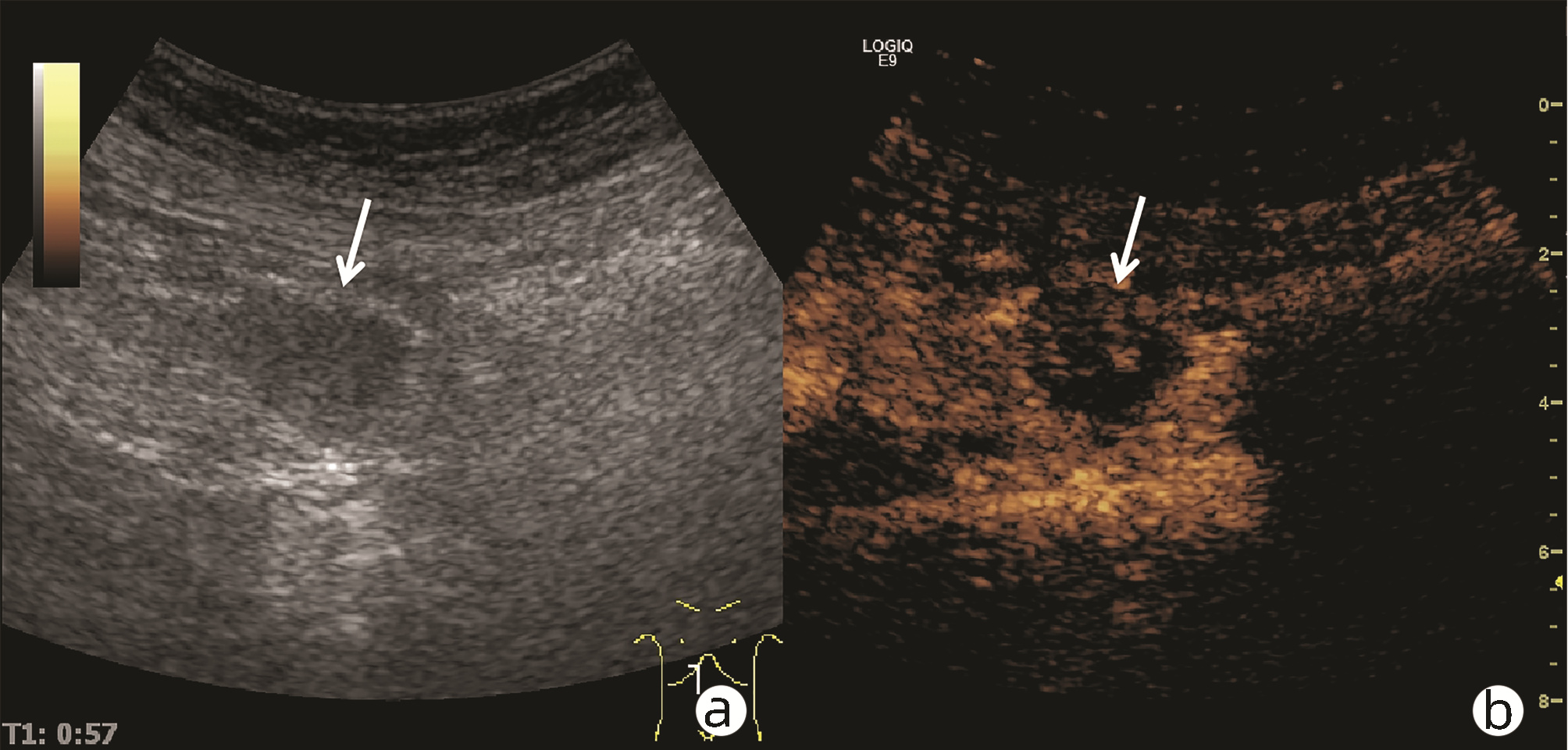
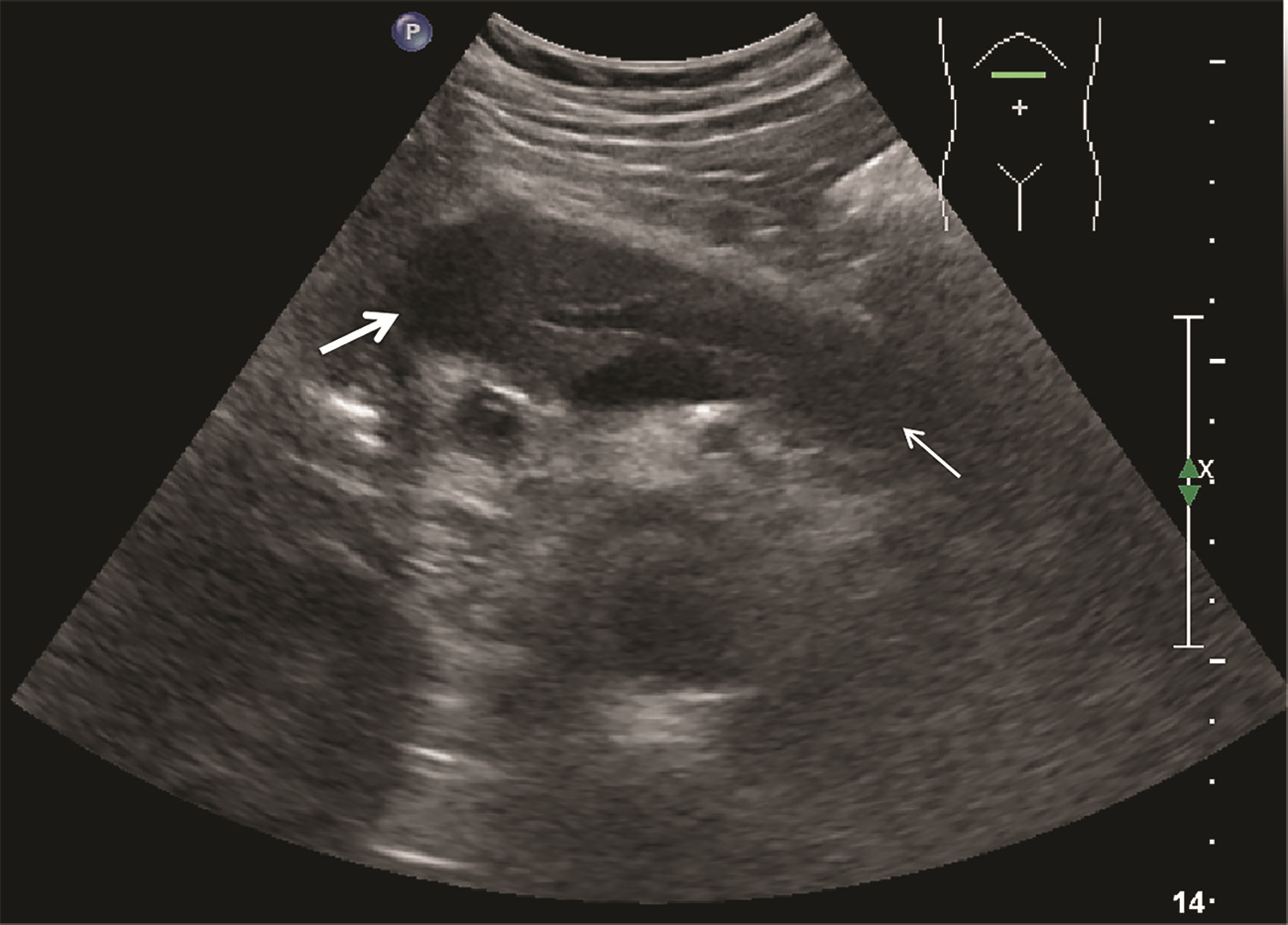
    ZHANG Q, YANG DH, YU LY, et al. Clinical value of contrast-enhanced ultrasound in diagnosis of isolated autoimmune pancreatitis[J]. Chin J Ultrasound Med, 2019, 35(1): 35-38. DOI: 10.3969/j.issn.1002-0101.2019.01.013.

    张琪, 杨道辉, 于凌云, 等. 超声造影在诊断肿块型自身免疫性胰腺炎中的临床价值[J]. 中国超声医学杂志, 2019, 35(1): 35-38. DOI: 10.3969/j.issn.1002-0101.2019.01.013.
    [6]
    NAITOH I, KAMISAWA T, TANAKA A, et al. Clinical characteristics of immunoglobulin IgG4-related sclerosing cholangitis: Comparison of cases with and without autoimmune pancreatitis in a large cohort[J]. Dig Liver Dis, 2021, 53(10): 1308-1314. DOI: 10.1016/j.dld.2021.02.009.
    [7]
    CHEN ZK, ZHANG XJ, QIAN QF, et al. Ultrasonic features and diagnosis of autoimmune pancreatitis[J]. Chin J Med Imaging Technol, 2019, 35(2): 310-311. DOI: 10.13929/j.1003-3289.201807102.

    陈志奎, 张秀娟, 钱清富, 等. 自身免疫性胰腺炎的超声表现与诊断[J]. 中国医学影像技术, 2019, 35(2): 310-311. DOI: 10.13929/j.1003-3289.201807102.
    [8]
    ZHANG XL, YANG PP, QIAN LX. Ultrasound manifestations and characteristics analysis of IgG4-related autoimmune pancreatitis[J]. J Clin Exp Med, 2019, 18(19): 2127-2129. DOI: 10.3969/j.issn.1671-4695.2019.19.034.

    张晓丽, 杨沛沛, 钱林学. IgG4相关自身免疫性胰腺炎的超声表现及特征分析[J]. 临床和实验医学杂志, 2019, 18(19): 2127-2129. DOI: 10.3969/j.issn.1671-4695.2019.19.034.
    [9]
    MACINGA P, BAJER L, DEL CHIARO M, et al. Pancreatic cancer in patients with autoimmune pancreatitis: A scoping review[J]. Pancreatology, 2021, 21(5): 928-937. DOI: 10.1016/j.pan.2021.03.007.
    [10]
    ZHOU CX, ZHU XH, HUANG Y, et al. Value of contrast-enhance ultrasound and color doppler ultrasonography in the diagnosis of pancreatic tumors[J]. Pract J Cancer, 2017, 32(5): 841-843. DOI: 10.3969/j.issn.1001-5930.2017.05.045.

    周成香, 朱小虎, 黄杨, 等. 超声造影联合彩色多普勒超声对胰腺肿瘤的诊断价值[J]. 实用癌症杂志, 2017, 32(5): 841-843. DOI: 10.3969/j.issn.1001-5930.2017.05.045.
    [11]
    HAN YC. Comparison of ultrasonography characteristics and clinical manifestations between autoimmune pancreatitis and pancreatic cancer[J]. Clin Res Pract, 2019, 4(8): 98-99. DOI: 10.19347/j.cnki.2096-1413.201908038.

    韩玉彩. 自身免疫性胰腺炎与胰腺癌的超声特点及临床表现比较[J]. 临床医学研究与实践, 2019, 4(8): 98-99. DOI: 10.19347/j.cnki.2096-1413.201908038.
    [12]
    CONTI CB, CEREATTI F, DRAGO A, et al. Focal autoimmune pancreatitis: A simple flow chart for a challenging diagnosis[J]. Ultrasound Int Open, 2020, 6(3): E67-E75. DOI: 10.1055/a-1323-4906.
    [13]
    GAO F, DU LF. The review of ultrasound mediated micrbubble for pancreatic cancer diagnosis and treatment[J]. Chin J Ultrasonogr, 2020, 29(10): 911-915. DOI: 10.3760/cma.j.cn131148-20200514-00397.

    高峰, 杜联芳. 超声介导微泡在胰腺癌诊疗中的研究进展[J]. 中华超声影像学杂志, 2020, 29(10): 911-915. DOI: 10.3760/cma.j.cn131148-20200514-00397.
    [14]
    ZHANG LP. Differential diagnosis of pancreatic cancer and mass pancreatitis by contrast-enhanced ultrasound[J]. Modern Med Imagel Bimonthly, 2019, 28(12): 2712-2713. https://www.cnki.com.cn/Article/CJFDTOTAL-XDYY201912062.htm

    张丽萍. 超声造影对胰腺癌与肿块型胰腺炎的鉴别诊断效果分析[J]. 现代医用影像学, 2019, 28(12): 2712-2713. https://www.cnki.com.cn/Article/CJFDTOTAL-XDYY201912062.htm
    [15]
    WANG Y, AIDIBAI MHMT, GENG C, et al. Differential diagnostic value of pancreatic cancer and mass pancreatitis by contrast-enhanced ultrasound[J]. J Clin Ultrasound in Med, 2017, 19(8): 526-530. https://www.cnki.com.cn/Article/CJFDTOTAL-LCCY201708006.htm

    王燕, 艾迪拜·木合买提, 耿诚, 等. 超声造影对胰腺癌与肿块型胰腺炎的鉴别诊断价值[J]. 临床超声医学杂志, 2017, 19(8): 526-530. https://www.cnki.com.cn/Article/CJFDTOTAL-LCCY201708006.htm
    [16]
    XU XH, GUO WH, LI Y. Diagnostic value of EUS-EG combined with CEH-EUS in pancreatic space-occupying lesions[J]. Guangdong Med J, 2020, 41(20): 2146-2149. DOI: 10.13820/j.cnki.gdyx.20192312.

    许晓辉, 郭卫红, 李艳. 超声内镜弹性成像联合谐波造影增强超声内镜在胰腺占位性病变中的诊断价值[J]. 广东医学, 2020, 41(20): 2146-2149. DOI: 10.13820/j.cnki.gdyx.20192312.
  • Relative Articles

    [1]Weiming DAI, Lungen LU, Xiaobo CAI. Association between liver sinusoidal endothelial cells and liver fibrosis[J]. Journal of Clinical Hepatology, 2023, 39(2): 419-423. doi: 10.3969/j.issn.1001-5256.2023.02.027
    [2]Yu ZHANG, Huali WANG, Shuming LU. Three cases of hepatic epithelioid hemangioendothelioma[J]. Journal of Clinical Hepatology, 2022, 38(9): 2116-2119. doi: 10.3969/j.issn.1001-5256.2022.09.031
    [3]Hao XU, Bai RUAN, Zhiwen LI, Zhiqiang FANG, Lin WANG, Kefeng DOU. Establishment of a mouse model of vascular endothelial-mesenchymal transdifferentiation genetic tracing and its role in liver fibrosis studies[J]. Journal of Clinical Hepatology, 2022, 38(4): 832-836. doi: 10.3969/j.issn.1001-5256.2022.04.018
    [4]Jin XU, Yan PENG, Chuankang TANG. Value of neutrophil-lymphocyte ratio combined with apolipoprotein A-I level in predicting the severity of acute pancreatitis in the early stage after admission[J]. Journal of Clinical Hepatology, 2021, 37(3): 660-665. doi: 10.3969/j.issn.1001-5256.2021.03.030
    [5]Wei BiWei, Gong YaHui, Liang ZhiHai. Role of pyroptosis in the pathogenesis of acute pancreatitis[J]. Journal of Clinical Hepatology, 2020, 36(8): 1905-1908. doi: 10.3969/j.issn.1001-5256.2020.08.050
    [6]Qi QinQin, Yang Bin, Li HuiHui, Bao JunJun, Li HongYe, Wang BingBing, Mei Qiao. Level of platelet microparticles in acute pancreatitis and their role in the formation of neutrophil extracellular traps[J]. Journal of Clinical Hepatology, 2020, 36(4): 870-873. doi: 10.3969/j.issn.1001-5256.2020.04.032
    [7]Jiang Zhao, Ruan Bo. The role of liver sinusoidal endothelial cells in the progression of nonalcoholic fatty liver disease[J]. Journal of Clinical Hepatology, 2020, 36(3): 684-686. doi: 10.3969/j.issn.1001-5256.2020.03.047
    [8]Zhu ZiYi, Liu Yuan, Wang WenBo, Jiang ZhongYong, Chang Kai, Ye YuSheng, Xiong Jie. Mechanism of HCV stimulation of human umbilical vein endothelial cells in the pathogenesis of atherosclerosis[J]. Journal of Clinical Hepatology, 2019, 35(6): 1313-1317. doi: 10.3969/j.issn.1001-5256.2019.06.026
    [9]Yu ZiYue, Lin Xin, Han Ying, Cui LiNa. Role of liver sinusoidal endothelial cells in liver regeneration and the development of liver fibrosis[J]. Journal of Clinical Hepatology, 2019, 35(9): 2072-2074. doi: 10.3969/j.issn.1001-5256.2019.09.041
    [10]Wang Lin, Liu XueEn, Zhuang Hui. Cellular mechanisms in liver fibrosis regression[J]. Journal of Clinical Hepatology, 2018, 34(4): 862-866. doi: 10.3969/j.issn.1001-5256.2018.04.036
    [11]Zhu ZeMin, Xie ZhiQin, Zhao ZhiJian, Li HongXia, Xu Tao, Tang CaiXi. Pancreatic acinar cell carcinoma with acute pancreatitis as the first symptom: A case report[J]. Journal of Clinical Hepatology, 2018, 34(12): 2662-2664. doi: 10.3969/j.issn.1001-5256.2018.12.032
    [12]Hu QingMei, Niu ChunYan. Predictive value of combined measurement of D-dimer, fibrinogen, and peripheral blood leukocyte count for severity of acute pancreatitis[J]. Journal of Clinical Hepatology, 2017, 33(8): 1522-1526. doi: 10.3969/j.issn.1001-5256.2017.08.023
    [13]Wang Yan, Cui LiJian, Xiao HongLi, Wang GuoXing, Yin ChengHong. Effect of angiotensin 1-7 on the TLR4/NF-κB pathway in cerulein-induced pancreatic acinar cells[J]. Journal of Clinical Hepatology, 2016, 32(11): 2146-2150. doi: 10.3969/j.issn.1001-5256.2016.11.029
    [14]Jiao ShuHua, Liu PengLiang, Wen YanHui. Combined determination of serum C-reactive protein,lipase,interleukin-1β,and intercellular adhesion molecule1 in early identification of severity of acute pancreatitis[J]. Journal of Clinical Hepatology, 2016, 32(1): 131-134. doi: 10.3969/j.issn.1001-5256.2016.01.025
    [15]Hao TingTing, Ma XiaoPeng, Wen YanLi, Dai GuangRong, Feng YiChao, Zhang Li. Changes in serum levels of M30,M65,and IL-17 and their clinical significance in patients with acute pancreatitis[J]. Journal of Clinical Hepatology, 2016, 32(2): 329-332. doi: 10.3969/j.issn.1001-5256.2016.02.027
    [16]Lu Wang, Zhang Rui, Jiang HongLi, Hu YuLin, Xin GuiJie. One case of hepatic epithelioid hemangioendothelioma[J]. Journal of Clinical Hepatology, 2015, 31(8): 1330-1331. doi: 10.3969/j.issn.1001-5256.2015.08.038
    [17]Wu Gang, Zheng Jie, Hu GuoXin, Peng YanZhong. Role of liver nonparenchymal cells in hepatic ischemia-reperfusion injury[J]. Journal of Clinical Hepatology, 2014, 30(12): 1366-1369. doi: 10.3969/j.issn.1001-5256.2014.12.033
    [18]Ren LiNan, Guo XiaoZhong, Zou DeLi. EG-VEGF expression and significance in pancreatic carcinoma[J]. Journal of Clinical Hepatology, 2012, 28(8): 589-591.
  • Cited by

    Periodical cited type(15)

    1. 许娟,闫涛涛,李严锋,侯静涛,樊研. ALBI评分联合PCT在肝硬化腹水患者自发性细菌性腹膜炎的预测作用. 胃肠病学和肝病学杂志. 2023(01): 85-89 .
    2. 周苏云,周影,程新月. 肝硬化患者发生肝性脑病的影响因素分析及对预检分诊意义的探讨. 中西医结合护理(中英文). 2023(03): 130-132 .
    3. 方翊天,吴若林,黄帆,王国斌,冯丽娟,余孝俊,侯刘进,叶征辉,耿小平,赵红川. 肝移植术后多重耐药菌感染危险因素的单中心临床研究. 器官移植. 2021(02): 197-202 .
    4. 李尚书,刘群,黄丽雯. 慢性肝病血小板减少的原因. 中国肝脏病杂志(电子版). 2021(02): 24-28 .
    5. 陆素芳,周红,王丹丹,陈益群,赵红利,张勇杨. MELD评分联合SII对失代偿期肝硬化患者预后的预测作用. 现代消化及介入诊疗. 2021(08): 973-977 .
    6. 黄敏,李冬冬,刘传苗. 乙肝相关慢加急性肝衰竭患者临床短期预后因素的分析. 中华全科医学. 2021(12): 2028-2030 .
    7. 贾亚男,李瀚,李先亮,贺强. 危重症肝病肝移植患者诊断和评估标准的临床进展. 器官移植. 2020(03): 326-331+368 .
    8. 姚运海,赵卫峰,甘建和. 苏州地区血吸虫性肝病患者临床特征及外周血T淋巴细胞亚群分布. 中国血吸虫病防治杂志. 2020(02): 154-158+167 .
    9. 张耀弟. △CTP评分评估肝硬化患者短期预后的价值分析. 甘肃科技. 2020(09): 117-118+152 .
    10. 刘小婷,杨欢,姚红,任小婵,罗选娟,王小闯. 碳青霉烯类耐药肺炎克雷伯菌感染死亡风险预测模型的建立及其对患者预后的预测价值研究. 中国全科医学. 2020(30): 3789-3797 .
    11. 韩志钧,韩柠泽,闫伟,马金彤. 肝血流动力学变化与肝储备功能相关性的研究. 中华医学超声杂志(电子版). 2020(06): 563-565 .
    12. 寇建涛,刘喆,朱继巧,马军,许文犁,李瀚,贾亚男,李先亮,贺强. 肝移植治疗危重症肝病的临床疗效研究. 器官移植. 2020(04): 482-486 .
    13. 宋梅,汪明星,李乐,何进伟,何丽丽. MELD评分结合降钙素原评估终末期肝病预后的临床分析. 中国医药科学. 2020(09): 254-256 .
    14. 李帆,成金英,刘贤贤,丁国锋. 乙肝肝硬化并发甲型流感及肺曲霉菌病1例并文献复习. 中国当代医药. 2020(34): 214-217 .
    15. 韩志钧,韩柠泽,闫伟,马金彤. 肝血流动力学变化与吲哚菁绿代谢试验评价肝储备功能的对比. 医学研究与教育. 2019(06): 28-31 .

    Other cited types(1)

  • 加载中
    Created with Highcharts 5.0.7Chart context menuAccess Class DistributionFULLTEXT: 24.8 %FULLTEXT: 24.8 %META: 69.6 %META: 69.6 %PDF: 5.5 %PDF: 5.5 %FULLTEXTMETAPDF
    Created with Highcharts 5.0.7Chart context menuAccess Area Distribution其他: 8.3 %其他: 8.3 %其他: 1.6 %其他: 1.6 %China: 0.8 %China: 0.8 %Russian Federation: 0.3 %Russian Federation: 0.3 %[]: 0.5 %[]: 0.5 %三亚: 0.2 %三亚: 0.2 %上海: 1.8 %上海: 1.8 %中卫: 0.2 %中卫: 0.2 %佛山: 0.2 %佛山: 0.2 %兰州: 0.3 %兰州: 0.3 %北京: 4.4 %北京: 4.4 %南京: 1.3 %南京: 1.3 %南宁: 0.3 %南宁: 0.3 %南阳: 0.2 %南阳: 0.2 %卡纳塔克: 0.2 %卡纳塔克: 0.2 %台州: 0.2 %台州: 0.2 %合肥: 1.5 %合肥: 1.5 %吉林: 0.5 %吉林: 0.5 %呼和浩特: 0.3 %呼和浩特: 0.3 %唐山: 0.2 %唐山: 0.2 %商丘: 0.2 %商丘: 0.2 %大连: 0.2 %大连: 0.2 %天津: 0.2 %天津: 0.2 %太原: 0.2 %太原: 0.2 %宁波: 0.2 %宁波: 0.2 %安康: 0.2 %安康: 0.2 %安阳: 0.2 %安阳: 0.2 %宣城: 0.3 %宣城: 0.3 %密蘇里城: 1.1 %密蘇里城: 1.1 %巴黎: 0.5 %巴黎: 0.5 %常德: 0.2 %常德: 0.2 %广州: 0.3 %广州: 0.3 %张家口: 4.4 %张家口: 4.4 %德罕: 0.2 %德罕: 0.2 %成都: 0.5 %成都: 0.5 %扬州: 0.2 %扬州: 0.2 %拉斯维加斯: 0.5 %拉斯维加斯: 0.5 %昆明: 0.2 %昆明: 0.2 %杭州: 0.2 %杭州: 0.2 %武汉: 1.1 %武汉: 1.1 %洛阳: 0.3 %洛阳: 0.3 %济南: 0.2 %济南: 0.2 %海得拉巴: 0.5 %海得拉巴: 0.5 %湖州: 0.2 %湖州: 0.2 %湘潭: 0.2 %湘潭: 0.2 %湛江: 0.2 %湛江: 0.2 %漯河: 0.2 %漯河: 0.2 %福州: 0.2 %福州: 0.2 %罗奥尔凯埃: 0.2 %罗奥尔凯埃: 0.2 %自贡: 0.2 %自贡: 0.2 %芒廷维尤: 43.0 %芒廷维尤: 43.0 %芝加哥: 0.5 %芝加哥: 0.5 %苏州: 0.2 %苏州: 0.2 %茂名: 0.2 %茂名: 0.2 %莫斯科: 3.6 %莫斯科: 3.6 %西宁: 10.4 %西宁: 10.4 %诺沃克: 0.6 %诺沃克: 0.6 %贵阳: 0.2 %贵阳: 0.2 %运城: 2.3 %运城: 2.3 %金华: 0.3 %金华: 0.3 %长春: 1.8 %长春: 1.8 %长沙: 0.5 %长沙: 0.5 %长治: 0.2 %长治: 0.2 %马湾: 0.2 %马湾: 0.2 %马鞍山: 0.2 %马鞍山: 0.2 %黔西南: 0.2 %黔西南: 0.2 %其他其他ChinaRussian Federation[]三亚上海中卫佛山兰州北京南京南宁南阳卡纳塔克台州合肥吉林呼和浩特唐山商丘大连天津太原宁波安康安阳宣城密蘇里城巴黎常德广州张家口德罕成都扬州拉斯维加斯昆明杭州武汉洛阳济南海得拉巴湖州湘潭湛江漯河福州罗奥尔凯埃自贡芒廷维尤芝加哥苏州茂名莫斯科西宁诺沃克贵阳运城金华长春长沙长治马湾马鞍山黔西南

Catalog

    通讯作者: 陈斌, bchen63@163.com
    • 1. 

      沈阳化工大学材料科学与工程学院 沈阳 110142

    1. 本站搜索
    2. 百度学术搜索
    3. 万方数据库搜索
    4. CNKI搜索

    Figures(6)  / Tables(2)

    Article Metrics

    Article views (792) PDF downloads(30) Cited by(16)
    Proportional views
    Related

    /

    DownLoad:  Full-Size Img  PowerPoint
    Return
    Return