中文English
ISSN 1001-5256 (Print)
ISSN 2097-3497 (Online)
CN 22-1108/R

Value of Stroop test in the diagnosis of minimal hepatic encephalopathy

DOI: 10.3969/j.issn.1001-5256.2022.04.017
More Information
  • Corresponding author: ZHAO Shousong, 1260345655@qq.com (ORCID: 0000-0002-8076-518X)
  • Received Date: 2021-08-25
  • Accepted Date: 2021-10-09
  • Published Date: 2022-04-20
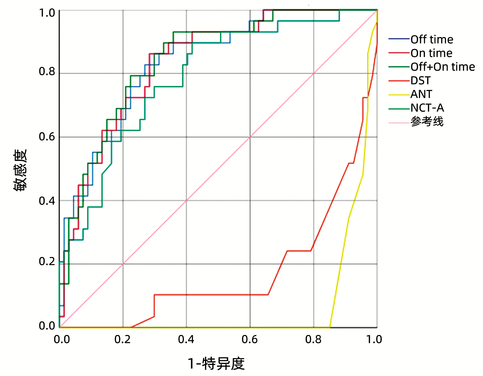
  •   Objective  To investigate the value of Stroop test, a neuropsychological test, in the diagnosis of minimal hepatic encephalopathy (MHE).  Methods  A total of 96 patients with liver cirrhosis who were hospitalized in Department of Infectious Diseases, The First Affiliated Hospital of Bengbu Medical College, from August 2020 to March 2021 were enrolled, and the number connection test-A (NCT-A), digit symbol test (DST), animal naming test (ANT), and Stroop test were performed for all patients. Test results were recorded and related clinical data were collected. The t-test was used for comparison of normally distributed continuous data between two groups, and the Mann-Whitney U test was used for comparison of non-normally distributed continuous data between two groups; the chi-square test was used for comparison of categorical data between groups. The receiver operating characteristic (ROC) curve was used to investigate the value of Stroop test in the diagnosis of MHE, and the Pearson correlation coefficient was used to analyze the correlation of the results of Stroop test with those of NCT-A, DST, and ANT.  Results  For the 96 patients with liver cirrhosis, the prevalence rate of MHE was 30.21% (29/96). The Off+On time of Stroop test had a cut-off value of 212.49 s in the diagnosis of MHE, with an area under the ROC curve of 0.845, a sensitivity of 93.10%, and a specificity of 64.20%. The Pearson correlation analysis showed that the On+Off time and On time of Stroop test were moderately correlated with NCT-A(r=0.580 and 0.590, both P < 0.01), the Off time of Stroop test was strongly correlated with NCT-A(r=0.620, P < 0.01), and the On+Off time, On time, and Off time of Stroop test were strongly correlated with DST(r=-0.650, -0.650, and -0.630, all P < 0.01).  Conclusion  In the diagnosis of MHE, Stroop test is a highly sensitive method with easy-to-read results and a high diagnostic value and does not require professional equipment.


  • 非酒精性脂肪性肝病(NAFLD)是指除外过量饮酒和其他明确的损肝因素所致的肝细胞内脂肪沉积,全球2型糖尿病(T2DM)患者NAFLD患病率约为55.5%。NAFLD与高血糖、胰岛素抵抗、血脂异常等密切相关,T2DM合并NAFLD患者心血管疾病、肾脏病变等风险明显增加且更为严重[1-3]。ALP是一种广泛存在于人体组织中的一种酶,研究[4]显示ALP与胰岛素抵抗(IR)呈正相关,可导致基础状态下葡萄糖过剩,T2DM患者血清ALP活性升高,ALP增高是T2DM的独立危险因素。ALP在肝脏和骨骼中活性较高,通常作为胆汁淤积和骨代谢的指标,其在NAFLD中也可出现升高。在T2DM人群中,有关血清ALP与NAFLD关系的报道甚少,本研究旨在探讨ALP是否为T2DM合并NAFLD的危险因素,为相关疾病的早期防治提供一定的依据。

    选取2016年7月—2018年12月在本院内分泌科就诊的T2DM患者599例(其中男320例,女279例),平均年龄(63.32±11.30)岁。T2DM诊断符合1999年世界卫生组织糖尿病的诊断标准及分型标准[5]。根据超声检查结果将患者分为NAFLD组和非NAFLD组,NAFLD的诊断符合《非酒精性脂肪性肝病防治指南(2018年更新版)》[2]。排除标准:(1)1型糖尿病、特殊类型糖尿病、糖尿病急性严重代谢紊乱、急性感染;(2)骨骼系统疾病、慢性肾脏疾病;(3)病毒性肝炎、自身免疫性肝炎、肝豆状核变性、肝硬化等慢性肝病及肝脏恶性肿瘤、胆道疾病;(4)过度饮酒:饮酒折合乙醇量男性>30 g/d(>210 g/周),女性>20 g/d(>140 g/周);(5)服用噻唑烷二酮类药物、糖皮质激素、降脂药、丙戊酸钠、奥氮平等;(6)垂体前叶功能减退、甲状腺功能减退、性腺功能减退、甲状旁腺功能亢进、炎症性肠病、全胃肠外营养等全身性疾病[2]

    (1) 肝区近场回声弥漫性增强(强于肾脏和脾脏)远场回声逐渐衰减;(2)肝内管道结构显示不清;(3)肝脏轻至中度肿大,边缘角圆钝;(4)彩色多普勒血流显象提示肝内彩色血流信号减少或不易显示,但肝内血管走向正常;(5)肝右叶包膜及横膈回声显示不清或不完整。具备上述第1项及第2~4项中一项者为轻度脂肪肝;具备上述第1项及第2~4项中两项者为中度脂肪肝;具备上述第1项以及2~4项中两项和第5项者为重度脂肪肝[6]

    1.3.1   一般资料测量

    采用统一标准测量T2DM患者一般临床资料包括身高、体质量、腰围(WC)、收缩压(SBP)和舒张压(DBP);收集患者性别、年龄、糖尿病病程、吸烟史、高血压病史。

    1.3.2   生化指标检测

    所有受试者于试验前一晚禁食12 h,于第2日清晨空腹状态下抽取静脉血检测血清ALP、糖化血红蛋白(HbA1c)、空腹血糖(FPG)、空腹胰岛素(FIns)、空腹C肽(FC-P)、尿酸(SUA)、GGT、ALT、AST、TC、TG、HDL-C、LDL-C。BMI=体质量/身高2(kg/m2);稳态模型评估胰岛素抵抗指数(HOMA-IR)=空腹血糖×空腹胰岛素/22.5。

    1.3.3   肝脏彩超检测

    使用LOGIQ-9全身彩色多普勒诊断仪器,由超声科专业医生对受试者行空腹状态下腹部超声检查,观察其大小形态、肝脏回声及血流情况等。

    采用SPSS 22.0对资料行统计分析。正态分布计量资料以x±s表示,两组间比较采用t检验,多组间比较使用单因素方差分析;非正态分布计量资料以M(P25~P75)表示,两组间比较采用非参数Mann Whitney U检验,多组间比较使用Kruskal-Wallis H秩和检验;计数资料组间比较采用χ2检验。采用Pearson相关分析法和Spearman相关分析法进行相关性分析。采用二元Logistic回归分析T2DM发生NAFLD的影响因素。P<0.05为差异具有统计学意义。

    NAFLD组高血压病史所占比例、SBP、DBP、BMI、WC、FIns、FC-P、SUA、LDL-C、TG、TC、HOMA-IR、ALT、AST、GGT、ALP均高于非NAFLD组(P值均<0.05)。NAFLD组年龄、HDL-C低于非NAFLD组(P值均<0.05)。两组间性别、病程、有吸烟史者所占比例、HbA1c、FPG比较,差异均无统计学意义(P值均>0.05)(表 1)。

    表  1  两组一般资料及生化指标比较
    Table  1.  Comparison of general data between groups
    项目 non-NAFLD组(n=313) NAFLD组(n=286) 统计值 P
    男性[例(%)] 179(57.2) 141(49.3) χ2= 3.737 0.053
    年龄(岁) 64.28±11.15 62.27±11.38 t=2.184 0.029
    病程(月) 120(36~192) 120(36~159) Z=-1.365 0.172
    BMI(kg/m2) 23.51±2.99 26.49±3.18 t=-11.842 <0.001
    吸烟史[例(%)] 67(21.4) 60(21.0) χ2=0.016 0.898
    高血压病史[例(%)] 174(55.6) 191(66.8) χ2=7.864 0.005
    SBP(mmHg) 133.63±15.27 136.44±15.58 t=-2.226 0.026
    DBP(mmHg) 78.62±8.77 81.47±9.58 t=-3.800 <0.001
    WC(cm) 87.46±9.88 94.54±8.98 t=-9.150 <0.001
    HbA1c(%) 8.89±2.13 8.93±1.73 t=-0.279 0.781
    FPG(mmol/L) 9.69±3.71 9.50±3.12 t=0.684 0.495
    FIns(μIU/L) 6.25(3.62~11.14) 10.05(5.41~16.72) Z=-6.173 <0.001
    FC-P(μg/L) 1.74±0.96 2.18±1.04 t=-5.419 <0.001
    SUA(μmol/L) 269.37±84.92 305.03±91.13 t=-4.957 <0.001
    LDL-C(mmol/L) 2.54±0.86 2.72±0.75 t=-2.702 0.007
    HDL-C(mmol/L) 1.09(0.88~1.34) 0.95(0.81~1.14) Z=-5.273 <0.001
    TG(mmol/L) 1.28(0.96~1.79) 1.86(1.43~2.69) Z=-9.376 <0.001
    TC(mmol/L) 4.61±1.10 4.86±0.98 t=-3.016 0.003
    HOMA-IR 2.77(1.41~4.44) 3.92(2.19~6.71) Z=-5.794 <0.001
    ALT(U/L) 14.80(11.05~21.90) 20.20(14.68~30.40) Z=-6.737 <0.001
    AST(U/L) 14.70(11.35~17.95) 16.90(12.38~23.33) Z=-4.389 <0.001
    GGT(U/L) 19.00(14.30~28.00) 27.60(20.00~43.55) Z=-7.764 <0.001
    ALP(U/L) 69.73±19.94 74.71±23.03 t=-2.833 0.005
    下载: 导出CSV 
    | 显示表格

    随着脂肪肝严重程度的加重,T2DM合并NAFLD患者BMI、WC、FIns、HOMA-IR、ALT、AST、GGT、ALP水平均明显升高(P值均<0.05)(表 2)。相关性分析显示,NAFLD的脂肪肝严重程度(轻、中、重)与年龄(rs=0.140,P=0.018)、BMI(rs=0.239,P<0.001)、WC(rs=0.222,P<0.001)、FIns(rs=0.191,P=0.001)、HOMA-IR(rs=0.218,P<0.001)、ALT(rs=0.188,P=0.001)、AST(rs=0.279,P<0.001)、GGT(rs=0.202,P=0.001)、ALP(rs=0.361,P<0.001)水平呈正相关。

    表  2  轻中重度NAFLD患者临床资料比较
    Table  2.  Comparison of general data among groups in patients with NAFLD
    项目 轻度NAFLD组(n=111) 中度NAFLD组(n=105) 重度NAFLD组(n=70) 统计值 P
    男性[例(%)] 58(52.3) 56(53.3) 27(38.6) χ2=4.294 0.117
    年龄(岁) 60.5±10.6 63.0±12.0 64.1±11.4 F=2.538 0.081
    病程(月) 120(36~168) 108(29~144) 120(33~156) χ2=0.442 0.802
    BMI(kg/m2) 25.48±2.59 26.88±2.991) 27.52±3.831) F=10.731 <0.001
    SBP(mmHg) 136.16±15.64 134.51±14.55 139.76±16.66 F=2.429 0.090
    DBP(mmHg) 81.10±9.33 81.57±9.68 81.91±9.92 F=0.163 0.849
    WC(cm) 92.17±7.37 95.33±8.381) 97.12±11.141) F=7.502 0.001
    HbA1c(%) 8.79±1.72 8.90±1.67 9.22±1.85 F=1.388 0.251
    FPG(mmol/L) 9.35±3.04 9.28±3.12 10.06±3.21 F=1.551 0.214
    FIns(μIU/L) 7.38(4.61~14.94) 10.25(5.69~16.22) 11.65(7.53~19.62)1) χ2=10.512 0.005
    FC-P(μg/L) 2.12±1.07 2.19±1.00 2.28±1.05 F=0.524 0.593
    SUA(μmol/L) 293.99±82.32 321.08±88.00 298.46±105.81 F=2.654 0.072
    LDL-C(mmol/L) 2.76±0.73 2.66±0.77 2.76±0.77 F=0.603 0.548
    HDL-C(mmol/L) 0.95(0.82~1.14) 0.93(0.80~1.11) 0.99(0.82~1.21) χ2=1.749 0.417
    TG(mmol/L) 1.84(1.37~2.87) 1.77(1.47~2.59) 1.92(1.45~3.16) χ2=0.679 0.712
    TC(mmol/L) 4.83±0.96 4.74±0.91 5.10±1.08 F=2.956 0.054
    HOMA-IR 3.32(1.83~5.55) 3.90(2.34~6.51) 5.47(2.96~9.53)1) χ2=14.183 0.001
    ALT(U/L) 18.00(13.60~27.60) 22.00(15.10~31.30) 23.95(16.00~42.23)1) χ2=10.453 0.005
    AST(U/L) 14.00(11.50~18.40) 18.40(13.00~25.30)1) 20.05(14.25~27.08)1) χ2=22.423 <0.001
    GGT(U/L) 23.00(17.00~38.80) 27.90(21.05~39.50) 34.50(21.65~55.43)1) χ2=11.791 0.003
    ALP(U/L) 65.72±16.63 73.71±20.941) 90.44±26.581)2) F=29.920 <0.001
    注:与轻度NAFLD组比较,1)P<0.01;与中度NAFLD组比较,2)P<0.01。
    下载: 导出CSV 
    | 显示表格

    相关分析显示,T2DM合并NAFLD患者ALP与HbA1c、FPG、HOMA-IR、TC、ALT、AST、GGT呈正相关(P值均<0.05)(表 3)。

    表  3  NAFLD患者ALP与临床各指标的相关性分析
    Table  3.  Correlation analysis between ALP and clinical indicators in patients with NAFLD
    项目 rrs P
    HbA1c 0.149 0.012
    FPG 0.146 0.014
    HOMA-IR 0.132 0.025
    TC 0.151 0.011
    ALT 0.210 <0.001
    AST 0.192 0.001
    GGT 0.297 <0.001
    下载: 导出CSV 
    | 显示表格

    以是否出现NAFLD为因变量,以年龄、BMI、高血压病史、FPG、FC-P、TC、TG、HDL-C、LDL-C、SUA、HOMA-IR、ALP为自变量,行二元Logistic回归分析,结果显示,年龄、BMI、FC-P、LDL-C、TG、ALP均为T2DM患者发生NAFLD的影响因素(表 4)。

    表  4  Logistic回归分析NAFLD的影响因素
    Table  4.  Logistic regression analysis of the influencing factors for NAFLD
    因素 β SE Wald OR 95%CI P
    年龄 -0.030 0.010 8.527 0.971 0.951~0.990 0.003
    BMI 0.298 0.037 63.596 1.347 1.252~1.450 <0.001
    FC-P 0.267 0.111 5.807 1.307 1.051~1.624 0.016
    LDL-C 1.332 0.470 8.023 3.787 1.507~9.516 0.005
    TG 0.685 0.167 16.803 1.984 1.430~2.752 <0.001
    ALP 0.013 0.005 7.444 1.013 1.004~1.023 0.006
    下载: 导出CSV 
    | 显示表格

    ALP是一种常见的广泛存在于微生物和动植物中的酶,在人体内有四种ALP同工酶,包括组织非特异性ALP(TNAP)、肠型ALP、生殖细胞型ALP以及胎盘型ALP。TNAP主要在肝脏、肾脏和骨骼组织中表达,占血清总ALP活性的90%以上[7]。在小鼠3T3-L1前脂肪细胞中的研究[8]发现,TNAP水平在脂肪生成过程中升高,但在脂肪生成开始加入TNAP抑制剂后,TNAP活性和脂质积累均减少;同时在原代培养的人前脂肪细胞中也观察到类似现象。此外,更多证据表明ALP活性存在于大鼠脂肪组织、兔前脂肪细胞和人脂肪细胞中,TNAP敲除小鼠也表现出脂肪量的减少。TNAP除了参与调节脂肪细胞内脂质积累,也在肝细胞和肾上腺细胞的脂质积累中发挥作用,提示TNAP可能参与了多种不同类型细胞中的脂质积累[9-10]。一项研究[11]纳入了14 393例无糖尿病的高血压病患者,平均随访4.5年后发现血清ALP升高与IR相关,并且增加了糖尿病的患病风险。本研究结果显示,在T2DM合并NAFLD的患者中,ALP与HbA1c、FPG、HOMA-IR、TC、ALT、AST、GGT呈正相关,提示ALP增加可能与IR、血脂异常等多种致NAFLD风险密切相关。

    据估计,2016年—2030年我国NAFLD的患病人数将从2.46亿人增加至约3.14亿人,NAFLD患病率的迅速增长导致高血糖、IR、血脂异常、肝硬化、肝癌和亚临床炎症的增加,加剧如糖尿病、肥胖、心血管疾病和癌症等非传染性疾病的负担[1, 12]。本研究发现,NAFLD组BMI、WC、FIns、FC-P、LDL-C、TG、TC、HOMA-IR、ALP水平均高于非NAFLD组。随着脂肪肝严重程度的加重,T2DM合并NAFLD患者ALP水平均明显升高;NAFLD的脂肪肝严重程度与ALP水平呈正相关。Logistic回归分析表明,在调整了相关的NAFLD危险因素之后,年龄、BMI、FC-P、LDL-C、TG、ALP均为T2DM患者发生NAFLD的影响因素。NAFLD是肝脏能量代谢不平衡的表现,肝脏有将能量氧化为CO2或输出为极低密度脂蛋白的能力,但是过多的能量主要以碳水化合物和脂肪的形式进入肝脏后,导致TG在肝脏中的积累,因此NAFLD在肥胖者中广泛存在[13]。迄今为止,IR被认为是NAFLD发病及进展的主要机制。在IR状态下,肝脏失去在胰岛素作用下抑制肝葡萄糖生成的能力并且通过激活Notch信号通路增强脂肪从头合成。脂肪在肝脏的积累是由于游离脂肪酸(FFA)的摄取、合成、输出和氧化受损造成的。在NAFLD患者中,因为脂肪组织抑制脂肪分解的功能受损,所以肝脏脂肪变性的数量与血浆游离FFA水平的增加相关[14]。FFA导致JNK信号通路的激活,从而引起细胞应激、炎症、凋亡和线粒体功能障碍。此外,JNK信号通路使PPARγ磷酸化,加剧了脂毒性和肝炎症[15]。近年研究[16]表明,C肽也与NAFLD密切相关,可能与IR和C肽本身生物功能有关。本研究中年龄较长者NAFLD的发生率较低,考虑随着年龄的增长,脂肪组织储存脂肪能力下降、肝纤维化比例升高以及接受更长久的T2DM治疗干预等原因对结果造成一定的影响[17]

    综上所述,血清ALP水平与血糖、IR、脂代谢紊乱密切相关,ALP是T2DM合并NAFLD的危险因素,ALP可能参与了T2DM和NAFLD的发生发展。能否进行预防性用药干预ALP水平使NAFLD得到缓解,值得后续进一步研究。另外,本研究样本量偏少,为单中心横断面研究,可能存在选择偏倚,可进一步扩大样本量证实,ALP在NAFLD中的具体作用机制也需更加深入的研究。

  • [1]
    Chinese Society of Hepatology, Chinese Medical Association. Guidelines on the management of hepatic encephalopathy in cirrhosis[J]. J Clin Hepatol, 2018, 34(10): 2076-2089. DOI: 10.3969/j.issn.1001-5256.2018.10.007.

    中华医学会肝病学分会. 肝硬化肝性脑病诊疗指南[J]. 临床肝胆病杂志, 2018, 34(10): 2076-2089. DOI: 10.3969/j.issn.1001-5256.2018.10.007.
    [2]
    AGRAWAL S, UMAPATHY S, DHIMAN RK. Minimal hepatic encephalopathy impairs quality of life[J]. J Clin Exp Hepatol, 2015, 5(Suppl 1): S42-S48. DOI: 10.1016/j.jceh.2014.11.006.
    [3]
    Chinese Society of Hepatology, Chinese Medical Association. Chinese guidelines on the management of liver cirrhosis[J]. J Clin Hepatol, 2019, 35(11): 2408-2425. DOI: 10.3969/j.issn.1001-5256.2019.11.006.

    中华医学会肝病学分会. 肝硬化诊治指南[J]. 临床肝胆病杂志, 2019, 35(11): 2408-2425. DOI: 10.3969/j.issn.1001-5256.2019.11.006.
    [4]
    WANG YL, SHAO LC, YANG Z. Diagnostic value of number connection test for minimal hepatic encephalopathy and related factors of cirrhosis complicated with hepatic encephalopathy[J]. Clin J Med Offic, 2020, 48(12): 1424-1426, 1429. DOI: 10.16680/j.1671-3826.2020.12.11.

    王雨丽, 邵丽春, 杨卓. 数字连接试验对轻微肝性脑病诊断价值及肝硬化并发肝性脑病相关因素分析[J]. 临床军医杂志, 2020, 48(12): 1424-1426, 1429. DOI: 10.16680/j.1671-3826.2020.12.11.
    [5]
    CAMPAGNA F, MONTAGNESE S, RIDOLA L, et al. The animal naming test: An easy tool for the assessment of hepatic encephalopathy[J]. Hepatology, 2017, 66(1): 198-208. DOI: 10.1002/hep.29146.
    [6]
    BAJAJ JS, THACKER LR, HEUMAN DM, et al. The Stroop smartphone application is a short and valid method to screen for minimal hepatic encephalopathy[J]. Hepatology, 2013, 58(3): 1122-1132. DOI: 10.1002/hep.26309.
    [7]
    LABENZ C, BARON JS, TOENGES G, et al. Prospective evaluation of the impact of covert hepatic encephalopathy on quality of life and sleep in cirrhotic patients[J]. Aliment Pharmacol Ther, 2018, 48(3): 313-321. DOI: 10.1111/apt.14824.
    [8]
    SAN MARTÍN-VALENZUELA C, BORRAS-BARRACHINA A, GALLEGO JJ, et al. Motor and cognitive performance in patients with liver cirrhosis with minimal hepatic encephalopathy[J]. J Clin Med, 2020, 9(7): 2154. DOI: 10.3390/jcm9072154.
    [9]
    LABENZ C, ADARKWAH CC, WÖRNS MA, et al. Management of hepatic encephalopathy in Germany: A survey among physicians[J]. Z Gastroenterol, 2020, 58(1): 49-56. DOI: 10.1055/a-1010-6974.
    [10]
    WANG JY, ZHANG NP, CHI BR, et al. Prevalence of minimal hepatic encephalopathy and quality of life evaluations in hospitalized cirrhotic patients in China[J]. World J Gastroenterol, 2013, 19(30): 4984-4991. DOI: 10.3748/wjg.v19.i30.4984.
    [11]
    GIMÉNEZ-GARZÓ C, GARCÉS JJ, URIOS A, et al. The PHES battery does not detect all cirrhotic patients with early neurological deficits, which are different in different patients[J]. PLoS One, 2017, 12(2): e0171211. DOI: 10.1371/journal.pone.0171211.
    [12]
    BAJAJ JS, DUARTE-ROJO A, XIE JJ, et al. Minimal hepatic encephalopathy and mild cognitive impairment worsen quality of life in elderly patients with cirrhosis[J]. Clin Gastroenterol Hepatol, 2020, 18(13): 3008-3016. e2. DOI: 10.1016/j.cgh.2020.03.033.
    [13]
    ZENG X, ZHANG LY, LIU Q, et al. Combined scores from the encephalapp stroop test, number connection test B, and serial dotting test accurately identify patients with covert hepatic encephalopathy[J]. Clin Gastroenterol Hepatol, 2020, 18(7): 1618-1625. e7. DOI: 10.1016/j.cgh.2019.11.018.
    [14]
    LABENZ C, BEUL L, TOENGES G, et al. Validation of the simplified Animal Naming Test as primary screening tool for the diagnosis of covert hepatic encephalopathy[J]. Eur J Intern Med, 2019, 60: 96-100. DOI: 10.1016/j.ejim.2018.08.008.
    [15]
    QU Y, LI T, LIN C, et al. Animal naming test for the assessment of minimal hepatic encephalopathy in Asian cirrhotic populations[J]. Clin Res Hepatol Gastroenterol, 2021, 45(5): 101729. DOI: 10.1016/j.clinre.2021.101729.
  • Relative Articles

    [1]Yi LE, Hao YANG, Hu LIU, Zhaoyang CUI, Xiangning ZHAO, Liang AN, Haizhu LI, Shaogeng ZHANG. Advances in the diagnosis and treatment of gallbladder carcinoma[J]. Journal of Clinical Hepatology, 2023, 39(11): 2738-2744. doi: 10.3969/j.issn.1001-5256.2023.11.032
    [2]Xiaomei QIN, Shuangjie LI. Etiology, diagnosis, and treatment of acute liver failure in neonates[J]. Journal of Clinical Hepatology, 2022, 38(2): 260-263. doi: 10.3969/j.issn.1001-5256.2022.02.002
    [3]Xuchu LIU, Shiyun QIN, Lijun CHEN, Juan CHEN, Xiaoguang YOU. Prognostic value of KRAS mutations in patients with advanced primary liver cancer treated with transcatheter arterial chemoembolization[J]. Journal of Clinical Hepatology, 2022, 38(11): 2514-2519. doi: 10.3969/j.issn.1001-5256.2022.11.015
    [4]Chenlu ZHAO, Wenxia ZHAO. Association of circadian rhythm and related genes with nonalcoholic fatty liver disease[J]. Journal of Clinical Hepatology, 2021, 37(9): 2225-2230. doi: 10.3969/j.issn.1001-5256.2021.09.045
    [5]Yu WANG, Min WANG, Guanhua ZHANG, Fuliang HE, Xiaojuan OU, Jidong JIA. Clinical diagnosis, staging, and therapeutic principles of liver cirrhosis[J]. Journal of Clinical Hepatology, 2021, 37(1): 17-21. doi: 10.3969/j.issn.1001-5256.2021.01.004
    [6]Yong ZHANG, Zhengfa MAO. Association of tuberous sclerosis gene 1/2 mutations with the progression of hepatocellular carcinoma and prognosis[J]. Journal of Clinical Hepatology, 2021, 37(1): 84-88. doi: 10.3969/j.issn.1001-5256.2021.01.017
    [7]Shiyan YAN, Jiangao FAN. Diagnosis and treatment of hepatocellular carcinoma associated with nonalcoholic fatty liver disease[J]. Journal of Clinical Hepatology, 2021, 37(8): 1748-1752. doi: 10.3969/j.issn.1001-5256.2021.08.002
    [8]Zhu HongMei, Wang QinYing. Advances in the diagnosis and treatment of nonalcoholic fatty liver disease-related liver fibrosis[J]. Journal of Clinical Hepatology, 2020, 36(1): 178-181. doi: 10.3969/j.issn.1001-5256.2020.01.042
    [9]YANG Xu, SUN WenBing, GAO Jun. Diagnosis and treatment of hepatic vascular tumors[J]. Journal of Clinical Hepatology, 2020, 36(11): 2569-2573. doi: 10.3969/j.issn.1001-5256.2020.11.039
    [10]Fu YiZhen, Xu Li. Advances in multimodality therapy for hepatocellular carcinoma[J]. Journal of Clinical Hepatology, 2020, 36(10): 2179-2183. doi: 10.3969/j.issn.1001-5256.2020.10.004
    [11]Xu XiaoLei, Gao CanCan, Wang ZhiXin, Wang Zhan, ZHOU Ying, Wang HaiJiu, Ye HaiWen, Pang MingQuan, Zhou Hu, Hou YaJun, Guo Bing, Fan HaiNing. Advances in the diagnosis and treatment of cystic space-occupying lesions in the liver[J]. Journal of Clinical Hepatology, 2019, 35(5): 1118-1122. doi: 10.3969/j.issn.1001-5256.2019.05.043
    [12]Liang Chen, Bai Li, Zheng SuJun. Research advances in the genotype-phenotype correlation, diagnosis, treatment, and screening of Wilson's disease[J]. Journal of Clinical Hepatology, 2019, 35(9): 2116-2119. doi: 10.3969/j.issn.1001-5256.2019.09.052
    [13]Chinese Society of Hepatology, Chinese Medical Association; Chinese Society of Gastroenterology, Chinese Medical Association; Chinese Society of Infectious Diseases, Chinese Medical Association. Consensus on the diagnosis and therapy of hepatic fibrosis(2019)[J]. Journal of Clinical Hepatology, 2019, 35(10): 2163-2172. doi: 10.3969/j.issn.1001-5256.2019.10.007
    [14]Zhang YuPeng, Wang Fan, Chen PengFei, Liu Shi, Liu Lan, Chang Ying, Zhao Qiu. Expression and significance of the ABAT gene in hepatocellular carcinoma:An analysis based on data mining[J]. Journal of Clinical Hepatology, 2019, 35(3): 553-558. doi: 10.3969/j.issn.1001-5256.2019.03.020
    [15]Zhou HuaBang, Hu HePing. Challenges in precise treatment for primary liver cancer based on gene mutation[J]. Journal of Clinical Hepatology, 2017, 33(7): 1209-1210. doi: 10.3969/j.issn.1001-5256.2017.07.001
    [16]Wang ShiMei, Wu Xia, Kang Peng, Meng QingYang. Diagnostic value of H19 gene in hepatocellular carcinoma[J]. Journal of Clinical Hepatology, 2016, 32(11): 2203-2205. doi: 10.3969/j.issn.1001-5256.2016.11.046
    [17]Zheng JunFu, Lu: Jun. The biological function of human cervical cancer oncogene and its potential value in the early diagnosis of hepatocellular carcinoma[J]. Journal of Clinical Hepatology, 2011, 27(8): 888-892.
    [18]Li ChangXiao, Wang ChaoXia. The progression of genetic susceptibility in infantile intrahepatic cholestasis[J]. Journal of Clinical Hepatology, 2011, 27(7): 694-699+708.
  • Cited by

    Periodical cited type(3)

    1. 赵梓硕,朱玉光,马燕山,李志伟,景永帅,谢英花. 不同高脂饲料配方对建立非酒精性脂肪肝大鼠模型的影响. 中国临床药理学与治疗学. 2024(05): 543-553 .
    2. 罗志勇,刘斌. 利拉鲁肽联合达格列净治疗2型糖尿病合并NAFLD的临床效果. 中国医学创新. 2024(26): 72-76 .
    3. 李非凡,徐岷. 血清碱性磷酸酶与高血压合并非酒精性脂肪性肝病的相关性分析. 热带医学杂志. 2024(10): 1444-1448 .

    Other cited types(4)

  • 加载中

Catalog

    通讯作者: 陈斌, bchen63@163.com
    • 1. 

      沈阳化工大学材料科学与工程学院 沈阳 110142

    1. 本站搜索
    2. 百度学术搜索
    3. 万方数据库搜索
    4. CNKI搜索

    Figures(1)  / Tables(2)

    Article Metrics

    Article views (746) PDF downloads(59) Cited by(7)
    Proportional views
    Related

    /

    DownLoad:  Full-Size Img  PowerPoint
    Return
    Return