中文English
ISSN 1001-5256 (Print)
ISSN 2097-3497 (Online)
CN 22-1108/R

Effect of hemihepatic vascular exclusion versus total hepatic vascular exclusion in hepatectomy for primary liver cancer: A Meta-analysis

DOI: 10.3969/j.issn.1001-5256.2021.01.015
  • Received Date: 2020-08-06
  • Accepted Date: 2020-08-21
  • Published Date: 2021-01-20
  •   Objective   To systematically evaluate the effect of Pringle's measure (PM) versus hemihepatic vascular occlusion (HVO) in hepatectomy for primary liver cancer.   Methods   Related Chinese and English databases were searched for control studies on HVI versus PM in the treatment of primary liver cancer published up to June 2020. After quality evaluation and data extraction of the included studies, RevMan5.3 software was used for the meta-analysis.   Results   A total of 10 studies were included in the Meta-analysis, with 1272 patients in total. On days 1 and 3 after surgery, the HVO group had a significantly lower level of alanine aminotransferase than the PM group (day 1: mean difference [MD]=-172.71, 95% confidence interval [CI]: -289.26 to -56.16, P=0.004; day 3: MD=-130.35, 95%CI: -221.25 to -39.45, P=0.005). On day 3 after surgery, the HVO group had a significantly lower level of aspartate aminotransferase than the PM group (MD=-84.56, 95%CI: -166.47 to -2.65, P=0.04), and on days 1 and 3 after surgery, the HVO group had a significantly higher level of albumin than the PM group (day 1: MD=1.31, 95%CI: 0.06-2.56, P=0.04; day 3: MD=1.81, 95%CI: 0.27-3.35, P=0.02). The HVO group had a significantly longer time of operation than the PM group (MD=8.95, 95%CI: 4.30-13.60, P < 0.01).   Conclusion   HVO is a safe and effective method for vascular occlusion, and compared with PM, it can effectively alleviate liver injury. However, surgeons should select a suitable method for occlusion based on their own personal experience, patients' conditions, and specific situation during surgery.


  • 慢性HBV感染的自然史划分为4个期,即免疫耐受期、免疫清除期、免疫控制期和再活动期[1]。目前,对于处于免疫清除期以及再活动期的慢性乙型肝炎(CHB)患者,各大指南均推荐抗病毒治疗,对于免疫耐受期则不推荐抗病毒治疗,建议长期随访[1-4]。然而,有研究[5-11]表明,10%~49%免疫耐受期CHB (Immune-tolerant CHB,IT-CHB) 患者经肝组织病理学检查证实存在明显的肝脏炎症和/或纤维化,若不积极治疗,发展至肝硬化及肝癌的风险增加。IT-CHB患者是否抗病毒治疗尚存在争议[12-18],而评估肝组织学显著肝脏炎症及纤维化对于抗病毒治疗具有重要意义,肝活检仍然是金标准,但其有创性及不易重复等缺点限制了临床应用。本研究通过分析IT-CHB患者显著肝损伤(≥G2/S2)的高危因素,构建无创的个体化列线图预测模型,旨在为指导IT-CHB抗病毒治疗提供参考依据。

    回顾性选取2002年8月—2017年12月在解放军总医院第五医学中心住院的IT-CHB患者。免疫耐受期的诊断标准符合2018年版美国肝病学会CHB指南[2]中的定义。纳入标准:(1)年龄>18岁;(2)HBsAg阳性及HBeAg阳性>1年;(3)ALT水平持续正常(男性35 U/L,女性25 U/L)>1年;(4)HBV DNA>1×10⁶ IU/ml;(5)接受肝活检。排除标准:(1)合并其他病毒感染;(2)其他类型肝脏疾病;(3)失代偿期肝硬化;(4)肝癌或其他恶性肿瘤病史;(5)严重的心脏、肾脏或者其他脏器的原发疾病或精神系统疾病。

    采用16G活检针进行超声引导下经皮肝活检,要求肝组织长度≥15 mm,至少包括11个汇管区[19]。由2名经验丰富的病理医师进行双盲法阅片,肝组织炎症分级和纤维化分期标准参照《慢性乙型肝炎防治指南(2015年版)》[20]。显著肝损伤(≥G2/S2)定义为肝组织学存在明显的肝脏炎症(≥G2)或纤维化(≥S2)。

    采用贝克曼库尔特AU5421全自动生化仪检测血清ALT、AST、TBil、PLT等。乙型肝炎血清学标志物采用罗氏E170电化学发光法检测。计算APRI指数和FIB-4指数,APRI = (AST/正常值上限×100)/PLT,FIB-4=(年龄×AST)/(PLT×ALT1/2)[21]

    本研究通过解放军总医院第五医学中心伦理委员会审批,批号:2020056D。

    采用SPSS 22.0进行统计分析。正态分布的计量数据以x±s表示,2组间比较采用独立样本t检验;非正态分布数据以M(P25~P75)表示,2组间比较采用Mann- Whitney U检验; 多组比较采用Kruskal-Wallis H检验;计数资料2组间比较采用χ2检验。相关性分析采用Spearman秩相关。通过多因素logistic回归模型进入法筛选显著肝损伤的相关因素,采用R语言(3.6.1)的RMS(Regression Modeling Strategies)程序包构建列线图模型,通过Bootstrap重抽样法对模型进行内部验证,用一致性指数(C-指数)、ROC曲线、校准曲线来评价列线图的区分度及校准度。P<0.05为差异有统计学意义。

    共纳入382例IT-CHB患者,其中82例(21.5%)存在显著肝损伤。肝组织炎症活动度分级: G0 29例(7.6%)、G1 301例(78.8%)、G2 50例(13.1%)、G3 2例(0.5%);肝组织纤维化分期: S0 57例(14.9%)、S1 251例(65.7%)、S2 39例(10.2%)、S3 23例(6.0%)、S4 12例(3.1%)。按照是否存在显著肝损伤(≥G2/S2)分为2组,2组年龄、HBV DNA载量、ALT、AST、PLT比较差异均有统计学意义(P值均<0.001)(表 1)。

    表  1  患者基线的一般资料
    指标 总体(n=382) 非显著肝损伤组(n=300) 显著肝损伤组(n=82) 统计值 P
    男性[例(%)] 261(68.3) 201(67.0) 60(73.2) χ2=1.133 0.287
    年龄(岁) 33.3±10.2 31.5±9.1 39.9±11.2 t=-7.071 <0.001
    年龄段[例(%)] χ2=56.472 <0.001
    <30岁 161(42.1) 147(49.0) 14(17.1)
    30~39岁 130(34.0) 106(35.3) 24(29.3)
    40~49岁 64(16.8) 35(11.7) 29(35.4)
    ≥50岁 27(7.1) 12(4.0) 15(18.3)
    乙型肝炎家族史[例(%)] 221(57.9) 173(57.7) 48(58.5) χ2=0.020 0.888
    BMI(kg/m2) 23.2±3.53 23.0±3.4 23.7±3.9 t=-1.021 0.308
    HBV DNA(log10IU/ml) 8.4(7.8~8.8) 8.4(8.0~8.8) 7.9(6.9~8.5) Z=-4.924 <0.001
    ALT(U/L) 23.0(18.0~28.0) 23.0(18.0~28.0) 25.5(21.0~32.0) Z=-3.693 <0.001
    AST(U/L) 23.0(19.0~27.0) 21.0(19.0~26.0) 28.0(23.0~34.0) Z=-6.945 <0.001
    TBil(μmol/L) 11.1(8.3~15.3) 10.9(8.3~15.3) 11.5(8.6~15.4) Z=-0.585 0.559
    PLT(×109/L) 202(164~234) 208(176~239) 161(137~209) Z=-5.723 <0.001
    下载: 导出CSV 
    | 显示表格

    为了评估年龄对IT-CHB显著肝损伤的影响,将患者分为4个年龄段,即<30岁、30~39岁、40~49岁和≥50岁。随年龄的增加,肝组织炎症及纤维化程度逐渐升高,趋势性检验结果表明差异均具有统计学意义(P值均<0.001)。Spearman等级相关分析显示,两者呈正相关(r值分别为0.222、0.275,P值均<0.001)(图 1)。Logistic单因素分析结果显示,较年龄<30岁组,30~39岁组、40~49岁组、年龄≥50岁组出现显著肝损伤的可能性分别为2.4倍(95%CI: 1.175~4.811)、8.7倍(95%CI: 4.165~18.175)、13.1倍(95%CI: 5.146~33.477)(P值均<0.05)。

    图  1  不同年龄段与肝组织病理炎症及纤维化的关系

    随着肝脏坏死性炎症的加剧,HBV DNA水平呈下降趋势(H=34.161,P<0.001),组间两两比较结果显示:G0组、G1组与≥G2组之间差异有统计学意义(H值分别为80.688、96.903,P值均<0.05),而GO组与G1组无差异(图 2)。伴随肝纤维化的进展,HBV DNA同样表现出下降的趋势(H=26.627,P<0.001),组间两两比较显示,S0与S4、S1与S4、S1与S3之间差异均有统计学意义(H值分别为112.287、125.953、74.354,P值均<0.05)(图 2)。

    图  2  HBV DNA载量与不同阶段肝组织病理炎症及纤维化的关系

    为进一步构建无创预测模型,基于无创参数中单因素分析P<0.05的变量作为自变量,以显著肝损伤作为因变量进行logistic回归分析。结果显示,年龄、HBV DNA水平、AST以及PLT是显著肝损伤的独立影响因素(P值均<0.01)(表 2)。

    表  2  显著肝损伤的logistic回归分析
    因素 单因素分析 多因素分析
    OR(95%CI) P OR(95%CI) P
    年龄 1.084 (1.057~1.113) <0.001 1.074(1.043~1.107) <0.001
    HBV DNA 0.437 (0.324~0.589) <0.001 0.442(0.314~0.624) <0.001
    ALT 1.076(1.036~1.119) <0.001 1.009(0.959~1.060) 0.736
    AST 1.132 (1.089~1.177) <0.001 1.096(1.051~1.142) <0.001
    PLT 0.985 (0.98~0.991) <0.001 0.992(0.986~0.998) 0.006
    下载: 导出CSV 
    | 显示表格

    基于logistic回归分析结果,将独立影响因素引入R软件建立预测显著肝损伤的个体化列线图预测模型,并绘制校准曲线和ROC曲线。结果显示,列线图模型预测IT-CHB发生显著肝损伤的C-指数的ROC曲线下面积(AUC)为0.845(95%CI: 0.795~0.895), 明显优于单独使用APRI(AUC=0.781, 95%CI: 0.723~0.840)以及FIB-4(AUC=0.802, 95%CI: 0.746~0.859),差异有统计学意义。校正曲线贴近于理想曲线(对角线),斜率为1.017,Hosmer-Lemeshow拟合优度检验χ2=8.224,P=0.412,提示模型预测值与实际观测值之间的差异无统计学意义,预测模型有良好的校准度。ROC曲线分析显示,列线图的AUC高于APRI、FIB-4,预测IT-CHB患者显著肝损伤的最佳界值为141.4,其敏感度、特异度分别为74.4%、84.7%,差异有统计学意义(P<0.05)(表 3, 图 3)。

    表  3  列线图、APRI、FIB-4诊断显著肝损伤的效能比较
    诊断参数 AUC 95%CI 界值 敏感度(%) 特异度(%) 阳性预测值(%) 阴性预测值(%) Youden指数
    列线图 0.845 0.795 ~ 0.895 141.4 74.4 84.7 57.0 92.4 0.59
    APRI 0.781 0.723 ~ 0.840 0.338 70.7 77.7 46.4 90.7 0.48
    FIB-4 0.802 0.746 ~ 0.859 0.882 78.1 72.3 43.5 92.3 0.50
    下载: 导出CSV 
    | 显示表格
    图  3  预测IT-CHB患者显著肝损伤的列线图

    全球慢性HBV感染者约2.92亿人,其中约5940万处于免疫耐受期,我国的IT-CHB患者约有1584万例[22]。目前国内外指南对于免疫耐受期的定义尚存在争议[1-4, 23-25],ALT正常上限的标准亦不同, 按照美国肝病学会的标准意味我国IT-CHB患者并非全部处于免疫耐受阶段。单纯用病毒学、ALT水平评估免疫耐受可能存在临床误判,“真正”的免疫耐受需在肝活检基础上进一步确诊,目前多项研究[5-9]表明,10%~49% IT-CHB患者存在明显的肝细胞炎症坏死和肝纤维化病理学改变, 此类患者是否应抗病毒治疗逐渐成为热点问题。

    本研究发现IT-CHB患者中21.5%(82/382)存在显著肝损伤,19.4%(74/382)呈显著肝纤维化,其中12例患者(3.1%)处于S4期,提示并不是全部IT-CHB患者均不需要治疗,如何筛选出需要治疗的患者尤为重要。本研究筛选出4个显著肝损伤的高危因素,包括年龄、HBV DNA水平、AST以及PLT,其中AST、PLT作为APRI、FIB-4的参数之一,已被充分证实与肝纤维化程度有关[21, 26]。既往研究[27-28]表明,年龄是CHB患者疾病进展的独立危险因素,尤其年龄>30岁时,HBV相关性肝纤维化、肝硬化、肝癌患者的比例显著增加。Xing等[6]发现年龄是影响肝组织炎症及纤维化的独立预测因子,这一点与本研究结果一致,将IT-CHB患者的年龄分为4个年龄亚组,结果表明,随年龄的增加,肝组织炎症及纤维化程度逐渐升高。关于HBV DNA,我国台湾的大样本研究[29]发现高HBV DNA水平CHB患者进展至肝硬化的风险增加,但其中81.6%(2923/3582) 为HBeAg阴性患者,不属于IT-CHB患者,因此该研究无法准确反映高HBV DNA水平与IT-CHB患者肝纤维化的关系。而本研究发现,IT-CHB患者随着肝脏炎症及纤维化程度的加重,HBV DNA呈下降趋势;并且轻度肝损伤(<G2/S2)的IT- CHB患者中位HBV DNA水平更高(8.4 log10 IU/ml),因此单纯HBV DNA水平并不能准确反映出IT-CHB患者纤维化程度。基于上述分析,本研究建立了无创的列线图模型用于预测IT-CHB患者的显著肝损伤,该模型具有无创的优势,并将多因素分析结果可视化、量化、个体化,具有可重复性,可作为肝活检的有效替代方式。根据该列线图模型,假设某40岁的IT-CHB患者,HBV DNA水平为6.56 log10 IU/ml,AST 35 U/L,PLT 166×109/L,则该患者总得分为188.6分,发生显著肝损伤的概率高达85%,需积极抗病毒治疗。

    综上所述,免疫耐受期具有显著肝损伤的患者比例并不少见,基于年龄、HBV DNA、AST、PLT 4个因素构建的列线图模型具有良好的预测准确性,可用于个体化预测IT-CHB患者的显著肝损伤,减少肝活检,为抗病毒的精准治疗提供参考。

  • [1]
    BRUIX J, REIG M, SHERMAN M. Evidence-based diagnosis, staging, and treatment of patients with hepatocellular carcinoma[J]. Gastroenterology, 2016, 150(4): 835-853. DOI: 10.1053/j.gastro.2015.12.041
    [2]
    TAN YH, SHANG YY, ZHANG T, et al. Current status and perspectives of multimodality therapy for hepatocellular carcinoma[J]. J Clin Hepatol, 2019, 35(8): 1858-1860. (in Chinese) DOI: 10.3969/j.issn.1001-5256.2019.08.047

    谭运华, 商阳阳, 张涛, 等. 肝细胞癌综合治疗的现状和前景[J]. 临床肝胆病杂志, 2019, 35(8): 1858-1860. DOI: 10.3969/j.issn.1001-5256.2019.08.047
    [3]
    KOOBY DA, STOCKMAN J, BEN-PORAT L, et al. Influence of transfusions on perioperative and long-term outcome in patients following hepatic resection for colorectal metastases[J]. Ann Surg, 2003, 237(6): 860-869; discussion 869-870.
    [4]
    TORZILLI G, MAKUUCHI M, INOUE K, et al. No-mortality liver resection for hepatocellular carcinoma in cirrhotic and noncirrhotic patients: Is there a way? A prospective analysis of our approach[J]. Arch Surg, 1999, 134(9): 984-992. DOI: 10.1001/archsurg.134.9.984
    [5]
    PRINGLE JH. V. Notes on the arrest of hepatic hemorrhage due to trauma[J]. Ann Surg, 1908, 48(4): 541-549. DOI: 10.1097/00000658-190810000-00005
    [6]
    TORZILLI G, PROCOPIO F, DONADON M, et al. Safety of intermittent Pringle maneuver cumulative time exceeding 120 minutes in liver resection: A further step in favor of the "radical but conservative" policy[J]. Ann Surg, 2012, 255(2): 270-280. DOI: 10.1097/SLA.0b013e318232b375
    [7]
    GLANEMANN M, VOLLMAR B, NUSSLER AK, et al. Ischemic preconditioning protects from hepatic ischemia/reperfusion-injury by preservation of microcirculation and mitochondrial redox-state[J]. J Hepatol, 2003, 38(1): 59-66. DOI: 10.1016/S0168-8278(02)00327-6
    [8]
    KUPIEC-WEGLINSKI JW, BUSUTTIL RW. Ischemia and reperfusion injury in liver transplantation[J]. Transpl Proc, 2005, 37(4): 1653-1656. DOI: 10.1016/j.transproceed.2005.03.134
    [9]
    MAN K, FAN ST, NG IO, et al. Tolerance of the liver to intermittent pringle maneuver in hepatectomy for liver tumors[J]. Arch Surg, 1999, 134(5): 533-539. DOI: 10.1001/archsurg.134.5.533
    [10]
    WU CC, HWANG CR, LIU TJ, et al. Effects and limitations of prolonged intermittent ischaemia for hepatic resection of the cirrhotic liver[J]. Br J Surg, 1996, 83(1): 121-124. DOI: 10.1002/bjs.1800830139
    [11]
    BISMUTH H. Surgical anatomy and anatomical surgery of the liver[J]. World J Surg, 1982, 6(1): 3-9. DOI: 10.1007/BF01656368
    [12]
    CLAUDIO L, BRENDON S, MARCO S, et al. Assessing the quality of studies in meta-analyses: Advantage and limitations of the Newcastie Ottawa Scale[J]. WJMA, 2017, 5(4): 80. DOI: 10.13105/wjma.v5.i4.80
    [13]
    HUANG Z, ZHANG P, WANG H, et al. Comparing outcomes of two vascular inflow occlusion techniques and treatment without vascular occlusion during major hepatectomy in patients with hepatitis B-related hepatocellular carcinoma[J]. PLoS One, 2014, 9(9): e107303. DOI: 10.1371/journal.pone.0107303
    [14]
    LI M, ZHANG T, WANG L, et al. Selective hemihepatic vascular occlusion versus pringle maneuver in hepatectomy for primary liver cancer[J]. Med Sci Monit, 2017, 23: 2203-2210. DOI: 10.12659/MSM.900859
    [15]
    NI JS, LAU WY, YANG Y, et al. A prospective randomized controlled trial to compare pringle manoeuvre with hemi-hepatic vascular inflow occlusion in liver resection for hepatocellular carcinoma with cirrhosis[J]. J Gastrointest Surg, 2013, 17(8): 1414-1421. DOI: 10.1007/s11605-013-2236-z
    [16]
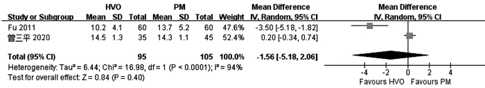
    FU SY, LAU WY, LI GG, et al. A prospective randomized controlled trial to compare Pringle maneuver, hemihepatic vascular inflow occlusion, and main portal vein inflow occlusion in partial hepatectomy[J]. Am J Surg, 2011, 201(1): 62-69. DOI: 10.1016/j.amjsurg.2009.09.029
    [17]
    CAI HY, SHAO BF, ZHOU Y. Comparative observation of selective hemihepatic blood flow occlusion and Pringle blood flow occlusion in the surgical treatment of HBV-related liver cancer[J]. Shandong Med J, 2019, 59(6): 53-55. (in Chinese) https://www.cnki.com.cn/Article/CJFDTOTAL-SDYY201906014.htm

    蔡鸿宇, 邵冰峰, 周元, 等. HBV相关性肝癌手术治疗中选择性半肝血流阻断与Pringle法血流阻断的应用对比观察[J]. 山东医药, 2019, 59(6): 53-55. https://www.cnki.com.cn/Article/CJFDTOTAL-SDYY201906014.htm
    [18]
    ZENG SP. Application effect comparison the first hepatic portal blood flow occlusion method and hemi-hepatic blood flow occlusion method in the treatment of primary hepatocellular carcinoma patients with hemi-hepatectomy[J]. Chin Mod Med, 2020, 27(3): 122-128. (in Chinese) DOI: 10.3969/j.issn.1674-4721.2020.03.036

    曾三平. 第一肝门血流阻断法与半肝血流阻断法在行半肝切除术治疗原发性肝癌患者中的应用效果比较[J]. 中国当代医药, 2020, 27(3): 122-128. DOI: 10.3969/j.issn.1674-4721.2020.03.036
    [19]
    HAN YL, MIAO J, YIN JJ. Comparison of different hepatic inflow occlusion in hepatectomy in treatm ent of patients with primary large liver cancer[J]. J Prac Hepatol, 2018, 21(1): 104-107. (in Chinese) DOI: 10.3969/j.issn.1672-5069.2018.01.025

    韩玉龙, 苗健, 尹家俊. 不同肝血流阻断方案对手术切除原发性大肝癌患者治疗效果比较[J]. 实用肝脏病杂志, 2018, 21(1): 104-107. DOI: 10.3969/j.issn.1672-5069.2018.01.025
    [20]
    QIAO HJ, SONG YC, ZHAO XY. Application of different blood flow occlusion methods of hepatectomy in the treatment of primary liver cancer[J]. J Hepatobiliary Surg, 2018, 26(4): 277-280. (in Chinese) https://www.cnki.com.cn/Article/CJFDTOTAL-GDWZ201804012.htm

    乔海军, 宋玉成, 赵小勇. 肝癌切除术中不同血流阻断方式在原发性肝癌治疗中的应用[J]. 肝胆外科杂志, 2018, 26(4): 277-280. https://www.cnki.com.cn/Article/CJFDTOTAL-GDWZ201804012.htm
    [21]
    WU ZY, FENG JY, WANG JB, et al. Clinical study of Glisson pedicle transverse hepatectomy in the treatment of liver cancer[J]. J Hepatopancreatobiliary Surg, 2014, 26(4): 342-344. (in Chinese) https://www.cnki.com.cn/Article/CJFDTOTAL-GDYW201404026.htm

    吴宗杨, 冯济业, 王金波, 等. Glisson蒂横断式肝切除术治疗肝癌的临床研究[J]. 肝胆胰外科杂志, 2014, 26(4): 342-344. https://www.cnki.com.cn/Article/CJFDTOTAL-GDYW201404026.htm
    [22]
    ZHUANG SW, LIN YY. Influence of different hepatic blood flow blocking methods on the curative effect of surgical resection of patients with primary massive liver cancer[J]. Chin Mod Doc, 2018, 56(29): 38-40. (in Chinese) https://www.cnki.com.cn/Article/CJFDTOTAL-ZDYS201829011.htm

    庄盛威, 林燕燕. 不同肝血流阻断方法对外科手术切除原发性大肝癌患者疗效的影响[J]. 中国现代医生, 2018, 56(29): 38-40. https://www.cnki.com.cn/Article/CJFDTOTAL-ZDYS201829011.htm
    [23]
    ZHANG NP, ZHANG XJ. Evaluation of different hepatic blood flow blocking methods in laparoscopic hepatectomy of patients with liver cancer[J/CD]. Chin J Liver Dis (Electronic Version), 2019, 11(3): 58-63. (in Chinese)

    张能平, 张雄杰. 不同肝血流阻断方式在肝癌患者腹腔镜肝切除术中的应用[J/CD]. 中国肝脏病杂志(电子版), 2019, 11(3): 58-63.
    [24]
    ZHU P, LAU WY, CHEN YF, et al. Randomized clinical trial comparing infrahepatic inferior vena cava clamping with low central venous pressure in complex liver resections involving the Pringle manoeuvre[J]. Br J Surg, 2012, 99(6): 781-788. DOI: 10.1002/bjs.8714
    [25]
    SELZNER N, RUDIGER H, GRAF R, et al. Protective strategies against ischemic injury of the liver[J]. Gastroenterology, 2003, 125(3): 917-936. DOI: 10.1016/S0016-5085(03)01048-5
    [26]
    GRACE PA. Ischaemia-reperfusion injury[J]. Br J Surg, 1994, 81(5): 637-647. DOI: 10.1002/bjs.1800810504
    [27]
    XIAOBIN F, ZIPEI L, SHUGUO Z, et al. The Pringle manoeuvre should be avoided in hepatectomy for cancer patients due to its side effects on tumor recurrence and worse prognosis[J]. Med Hypotheses, 2009, 72(4): 398-401.
    [28]
    BELGHITI J, NOUN R, MALAFOSSE R, et al. Continuous versus intermittent portal triad clamping for liver resection: A controlled study[J]. Ann Surg, 1999, 229(3): 369-375.
    [29]
    GUO T, XIAO Y, LIU Z, et al. The impact of intraoperative vascular occlusion during liver surgery on postoperative peak ALT levels: A systematic review and meta-analysis[J]. Int J Surg, 2016, 27: 99-104.
    [30]
    WANG HQ, YANG JY, YAN LN. Hemihepatic versus total hepatic inflow occlusion during hepatectomy: A systematic review and meta-analysis[J]. World J Gastroenterol, 2011, 17(26): 3158-3164.
    [31]
    DING HY, TAI QW, ZHOU CM, et al. Hemihepatic inflow occlusion versus total hepatic inflow occlusion in liver resection: A meta-analysis[J]. Chin J Evid-based Med, 2014, 14(4): 469-477. (in Chinese) https://www.cnki.com.cn/Article/CJFDTOTAL-ZZXZ201404019.htm

    丁贺义, 邰沁文, 周成明, 等. 肝切除术应用半肝与全肝入肝血流阻断法效果的Meta分析[J]. 中国循证医学杂志, 2014, 14(4): 469-477 https://www.cnki.com.cn/Article/CJFDTOTAL-ZZXZ201404019.htm
    [32]
    RAHBARI NN, KOCH M, MEHRABI A, et al. Portal triad clamping versus vascular exclusion for vascular control during hepatic resection: A systematic review and meta-analysis[J]. J Gastrointest Surg, 2009, 13(3): 558-568.
  • Relative Articles

    [1]Fei LIU, Hai LI, Qiang WU. Risk factors for bile leakage after hepatectomy without biliary reconstruction: A Meta - analysis[J]. Journal of Clinical Hepatology, 2022, 38(3): 594-600. doi: 10.3969/j.issn.1001-5256.2022.03.019
    [2]Lei WANG, Kangwei LIU, Yuling DUAN, Xinyao LI, Cijun PENG. Current status of the application of robot-assisted laparoscopic hepatectomy[J]. Journal of Clinical Hepatology, 2021, 37(11): 2732-2736. doi: 10.3969/j.issn.1001-5256.2021.11.054
    [3]Weiming YU, Wenqian HONG, Binglun SUN, Jingzhao HAN, Hongfang TUO. Value of albumin-bilirubin grade in predicting liver function changes and prognosis of hepatocellular carcinoma patients undergoing transarterial chemoembolization: A Meta-analysis[J]. Journal of Clinical Hepatology, 2021, 37(11): 2575-2583. doi: 10.3969/j.issn.1001-5256.2021.11.018
    [4]Yisheng PENG, Pan HE, Gang ZHU, Xinkai LI, Shunde TAN, Jianfei CHEN, Jun FAN, Bin LUO, Song SU, Bo LI, Xiaoli YANG. Efficacy and safety of CalliSpheres microsphere versus conventional transcatheter arterial chemoembolization in treatment of hepatocellular carcinoma: A Meta-analysis[J]. Journal of Clinical Hepatology, 2021, 37(8): 1841-1847. doi: 10.3969/j.issn.1001-5256.2021.08.019
    [5]Minjun LI, Zhujian DENG, Haotian LIU, Yuxian TENG, Rongrui HUO, Xiumei LIANG, Bangde XIANG, Lequn LI, Jianhong ZHONG. Clinical effect of re-hepatic resection versus radiofrequency ablation in treatment of recurrent hepatocellular carcinoma in Asia: A Meta-analysis[J]. Journal of Clinical Hepatology, 2021, 37(5): 1103-1109. doi: 10.3969/j.issn.1001-5256.2021.05.025
    [6]ZHANG ZhongLin, YUAN YuFeng. Key operating points of laparoscopic hepatectomy for liver tumor in difficult locations[J]. Journal of Clinical Hepatology, 2020, 36(12): 2663-2666. doi: 10.3969/j.issn.1001-5256.2020.12.006
    [7]Ren XiaoYing, Wu YongBiao, Ding XiangYuan, Li Xue, Yan HuiMing. Clinical effect of endoscopic sphincterotomy with balloon dilation versus sphincterotomy alone in treatment of large common bile duct stones: A Meta-analysis[J]. Journal of Clinical Hepatology, 2020, 36(4): 850-854. doi: 10.3969/j.issn.1001-5256.2020.04.028
    [8]Yu PeiHe, Su Song, Chen Shi, Wang JinCheng, Chen XinPei, Luo De. Clinical effect of pancreaticoduodenectomy with total mesopancreas excision versus traditional pancreaticoduodenectomy in treatment of pancreatic head carcinoma and periampullary cancer:A Meta-analysis[J]. Journal of Clinical Hepatology, 2020, 36(8): 1811-1815. doi: 10.3969/j.issn.1001-5256.2020.08.026
    [9]Tang JinQuan, He Pan, Su Song, Luo Bin, Fang Cheng, Li Bo, Xia XianMing, Li YuWei. Short-and long-term effects of radiofrequency ablation versus minimally invasive hepatectomy in treatment of small hepatocellular carcinoma: A Meta-analysis[J]. Journal of Clinical Hepatology, 2020, 36(2): 358-362. doi: 10.3969/j.issn.1001-5256.2020.02.025
    [10]Gao BenJian, Luo Jia, Liu Ying, Yang XiaoLi, Su Song, Li Bo. Efficacy and safety of laparoscopic versus open liver resection in treatment of colorectal cancer liver metastasis: A Meta-analysis[J]. Journal of Clinical Hepatology, 2020, 36(3): 573-579. doi: 10.3969/j.issn.1001-5256.2020.03.021
    [11]Sun DongXue, Yang Zhu, Long FengXi, Wei XianMan, Tang DongXin. Efficacy and safety of Kanglaite injection combined with transarterial chemoembolization in treatment of advanced liver cancer: A Meta-analysis[J]. Journal of Clinical Hepatology, 2020, 36(2): 363-368. doi: 10.3969/j.issn.1001-5256.2020.02.026
    [12]Zhang Bin, Luo De, Peng FangYi, Fang Cheng, Gan Yu, He Kai, Li Bo, Xia XianMing, Su Song. Clinical effect of robot-assisted laparoscopic hepatectomy versus open hepatectomy in treatment of liver diseases:A Meta-analysis[J]. Journal of Clinical Hepatology, 2020, 36(8): 1778-1782. doi: 10.3969/j.issn.1001-5256.2020.08.020
    [13]Lai Li, Peng FangYi, Su Song, Fang Cheng, Zhang MengYu, He Kai, Li Bo, He Pan, Xia XianMing. Clinical effect of robotic versus laparoscopic splenectomy in treatment of nontraumatic splenic diseases:A Meta-analysis[J]. Journal of Clinical Hepatology, 2019, 35(10): 2281-2285. doi: 10.3969/j.issn.1001-5256.2019.10.030
    [14]Ren Long, Zhang Miao, Zhang Yun. Application of enhanced recovery after surgery in perioperative period of hepatectomy: a Meta-analysis[J]. Journal of Clinical Hepatology, 2018, 34(3): 573-578. doi: 10.3969/j.issn.1001-5256.2018.03.025
    [15]Li Le, Chen JinMing, Liu ZhongHua, Li Qiang, Shi Ying. Application of enhanced recovery after surgery versus traditional rehabilitation program in hepatectomy: a Meta-analysis[J]. Journal of Clinical Hepatology, 2018, 34(2): 303-308. doi: 10.3969/j.issn.1001-5256.2018.02.017
    [16]Chen Kai, Li XiangNong, Liu Kun, Nan YunGuang, Yang Jun. Clinical effect of hepatectomy versus extracapsular peeling in treatment of hepatic hemangioma[J]. Journal of Clinical Hepatology, 2017, 33(4): 699-704. doi: 10.3969/j.issn.1001-5256.2017.04.020
    [17]Wang JinWei, Zhang YaMin. Research advances in methods for determination of tumor boundary in hepatectomy[J]. Journal of Clinical Hepatology, 2016, 32(2): 374-377. doi: 10.3969/j.issn.1001-5256.2016.02.039
    [18]Huang DongHai, Shi JianShe, Zhang ChengHua. Usage analysis of human serum albumin in patients with liver cancer and liver cirrhosis after hepatectomy[J]. Journal of Clinical Hepatology, 2015, 31(6): 910-914. doi: 10.3969/j.issn.1001-5256.2015.06.020
    [19]Shen YiNan, Huang ShengYu, Hu XianRong, Wang Jie, Yu HongMing, Lu Wen, Lu JunHua, Yang GuangShun, Wu MengChao. Clinical effect of laparoscopic hepatectomy in treating liver metastasis of colon cancer: a systematic review and meta-analysis[J]. Journal of Clinical Hepatology, 2014, 30(12): 1287-1291. doi: 10.3969/j.issn.1001-5256.2014.12.013
    [20]Chen Kai, Qiu FuNan, Tian YiFeng, Lai ZhiDe, Zhou SongQiang, Yan MaoLin, Wang YaoDong. Application of hepatectomy without hepatic blood flow occlusion in treatment of primary liver cancer[J]. Journal of Clinical Hepatology, 2014, 30(10): 1060-1063. doi: 10.3969/j.issn.1001-5256.2014.10.021
  • Cited by

    Periodical cited type(9)

    1. 李晓蓉,姚家喜,施志斌. SAA、GRP78、miR-21-3p与老年急性胰腺炎Ranson和APACHEⅡ评分的关联性. 中国老年学杂志. 2025(05): 1092-1095 .
    2. 赵永红,陈爱荣,胡梦茹,王燚鑫,衣桂荣. 急性胰腺炎伴代谢综合征的临床特点和危险因素分析. 现代消化及介入诊疗. 2024(06): 664-669 .
    3. 宋小利,陈璐. 大剂量维生素C联合乌司他丁、生长抑素治疗急性胰腺炎的效果及对肝肾功能的影响. 临床医学研究与实践. 2024(28): 39-42 .
    4. 卢景涛,彭琼. 非酒精性脂肪性肝病诊断联合BISAP评分对急性胰腺炎严重程度的预测价值. 医学信息. 2023(04): 118-122 .
    5. 黄雯雪,陈春洁,孙艳. 急性胰腺炎相关危险因素、严重程度评估及临床护理研究进展. 中国基层医药. 2022(03): 473-476 .
    6. 刘国雄,匡桥贵,喻欣荷,刘访,王宇,俞洋,陈杨. 加味大承气汤治疗湿热蕴结型高脂血症性急性胰腺炎的疗效及作用机制. 中国实验方剂学杂志. 2021(05): 91-97 .
    7. 王婷婷,何家俊,杨楚婷,李圆浩,陈炜炜,刘军. 非酒精性脂肪性肝病与急性胰腺炎的关系. 临床肝胆病杂志. 2021(03): 729-732 . 本站查看
    8. 赵冬雨,成丽娅,邵伟,马程,沈宏. 胰胆舒胶囊联合乌司他丁治疗急性胰腺炎的临床研究. 现代药物与临床. 2021(04): 712-716 .
    9. 黄莹,苗雨,林晚,刘昊,张飞雄,阮继刚. 急性复发性胰腺炎合并代谢综合征的临床特点及预后分析. 宁夏医学杂志. 2021(11): 968-971 .

    Other cited types(7)

  • 加载中

Catalog

    通讯作者: 陈斌, bchen63@163.com
    • 1. 

      沈阳化工大学材料科学与工程学院 沈阳 110142

    1. 本站搜索
    2. 百度学术搜索
    3. 万方数据库搜索
    4. CNKI搜索

    Figures(10)  / Tables(1)

    Article Metrics

    Article views (969) PDF downloads(53) Cited by(16)
    Proportional views
    Related

    /

    DownLoad:  Full-Size Img  PowerPoint
    Return
    Return