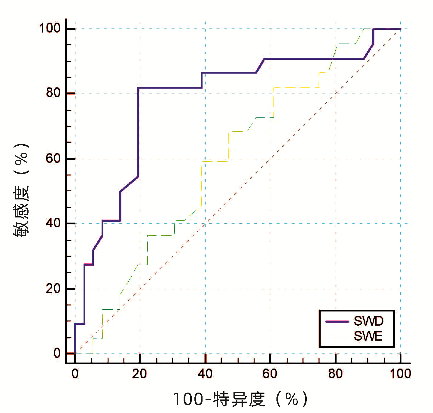
中文English
ISSN 1001-5256 (Print)
ISSN 2097-3497 (Online)
CN 22-1108/R

Value of ultrasonic shear-wave dispersion imaging in diagnosis of high-risk esophageal and gastric varices in compensated cirrhosis

DOI: 10.3969/j.issn.1001-5256.2022.07.018
Research funding:

Tianjin Science and Technology Major Project and Critical Disease Prevention and Control Project 2019 (19ZXDBSY00030)

More Information
  • Corresponding author: XIANG Huiling, huilingxiang@163.com(ORCID: 0000-0003-3678-4225)
  • Received Date: 2021-10-30
  • Accepted Date: 2021-12-17
  • Published Date: 2022-07-20
  •   Objective  To investigate the clinical value of Canon two-dimensional ultrasound shear wave elastography (SWE) and shear wave dispersion (SWD) in the diagnosis of high-risk esophageal and gastric varices in compensated cirrhosis.  Methods  A total of 58 patients with compensated cirrhosis of various etiologies who received electronic gastroscopy in Tianjin Third Central Hospital from February 2020 to February 2021, and Canon Aplio i800 color ultrasound instrument was used to perform SWE and SWD of the liver. According to the results of gastroscopy, the patients were divided into high-risk esophageal and gastric varices group (HREGV group) with 22 patients and non-high-risk esophageal and gastric varices group (NHREGV group) with 36 patients. The independent samples t-test was used for comparison of normally distributed continuous data between two groups, and the non-parametric Mann-Whitney U test was used for comparison of non-normally distributed continuous data between two groups; the chi-square test was used for comparison of categorical data between two groups. A multivariate logistic regression analysis was used to analyze and screen out the noninvasive indicators for predicting high-risk esophageal and gastric varices, and the receiver operating characteristic (ROC) curve was used to evaluate the clinical value of liver SWD and SWE in the diagnosis of high-risk esophageal and gastric varices.  Results  There were significant differences between the HREGV group and the NHREGV group in SWD (t=-3.84, P < 0.001), etiology (χ2=9.67, P=0.022), total bilirubin (Z=-2.00, P=0.045), spleen diameter (t=-2.44, P=0.018), and portal vein diameter (Z=-1.96, P=0.005). The patients with compensated cirrhosis had a mean liver SWD of 15.17±2.45 m·s-1·kHz-1, and the HREGV group had a significantly higher liver SWD than the NHREGV group (16.59±2.66 m·s-1·kHz-1 vs 14.31±1.86 m·s-1·kHz-1, t=-3.84, P < 0.001), while there was no significant difference in SWE between the two groups (Z=-1.21, P=0.223). SWD was an independent risk factor for high-risk esophageal and gastric varices in patients with compensated liver cirrhosis (odds ratio=1.67, 95% confidence interval: 1.17-2.39, P=0.005). In the diagnosis of high-risk esophageal and gastric varices, SWD had an area under the ROC curve (AUC) of 0.786, with a specificity of 80.56% and a sensitivity of 81.82% at the optimal cut-off value of 15.35 m·s-1·kHz-1; SWE had an AUC of 0.596, with a specificity of 52.78% and a sensitivity of 68.18% at the optimal cut-off value of 9.25 kPa.  Conclusion  Liver SWD measured by Canon Aplio i800 color ultrasound is excepted to become a new noninvasive method to predict the presence of high-risk esophageal and gastric varices in patients with compensated cirrhosis, while SWE has a limited diagnostic value.


  • [1]
    de FRANCHIS R, Baveno VI Faculty. Expanding consensus in portal hypertension: Report of the Baveno VI Consensus Workshop: Stratifying risk and individualizing care for portal hypertension[J]. J Hepatol, 2015, 63(3): 743-752. DOI: 10.1016/j.jhep.2015.05.022.
    [2]
    Chinese Society of Hepatology, Chinese Medical Association, Chinese Society of Gastroenterology, Chinese Medical Association, Chinese Society of Endoscopy, Chinese Medical Association. Guidelines for the diagnosis and treatment of esophageal and gastric variceal bleeding in cirrhotic portal hypertension[J]. J Clin Hepatol, 2016, 32(2): 203-219. DOI: 10.3969/j.issn.1001-5256.2016.02.002.

    中华医学会肝病学分会, 中华医学会消化病学分会, 中华医学会内镜学分会. 肝硬化门静脉高压食管胃静脉曲张出血的防治指南[J]. 临床肝胆病杂志, 2016, 32(2): 203-219. DOI: 10.3969/j.issn.1001-5256.2016.02.002.
    [3]
    GARCIA-TSAO G, ABRALDES JG, BERZIGOTTI A, et al. Portal hypertensive bleeding in cirrhosis: Risk stratification, diagnosis, and management: 2016 practice guidance by the American Association for the study of liver diseases[J]. Hepatology, 2017, 65(1): 310-335. DOI: 10.1002/hep.28906.
    [4]
    SHIHA G, IBRAHIM A, HELMY A, et al. Asian-Pacific Association for the Study of the Liver (APASL) consensus guidelines on invasive and non-invasive assessment of hepatic fibrosis: A 2016 update[J]. Hepatol Int, 2017, 11(1): 1-30. DOI: 10.1007/s12072-016-9760-3.
    [5]
    MANATSATHIT W, SAMANT H, KAPUR S, et al. Accuracy of liver stiffness, spleen stiffness, and LS-spleen diameter to platelet ratio score in detection of esophageal varices: Systemic review and meta-analysis[J]. J Gastroenterol Hepatol, 2018, 33(10): 1696-1706. DOI: 10.1111/jgh.14271.
    [6]
    SUGIMOTO K, MORIYASU F, OSHIRO H, et al. Viscoelasticity measurement in rat livers using shear-wave US elastography[J]. Ultrasound Med Biol, 2018, 44(9): 2018-2024. DOI: 10.1016/j.ultrasmedbio.2018.05.008.
    [7]
    LEE DH, CHO EJ, BAE JS, et al. Accuracy of two-dimensional shear wave elastography and attenuation imaging for evaluation of patients with nonalcoholic steatohepatitis[J]. Clin Gastroenterol Hepatol, 2021, 19(4): 797-805. e7. DOI: 10.1016/j.cgh.2020.05.034.
    [8]
    TANG Y, KONG W, ZHAO J, et al. Can viscoelasticity measurements obtained through shear-wave US elastography be used to monitor hepatic ischemia-reperfusion injury and treatment response? An animal study[J]. Ultrasound Med Biol, 2020, 46(9): 2464-2471. DOI: 10.1016/j.ultrasmedbio.2020.04.021.
    [9]
    Chinese Society of Hepatology, Chinese Medical Association. Chinese guidelines on the management of liver cirrhosis[J]. J Clin Hepatol, 2019, 35(11): 2408-2425. DOI: 10.3969/j.issn.1001-5256.2019.11.006.

    中华医学会肝病学分会. 肝硬化诊治指南[J]. 临床肝胆病杂志, 2019, 35(11): 2408-2425. DOI: 10.3969/j.issn.1001-5256.2019.11.006.
    [10]
    JIANG SL, XIONG JB, LOU X, et al. Influence of endoscopic dense ligation combined with sclerosing agent on hepatic synthesis function in treatment of severe esophageal and gastric varices in liver cirrhosis[J]. Clin J Med Offic, 2020, 48(9): 1097-1098. DOI: 10.16680/j.1671-3826.2020.09.38.

    蒋树林, 熊加彬, 娄晓, 等. 内镜下密集套扎结合硬化剂治疗肝硬化食管胃重度静脉曲张对患者肝合成功能影响[J]. 临床军医杂志, 2020, 48(9): 1097-1098. DOI: 10.16680/j.1671-3826.2020.09.38.
    [11]
    SHI XJ, SHEN JH, NIU HM, et al. Clinical effect and prognostic value of endoscopic ultrasound-guided treatment of esophageal varices in liver cirrhosis[J]. Chin J Gerontol, 2021, 41(8): 1634-1638. DOI: 10.3969/j.issn.1005-9202.2021.08.021.

    石小静, 申军华, 牛红梅, 等. 超声内镜指导下对肝硬化并发食管静脉曲张内镜下治疗的临床疗效及预后价值[J]. 中国老年学杂志, 2021, 41(8): 1634-1638. DOI: 10.3969/j.issn.1005-9202.2021.08.021.
    [12]
    QI X, BERZIGOTTI A, CARDENAS A, et al. Emerging non-invasive approaches for diagnosis and monitoring of portal hypertension[J]. Lancet Gastroenterol Hepatol, 2018, 3(10): 708-719. DOI: 10.1016/S2468-1253(18)30232-2.
    [13]
    WANG L, FENG Y, MA X, et al. Diagnostic efficacy of noninvasive liver fibrosis indexes in predicting portal hypertension in patients with cirrhosis[J]. PLoS One, 2017, 12(8): e0182969. DOI: 10.1371/journal.pone.0182969.
    [14]
    DENG H, QI X, ZHANG T, et al. Supersonic shear imaging for the diagnosis of liver fibrosis and portal hypertension in liver diseases: A meta-analysis[J]. Expert Rev Gastroenterol Hepatol, 2018, 12(1): 91-98. DOI: 10.1080/17474124.2018.1412257.
    [15]
    LI Q, WANG R, GUO X, et al. Contrast-enhanced CT may be a diagnostic alternative for gastroesophageal varices in cirrhosis with and without previous endoscopic variceal therapy[J]. Gastroenterol Res Pract, 2019, 2019: 6704673. DOI: 10.1155/2019/6704673.
    [16]
    QI X, AN W, LIU F, et al. Virtual hepatic venous pressure gradient with CT angiography (CHESS 1601): A prospective multicenter study for the noninvasive diagnosis of portal hypertension[J]. Radiology, 2019, 290(2): 370-377. DOI: 10.1148/radiol.2018180425.
    [17]
    ABRALDES JG, BUREAU C, STEFANESCU H, et al. Noninvasive tools and risk of clinically significant portal hypertension and varices in compensated cirrhosis: The "Anticipate" study[J]. Hepatology, 2016, 64(6): 2173-2184. DOI: 10.1002/hep.28824.
    [18]
    Chinese Foundation for Hepatitis Prevention and Control, Chinese Society of Infectious Disease, Chinese Society of Hepatology and Chinese Medical Association Liver Disease Committee of Chinese Research Hospital Association. Consensus on clinical application of transient alastography detecting liver fibrosis: A 2018 update[J]. Chin J Hepatol, 2019, 27(3): 182-191. DOI: 10.3760/cma.j.issn.1007-3418.2019.03.004.

    中国肝炎防治基金会, 中华医学会感染病学分会, 中华医学会肝病学分会和中国研究性医院学会肝病专业委员会. 瞬时弹性成像技术诊断肝纤维化专家共识(2018年更新版)[J]. 中华肝脏病杂志, 2019, 27(3): 182-191. DOI: 10.3760/cma.j.issn.1007-3418.2019.03.004
    [19]
    XIE LT, YAN CH, ZHAO QY, et al. Quantitative and noninvasive assessment of chronic liver diseases using two-dimensional shear wave elastography[J]. World J Gastroenterol, 2018, 24(9): 957-970. DOI: 10.3748/wjg.v24.i9.957.l.
    [20]
    Panel of Elastography Assessment of Liver Fibrosis, Study Group of Interventional Ultrasound, Society of Ultrasound in Medicine of Chinese Medical Association. Guidelines for clinical application of two-dimensional shear wave elastography in assessment of liver fibrosis in chronic hepatitis B[J]. J Clin Hepatol, 2018, 34(2): 255-261. DOI: 10.3969/j.issn.1001-5256.2018.02.008.

    中华医学会超声医学分会介入超声学组弹性成像评估肝纤维化专家组. 二维剪切波弹性成像评估慢性乙型肝炎肝纤维化临床应用指南[J]. 临床肝胆病杂志, 2018, 34(2): 255-261. DOI: 10.3969/j.issn.1001-5256.2018.02.008.
    [21]
    CHEN S, URBAN MW, PISLARU C, et al. Liver elasticity and viscosity quantification using shearwave dispersion ultrasound vibrometry (SDUV)[J]. Annu Int Conf IEEE Eng Med Biol Soc, 2009, 2009: 2252-2255. DOI: 10.1109/IEMBS.2009.5334992.
    [22]
    LEE DH, CHO EJ, BAE JS, et al. Accuracy of two-dimensional shear wave elastography and attenuation imaging for evaluation of patients with nonalcoholic steatohepatitis[J]. Clin Gastroenterol Hepatol, 2021, 19(4): 797-805. e7. DOI: 10.1016/j.cgh.2020.05.034.
    [23]
    LEE DH, LEE JY, BAE JS, et al. Shear-wave dispersion slope from US shear-wave elastography: Detection of allograft damage after liver transplantation[J]. Radiology, 2019, 293(2): 327-333. DOI: 10.1148/radiol.2019190064.
    [24]
    NIGHTINGALE KR, ROUZE NC, ROSENZWEIG SJ, et al. Derivation and analysis of viscoelastic properties in human liver: Impact of frequency on fibrosis and steatosis staging[J]. IEEE Trans Ultrason Ferroelectr Freq Control, 2015, 62(1): 165-175. DOI: 10.1109/TUFFC.2014.006653.
    [25]
    DEFFIEUX T, GENNISSON JL, BOUSQUET L, et al. Investigating liver stiffness and viscosity for fibrosis, steatosis and activity staging using shear wave elastography[J]. J Hepatol, 2015, 62(2): 317-324. DOI: 10.1016/j.jhep.2014.09.020.
    [26]
    VIZZUTTI F, ARENA U, ROMANELLI RG, et al. Liver stiffness measurement predicts severe portal hypertension in patients with HCV-related cirrhosis[J]. Hepatology, 2007, 45(5): 1290-1297. DOI: 10.1002/hep.21665.
    [27]
    JEON SK, LEE JM, JOO I, et al. Two-dimensional shear wave elastography with propagation maps for the assessment of liver fibrosis and clinically significant portal hypertension in patients with chronic liver disease: A prospective study[J]. Acad Radiol, 2020, 27(6): 798-806. DOI: 10.1016/j.acra.2019.08.006.
    [28]
    YOU MW, KIM KW, PYO J, et al. A Meta-analysis for the diagnostic performance of transient elastography for clinically significant portal hypertension[J]. Ultrasound Med Biol, 2017, 43(1): 59-68. DOI: 10.1016/j.ultrasmedbio.2016.07.025.
    [29]
    GILLIGAN LA, TROUT AT, BENNETT P, et al. Repeatability and agreement of shear wave speed measurements in phantoms and human livers across 6 ultrasound 2-dimensional shear wave elastography systems[J]. Invest Radiol, 2020, 55(4): 191-199. DOI: 10.1097/RLI.0000000000000627.
    [30]
    DENG H, QI X, ZHANG T, et al. Supersonic shear imaging for the diagnosis of liver fibrosis and portal hypertension in liver diseases: A meta-analysis[J]. Expert Rev Gastroenterol Hepatol, 2018, 12(1): 91-98. DOI: 10.1080/17474124.2018.1412257.
    [31]
    BERZIGOTTI A. Non-invasive evaluation of portal hypertension using ultrasound elastography[J]. J Hepatol, 2017, 67(2): 399-411. DOI: 10.1016/j.jhep.2017.02.003.
    [32]
    SHARMA P, KIRNAKE V, TYAGI P, et al. Spleen stiffness in patients with cirrhosis in predicting esophageal varices[J]. Am J Gastroenterol, 2013, 108(7): 1101-1107. DOI: 10.1038/ajg.2013.119.
  • Relative Articles

    [1]Feixiang XIONG, Peipei MENG, Yuyong JIANG, Qin ZHANG, Yandan JIANG, Haoxin DUAN, Saisai ZHAO. Clinical value of C-reactive protein/albumin ratio in the diagnosis of alcoholic cirrhosis with spontaneous bacterial peritonitis[J]. Journal of Clinical Hepatology, 2023, 39(8): 1880-1885. doi: 10.3969/j.issn.1001-5256.2023.08.017
    [2]Shunxiao ZHANG, Sheng ZHANG, Yan ZHANG, Yuanyuan LI, Yue CHEN, Mingyu SUN. Influencing factors for nonalcoholic fatty liver disease in patients with type 2 diabetes mellitus[J]. Journal of Clinical Hepatology, 2023, 39(5): 1081-1088. doi: 10.3969/j.issn.1001-5256.2023.05.013
    [3]Fangfang QIAN, Meiqing DAI, Li ZHAO, Xia DENG, Ling YANG, Jue JIA, Jifang WANG, Dong WANG, Guoyue YUAN. Association between serum alkaline phosphatase and type 2 diabetes mellitus with nonalcoholic fatty liver disease[J]. Journal of Clinical Hepatology, 2023, 39(1): 83-88. doi: 10.3969/j.issn.1001-5256.2023.01.013
    [4]Mengya GAO, Qian QIN, Shoujun WANG. Value of atherogenic index of plasma in predicting metabolic associated fatty liver disease in patients with type 2 diabetes mellitus[J]. Journal of Clinical Hepatology, 2023, 39(6): 1325-1331. doi: 10.3969/j.issn.1001-5256.2023.06.012
    [5]Dongxu WANG, Nan NAN, Hao BING, Lianjie LIN. Value of triglyceride-glucose index and body mass index in predicting nonalcoholic fatty liver disease in patients with type 2 diabetes mellitus[J]. Journal of Clinical Hepatology, 2022, 38(5): 1064-1068. doi: 10.3969/j.issn.1001-5256.2022.05.017
    [6]Ping JIANG, Rencheng DOU, Zijin CUI, Yinni TONG, Chen LI, Shanshan JIANG, Yun BAI, Jimin ZHENG, Cunkai WANG, Yuzhen WANG. Value of albumin-bilirubin score combined with neutrophil count and procalcitonin in ascites in predicting spontaneous bacterial peritonitis in patients with cirrhotic ascites[J]. Journal of Clinical Hepatology, 2021, 37(9): 2097-2101. doi: 10.3969/j.issn.1001-5256.2021.09.019
    [7]Chaomei QIN, Yongming LIU, Heling SU. Association of hepatitis B and hepatitis C with type 2 diabetes mellitus[J]. Journal of Clinical Hepatology, 2021, 37(4): 921-924. doi: 10.3969/j.issn.1001-5256.2021.04.042
    [8]Renshu MA, Tong WU, Ge YU, Ying CONG, Guijie XIN. A case of type 2 diabetes with glycogenic hepatopathy[J]. Journal of Clinical Hepatology, 2021, 37(9): 2183-2184. doi: 10.3969/j.issn.1001-5256.2021.09.035
    [9]Peiru LEI, Yingjie LI, Jing LI. Clinical significance of homocysteine and neutrophil-to-lymphocyte ratio in patients with nonalcoholic fatty liver disease and type 2 diabetes mellitus[J]. Journal of Clinical Hepatology, 2021, 37(10): 2352-2356. doi: 10.3969/j.issn.1001-5256.2021.10.019
    [10]Zhenzhen ZHANG, Yaoyi ZHANG, Kai WANG, Chenguang TIAN. Effect of dapagliflozin on metabolic markers and autonomic function in patients with type 2 diabetes mellitus and metabolic associated fatty liver disease[J]. Journal of Clinical Hepatology, 2021, 37(12): 2849-2853. doi: 10.3969/j.issn.1001-5256.2021.12.023
    [11]Jiao LIU, Caiqin REN, Yali FENG, Jirui HE. Association between liver fibrosis and C-peptide in patients with type 2 diabetes and nonalcoholic fatty liver disease[J]. Journal of Clinical Hepatology, 2021, 37(5): 1132-1136. doi: 10.3969/j.issn.1001-5256.2021.05.030
    [12]Liu Li, Jiang YingYi, Liu Jing. Glucose metabolism and insulin secretion in patients with chronic hepatitis B cirrhosis of different Child-Pugh grades and their association with inflammatory response[J]. Journal of Clinical Hepatology, 2020, 36(2): 324-328. doi: 10.3969/j.issn.1001-5256.2020.02.018
    [13]Yang LiXia, Zhang LunLi, Lai LingLing, Fang Ming. Value of heparin-binding protein in ascites and serum procalcitonin in diagnosis of spontaneous bacterial peritonitis[J]. Journal of Clinical Hepatology, 2019, 35(6): 1266-1269. doi: 10.3969/j.issn.1001-5256.2019.06.017
    [14]Song YuanYuan, Jiang YuYong. Research advances in diagnosis and treatment of spontaneous bacterial peritonitis[J]. Journal of Clinical Hepatology, 2016, 32(6): 1188-1191. doi: 10.3969/j.issn.1001-5256.2016.06.040
    [15]Zhan GuoQing, Li Fang, Li RuGui, Guo Peng, Liu Xiang, Zhang WeiWei, Tan HuaBing. Efficacy of terlipressin therapy for refractory ascites in cirrhosis and type-2 hepatorenal syndrome[J]. Journal of Clinical Hepatology, 2015, 31(8): 1287-1290. doi: 10.3969/j.issn.1001-5256.2015.08.025
    [16]Wu HaiQing, Wang Hui. Diagnosis and treatment of spontaneous bacterial peritonitis in patients with liver ascites[J]. Journal of Clinical Hepatology, 2012, 28(9): 651-653.
    [17]Zhang Wei, Gao ZhanQi, Zhang ShiLan. Clinical significance of detection of plasma and ascitic procalcitonin in cirrhotic patients with spontaneous bacterial peritonitis[J]. Journal of Clinical Hepatology, 2004, 20(3): 174-175.
    [19]Zhang Mei, Hu ShuiQing, Lu MeiHua, Zhou MeiZhi, Li YongWei, Sun LiDong. The effect of insulin resistance and secreting functions on glucose metabolism of patients with Cirrhosis.[J]. Journal of Clinical Hepatology, 2000, 16(1): 39-40.
  • Created with Highcharts 5.0.7Amount of accessChart context menuAbstract Views, HTML Views, PDF Downloads StatisticsAbstract ViewsHTML ViewsPDF Downloads2024-052024-062024-072024-082024-092024-102024-112024-122025-012025-022025-032025-040102030
    Created with Highcharts 5.0.7Chart context menuAccess Class DistributionFULLTEXT: 22.1 %FULLTEXT: 22.1 %META: 72.5 %META: 72.5 %PDF: 5.4 %PDF: 5.4 %FULLTEXTMETAPDF
    Created with Highcharts 5.0.7Chart context menuAccess Area Distribution其他: 8.6 %其他: 8.6 %其他: 2.5 %其他: 2.5 %China: 0.2 %China: 0.2 %Fairborn: 0.3 %Fairborn: 0.3 %Hollywood: 0.3 %Hollywood: 0.3 %India: 0.1 %India: 0.1 %Jamaica Plain: 0.3 %Jamaica Plain: 0.3 %Kao-sung: 0.1 %Kao-sung: 0.1 %Koesan: 0.1 %Koesan: 0.1 %Osaka: 0.1 %Osaka: 0.1 %Russian Federation: 0.1 %Russian Federation: 0.1 %Valencia: 0.5 %Valencia: 0.5 %[]: 1.7 %[]: 1.7 %三亚: 0.1 %三亚: 0.1 %上海: 1.9 %上海: 1.9 %中卫: 0.2 %中卫: 0.2 %丽水: 0.1 %丽水: 0.1 %乌鲁木齐: 0.1 %乌鲁木齐: 0.1 %伦敦: 0.1 %伦敦: 0.1 %内萨瓦尔科约特尔城: 0.3 %内萨瓦尔科约特尔城: 0.3 %北京: 3.6 %北京: 3.6 %十堰: 0.1 %十堰: 0.1 %南京: 0.1 %南京: 0.1 %南宁: 0.2 %南宁: 0.2 %南阳: 0.1 %南阳: 0.1 %卡纳塔克: 0.2 %卡纳塔克: 0.2 %厦门: 0.1 %厦门: 0.1 %台北: 0.1 %台北: 0.1 %台州: 0.2 %台州: 0.2 %合肥: 0.2 %合肥: 0.2 %吉林: 1.6 %吉林: 1.6 %哥印拜陀: 0.1 %哥印拜陀: 0.1 %商丘: 0.1 %商丘: 0.1 %嘉兴: 0.1 %嘉兴: 0.1 %埃克塞特: 0.5 %埃克塞特: 0.5 %多伦多: 0.1 %多伦多: 0.1 %大连: 0.4 %大连: 0.4 %天津: 0.9 %天津: 0.9 %安阳: 0.1 %安阳: 0.1 %宣城: 0.2 %宣城: 0.2 %山梨县: 0.2 %山梨县: 0.2 %常德: 0.1 %常德: 0.1 %平顶山: 0.1 %平顶山: 0.1 %广州: 0.6 %广州: 0.6 %延边: 0.1 %延边: 0.1 %弗吉尼亚州: 0.1 %弗吉尼亚州: 0.1 %张家口: 2.6 %张家口: 2.6 %德宏: 0.1 %德宏: 0.1 %惠州: 0.1 %惠州: 0.1 %成都: 0.3 %成都: 0.3 %扬州: 0.1 %扬州: 0.1 %无锡: 0.4 %无锡: 0.4 %旧金山: 0.3 %旧金山: 0.3 %昆卡: 0.1 %昆卡: 0.1 %昆明: 0.5 %昆明: 0.5 %曼谷: 0.1 %曼谷: 0.1 %朝阳: 0.5 %朝阳: 0.5 %杭州: 0.5 %杭州: 0.5 %柳州: 0.4 %柳州: 0.4 %森尼韦尔: 0.5 %森尼韦尔: 0.5 %横滨: 0.3 %横滨: 0.3 %武汉: 0.5 %武汉: 0.5 %沈阳: 0.1 %沈阳: 0.1 %泉州: 0.2 %泉州: 0.2 %洛杉矶: 0.3 %洛杉矶: 0.3 %济南: 0.3 %济南: 0.3 %海得拉巴: 0.3 %海得拉巴: 0.3 %深圳: 0.4 %深圳: 0.4 %湖州: 0.3 %湖州: 0.3 %湘潭: 0.1 %湘潭: 0.1 %漯河: 0.2 %漯河: 0.2 %烟台: 0.2 %烟台: 0.2 %盐城: 0.1 %盐城: 0.1 %石家庄: 0.2 %石家庄: 0.2 %福州: 0.1 %福州: 0.1 %秦皇岛: 0.1 %秦皇岛: 0.1 %罗奥尔凯埃: 0.4 %罗奥尔凯埃: 0.4 %芒廷维尤: 33.8 %芒廷维尤: 33.8 %芝加哥: 0.4 %芝加哥: 0.4 %苏州: 0.2 %苏州: 0.2 %莫斯科: 1.9 %莫斯科: 1.9 %菏泽: 0.1 %菏泽: 0.1 %葵涌: 0.1 %葵涌: 0.1 %蔚山: 0.1 %蔚山: 0.1 %衢州: 0.1 %衢州: 0.1 %西宁: 19.8 %西宁: 19.8 %西安: 0.3 %西安: 0.3 %诺沃克: 0.1 %诺沃克: 0.1 %贵阳: 0.1 %贵阳: 0.1 %赤峰: 0.2 %赤峰: 0.2 %运城: 1.5 %运城: 1.5 %郑州: 0.2 %郑州: 0.2 %都伯林: 0.6 %都伯林: 0.6 %重庆: 0.4 %重庆: 0.4 %锦州: 0.4 %锦州: 0.4 %长春: 0.6 %长春: 0.6 %长沙: 0.3 %长沙: 0.3 %长治: 0.1 %长治: 0.1 %雅西: 0.1 %雅西: 0.1 %青岛: 0.1 %青岛: 0.1 %马哈拉施特拉邦: 0.5 %马哈拉施特拉邦: 0.5 %鹿儿岛: 0.3 %鹿儿岛: 0.3 %其他其他ChinaFairbornHollywoodIndiaJamaica PlainKao-sungKoesanOsakaRussian FederationValencia[]三亚上海中卫丽水乌鲁木齐伦敦内萨瓦尔科约特尔城北京十堰南京南宁南阳卡纳塔克厦门台北台州合肥吉林哥印拜陀商丘嘉兴埃克塞特多伦多大连天津安阳宣城山梨县常德平顶山广州延边弗吉尼亚州张家口德宏惠州成都扬州无锡旧金山昆卡昆明曼谷朝阳杭州柳州森尼韦尔横滨武汉沈阳泉州洛杉矶济南海得拉巴深圳湖州湘潭漯河烟台盐城石家庄福州秦皇岛罗奥尔凯埃芒廷维尤芝加哥苏州莫斯科菏泽葵涌蔚山衢州西宁西安诺沃克贵阳赤峰运城郑州都伯林重庆锦州长春长沙长治雅西青岛马哈拉施特拉邦鹿儿岛

Catalog

    通讯作者: 陈斌, bchen63@163.com
    • 1. 

      沈阳化工大学材料科学与工程学院 沈阳 110142

    1. 本站搜索
    2. 百度学术搜索
    3. 万方数据库搜索
    4. CNKI搜索

    Figures(2)  / Tables(3)

    Article Metrics

    Article views (667) PDF downloads(61) Cited by()
    Proportional views
    Related

    /

    DownLoad:  Full-Size Img  PowerPoint
    Return
    Return