中文English
ISSN 1001-5256 (Print)
ISSN 2097-3497 (Online)
CN 22-1108/R

Expression and significance of response gene to complement 32 in liver regeneration after partial hepatectomy in mice

DOI: 10.3969/j.issn.1001-5256.2023.10.018
Research funding:

National Natural Science Foundation of China (31760339);

Scientific Research Program of Education Department of Hubei Province (Q20212107);

Guizhou Provincial Science and Technology Plan Project (Qiankehe Foundation-ZK [2022] General 594)

More Information
  • Corresponding author: KONG Deying, vsysongyer@126.com (ORCID: 0000-0002-6851-955X)
  • Received Date: 2022-12-19
  • Published Date: 2023-10-30
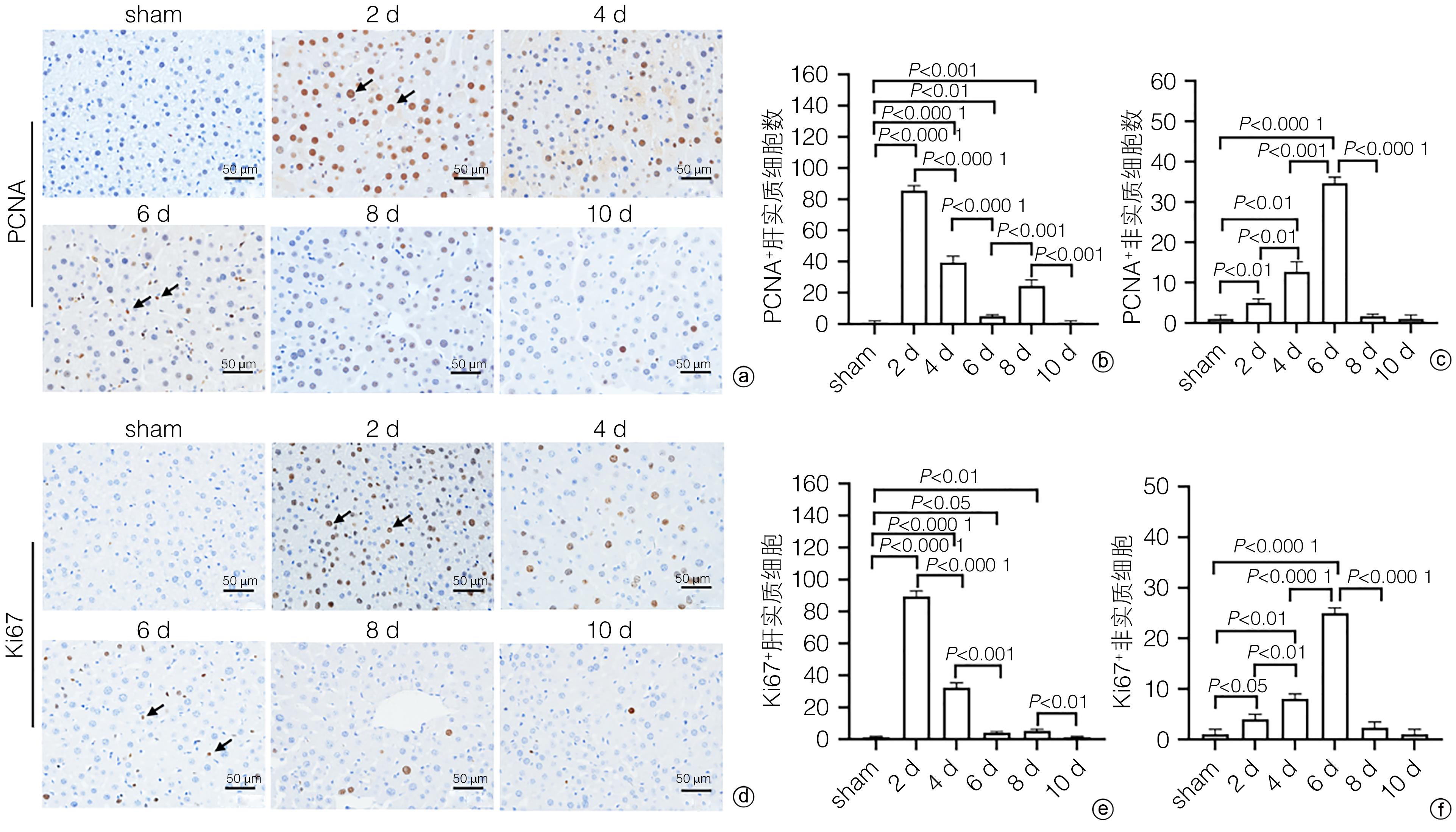
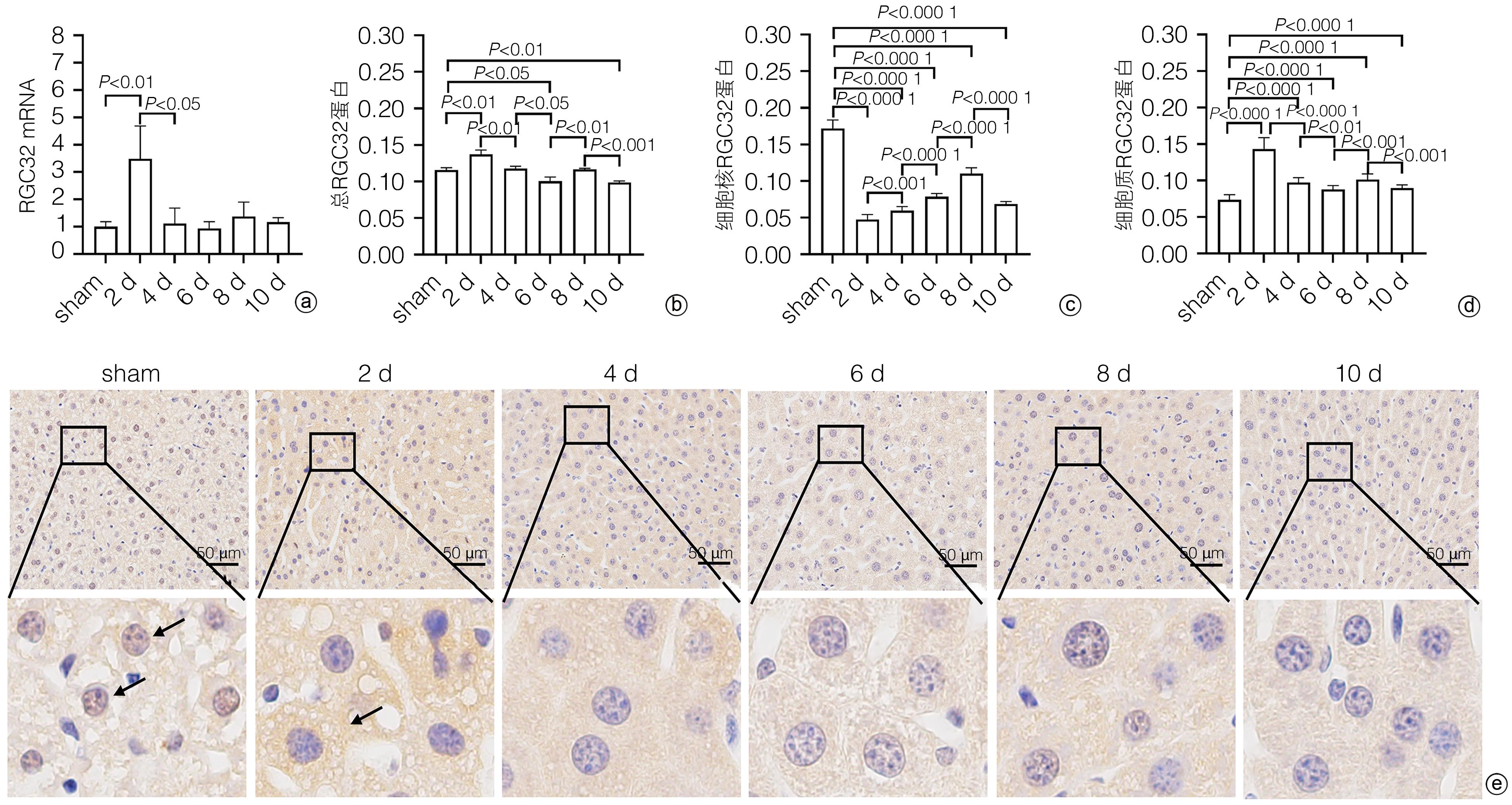
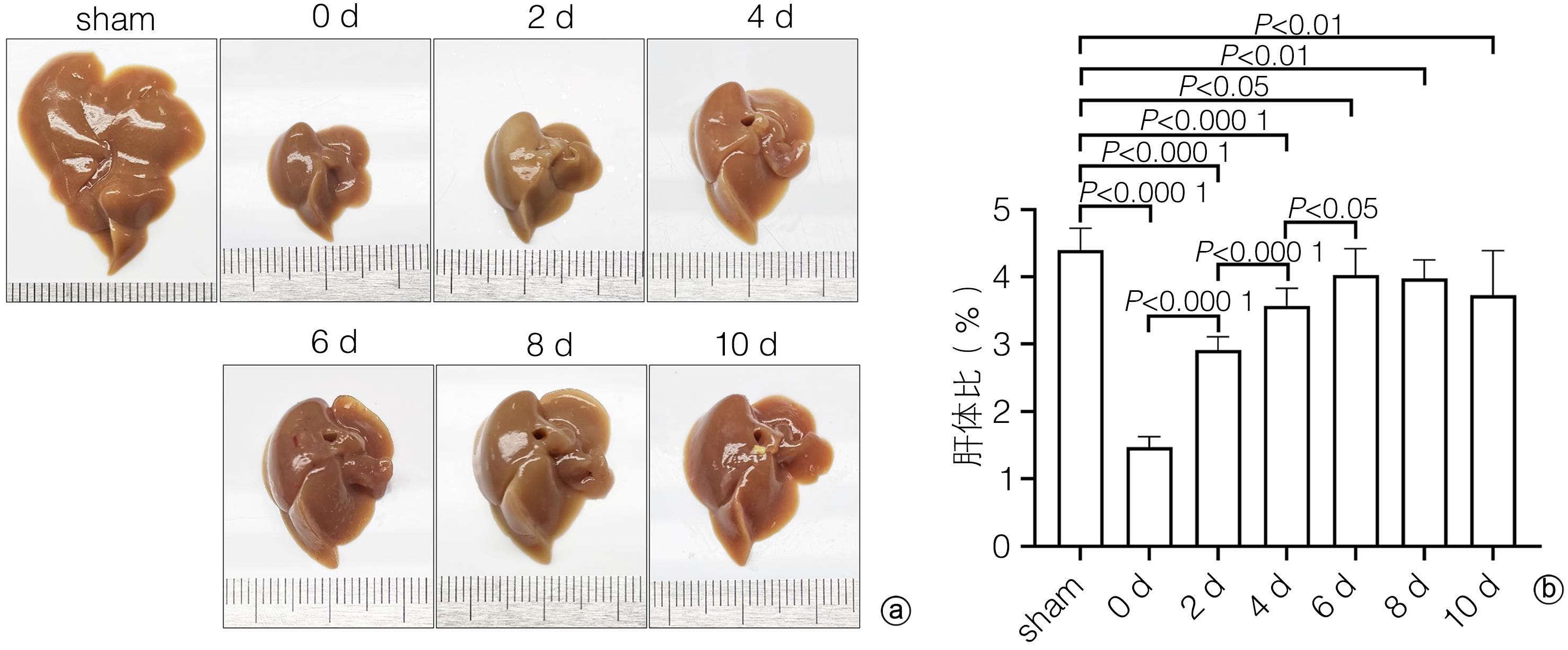
  •   Objective  To investigate the expression and role of response gene to complement 32 (RGC32) in liver regeneration after partial hepatectomy (PH).  Methods  A total of 42 male C57BL/6 mice, aged 10 weeks, were randomly divided into control group, postoperative day 1 group (1-d group), postoperative day 2 group (2-d group), postoperative day 4 group (4-d group), postoperative day 6 group (6-d group), postoperative day 8 group (8-d group), and postoperative day 10 group (10-d group), with 6 mice in each group. In the control group, the complete liver of the mice was resected for weighing and photography as the normal control group (sham group); further, the left and middle lobes of the liver were resected for weighing and photography as the surgical control group (0-day group); the sham group and the 0-day group shared the same group of mice. After successful modeling by PH, the mice were sacrificed on days 1, 2, 4, 6, 8, and 10 after surgery, and the liver was collected to measure the change in size. HE staining and oil red O staining were used to evaluate liver histomorphological changes; serum alanine aminotransferase (ALT) and aspartate aminotransferase (AST) were measured to evaluate the changes in liver function; immunohistochemical staining was used to measure the expression of proliferating cell nuclear antigen (PCNA) and Ki67 and analyze the change in cell proliferation during liver regeneration; quantitatie real-time PCR and immunohistochemical staining were uused to measure the expression and subcellular distribution of RGC32 during liver regeneration; EdU cell proliferation assay was used to analyze the effect of RGC32 overexpression or knocknout on hepatocyte proliferation in L02 cells. For continuous data, comparison between multiple groups was made by analysis of variance, and further pairwise comparisons were conducted using the LSD-t test. The independent samples t-test was used for comparison of continuous data between two groups. A Pearson correlation analysis was performed.  Results  The liver gradually enlarged after PH, and the liver/body weight ratio rose to the peak from days 0 to 6, with significant differences between different time points (all P<0.05), while there was no significant change in liver size from days 6 to 10. The number of liver lipid droplets significantly increased after PH surgery and gradually decreased with liver regeneration, with a significant difference between the portal vein region and the central vein region (all P<0.05). Compared with the sham group, the 1d group had significant increases in the serum levels of ALT and AST (all P<0.05), which gradually returned to the levels of the sham group on day 6 and day 2 after surgery, respectively (P>0.05). Immunohistochemical staining showed that there were rapid increases in the numbers of PCNA- and Ki67-positive liver parenchymal cells after PH surgery, with the highest numbers of 86±5 and 89±5, respectively, on day 2, which then gradually decreased; however, there were gradual increases in the numbers of PCNA- and Ki67-positive nonparenchymal cells, with the peak numbers of 34±5 and 25±3, respectively, on day 6, which then gradually decreased. The total expression of RGC32 increased to the highest level on day 2 after PH surgery and then gradually decreased, and the changing trend of RGC32 expression in cytoplasm was consistent with that of total RGC32 expression; however, the expression of RGC32 in nucleus decreased to the lowest level on day 2 after PH surgery and then increased gradually. The correlation analysis showed that the expression of RGC32 in nucleus was negatively correlated with the proliferation of liver parenchymal cells (R2=0.308 3, P=0.016 7), and the expression of RGC32 in cytoplasm was positively correlated with the proliferation of liver parenchymal cells (R2=0.808 6, P<0.000 1). Cell experiments showed that compared with the control group, the EdU-positive rate was reduced by 15.6% after RGC32 overexpression (P<0.01) and was increased by 19.2% after RGC32 knockdown (P<0.01).  Conclusion  Liver parenchymal cells and nonparenchymal cells show asynchronous proliferation and participate in liver regeneration together. During liver regeneration after hepatectomy, there are differences in the expression of RGC32 between nucleus and cytoplasm, and RGC32 in nucleus may inhibit hepatocyte proliferation.


  • 非酒精性脂肪性肝病(NAFLD)是指除外酒精和其他明确的损肝因素所致肝脏细胞内脂肪过度沉积(>5%)为主要特征的一种临床病理综合征[1]。NAFLD是与胰岛素抵抗和遗传易感性密切相关的一种获得性代谢应激性肝损伤[2],随着肥胖及其相关代谢综合征全球化的流行趋势,NAFLD现已成为欧美等发达国家和我国相对富裕地区慢性肝病的重要病因。NAFLD疾病谱包括非酒精性肝脂肪变、非酒精性脂肪性肝炎(NASH)、肝硬化和肝细胞癌(HCC)[3]

    尿酸是内源性和外源性嘌呤核苷酸代谢的终产物,血清尿酸(serum uric acid,sUA)产生过多或排泄减少可导致sUA升高,即为高尿酸血症(hyperuricemia,HUA)。HUA可引起全身系统的损害,并与代谢综合征密切相关。研究[4]显示,sUA/肌酐(serum creatinine,Cr)比值与代谢综合征显著相关。本研究着眼于sUA/Cr与NAFLD发生的相关性,以期对NAFLD的诊疗提供进一步的依据。

    回顾性选取2020年1月—2020年12月就诊于本院的97例NAFLD患者(NAFLD组)。另选取同期于本院体检的36例健康成人作为对照组。NAFLD诊断标准:根据《非酒精性脂肪性肝病防治指南(2018年更新版)》[2]中相关诊断标准,年龄≥18岁。排除标准:(1)已确诊的糖尿病、高血压患者,慢性肾功能衰竭(肾小球滤过率<30 mL/min)、肿瘤等严重的急慢性病患者;(2)酒精性、病毒性、药物性、自身免疫性、全胃肠外营养等明确病因所致肝脏疾病患者。

    (1) 肝肾对比或肝肾回声差异,肝实质回声明显大于肾实质回声;(2)肝脏近场和远场回声差异,近场回声增强,远场则衰减;(3)肝内管道结构特别是静脉内径变细,显示不清;(4)肝脏各径线测值存在不同程度增大;(5)彩色血流显示肝脏中血流信号不同程度存在减少;(6)肝右叶包膜及横膈回声显示不清晰。轻度脂肪肝:满足(1)与其余任何一项的情况。中度脂肪肝:满足(1)以及其余任何2项的情况。重度脂肪肝:满足(1)及其余任何3项的情况[5]

    收集所有研究对象的性别、年龄、空腹血糖(fasting blood sugar,FBS)、ALT、AST、TC、TG、高密度脂蛋白胆固醇(HDL)、低密度脂蛋白胆固醇(LDL)、GGT、sUA、sCr、sUA/Cr,以及腹部超声结果。

    本研究方案经由首都医科大学附属北京天坛医院伦理委员会审批,批号:KY2016-042-01。

    采用SPSS 18.0软件对所有数据进行统计学分析。正态分布的计量资料以x ±s表示,2组间比较采用独立样本t检验,3组间比较采用方差分析;非正态分布数据以M(P25~P75)表示,组间比较采用Mann-Whitney U检验;计数资料组间比较采用χ2检验;相关性分析采用Spearman检验。采用logistic回归分析NAFLD相关的危险因素。P<0.05为差异有统计学意义。

    NAFLD组ALT、AST、TG、GGT、sUA、sUA/Cr水平均显著高于健康对照组,而HDL水平则显著低于健康对照组(P值均<0.05)(表 1)。

    表  1  NAFLD组与健康对照组临床资料比较
    项目 NAFLD组(n=97) 健康对照组(n=36) 统计值 P
    男性[例(%)] 62(63.9) 25(69.4) χ2=0.354 0.682
    年龄(岁) 44(34~55) 39(34~52) Z=-1.353 0.176
    FBS(mmol/L) 5.15(4.69~6.28) 5.05(4.78~5.39) Z=-1.473 0.141
    ALT(U/L) 36.7(21.5~58.6) 14.6(11.6~20.0) Z=-4.881 <0.001
    AST(U/L) 23.6(18.4~35.7) 15.6(13.5~17.8) Z=-4.616 <0.001
    TC(mmol/L) 4.53(3.66~5.37) 4.67(3.88~5.23) Z=-0.765 0.444
    TG(mmol/L) 1.71(1.03~2.72) 0.95(0.62~1.32) Z=-4.221 <0.001
    HDL(mmol/L) 1.14(1.00~1.38) 1.58(1.43~1.88) Z=-5.682 <0.001
    LDL(mmol/L) 2.70±0.97 2.69±0.93 t=0.003 0.958
    GGT(U/L) 39.46(20.31~55.72) 19.21(11.95~31.05) Z=-3.563 <0.001
    sUA(μmmol/L) 363.46±95.75 294.27±92.36 t=12.974 <0.001
    sCr(μmmol/L) 63.20±19.19 58.37±14.93 t=1.723 0.192
    sUA/Cr 6.04±1.60 5.05±1.11 t=10.710 0.001
    下载: 导出CSV 
    | 显示表格

    随着脂肪肝严重程度的加重,NAFLD患者ALT、AST、TC、TG、LDL、GGT水平均显著升高(P值均<0.05)(表 2)。相关性分析显示,NAFLD的脂肪肝严重程度(轻中重)与ALT(r=0.291,P=0.004)、TC(r=0.272,P=0.010)、LDL(r=0.253,P=0.017)水平呈正相关。

    表  2  轻中重度NAFLD患者临床资料比较
    项目 轻度NAFLD组(n=33) 中度NAFLD组(n=31) 重度NAFLD组(n=33) 统计值 P
    男性[例(%)] 23(69.7) 21(67.7) 18(54.5) χ2=1.931 0.381
    年龄(岁) 54(44~60) 42(34~61) 38(32~52) Z=7.574 0.023
    FBS(mmol/L) 5.11(4.39~6.12) 5.34(4.79~7.28) 5.29(4.76~6.12) Z=2.937 0.230
    ALT(U/L) 20.23(14.05~32.45) 45.30(24.20~76.51) 46.55(33.65~77.88) Z=19.181 <0.001
    AST(U/L) 19.20(15.25~25.65) 26.40(20.00~41.30) 29.55(21.58~46.83) Z=11.969 0.003
    TC(mmol/L) 4.07(3.44~4.73) 4.62(3.57~4.97) 5.03(4.27~5.64) Z=9.097 0.011
    TG(mmol/L) 1.32(0.85~2.39) 1.41(1.05~2.12) 2.16(1.25~2.89) Z=6.333 0.042
    HDL(mmol/L) 1.13(0.98~1.35) 1.14(1.00~1.38) 1.23(1.02~1.45) Z=1.100 0.577
    LDL(mmol/L) 2.51±1.00 2.48±0.85 3.10±0.93 F=4.175 0.019
    GGT(U/L) 24.30(16.05~47.80) 40.6(30.00~65.70) 42.15(30.05~57.45) Z=7.667 0.022
    sUA(μmmol/L) 349.55±109.80 368.60±100.52 372.28±76.64 F=0.466 0.629
    sCr(μmmol/L) 62.44±16.03 68.21±24.99 59.08±14.32 F=1.855 0.162
    sUA/Cr 5.72±1.48 5.95±1.86 6.45±1.42 F=1.648 0.199
    下载: 导出CSV 
    | 显示表格

    多因素logistic回归分析结果显示,年龄、HDL水平、sUA/Cr是发生NAFLD的独立影响因素(P值均<0.05)(表 3)。

    表  3  NAFLD发生的影响因素logistic回归分析
    因素 OR 95%CI B Wald P
    年龄(岁) 1.056 1.005~1.109 0.055 4.735 0.030
    HDL(mmol/L) 0.023 0.003~0.186 -3.788 12.423 <0.001
    sUA/Cr 1.885 1.162~3.060 0.634 6.584 0.010
    下载: 导出CSV 
    | 显示表格

    NAFLD是全球最常见的慢性肝病之一,世界范围内约有25%的人罹患NAFLD[6]。近几十年来,随着人们生活方式、饮食结构的改变,NAFLD发病率不断上升,已成为我国第一大慢性肝病以及健康体检肝生物化学指标异常的最常见原因。目前认为,NAFLD与代谢综合征、胰岛素抵抗、糖尿病以及心血管疾病的风险增加有关[7]。近10年间,NASH所致的肝移植率已从1.2%增加至9.7%[8]。因此,寻找高效的NAFLD发生风险预测工具成为目前研究的热点。

    尿酸主要由腺苷酸和鸟嘌呤分解后形成。研究[9]显示,高尿酸血症是高血压病、脑卒中、动脉粥样硬化的独立危险因素。高尿酸血症也是代谢综合征的重要组分之一。尿酸在脂肪代谢中具有重要作用。即使是正常范围内sUA的水平升高,也与高脂血症、动脉硬化等具有显著相关性[10]。Ali等[11]研究指出,高尿酸血症是脂肪组织中促炎性细胞分泌失衡的重要介质,sUA与TG、TC和LDL水平呈显著正相关,与HDL呈负相关。诸多研究提示血尿酸水平与NAFLD具有显著的相关性。Afzali等[12]研究指出,sUA与肝硬化的发展及肝酶升高具有显著相关性。尿酸的代谢与肾脏功能密切相关,尿酸经肾脏排出减少即会引起高尿酸血症。Kawamoto等[13]指出,sUA/Cr水平是代谢综合征的独立预测因子。目前,国内尚未有sUA/Cr与NAFLD相关性的研究。

    本研究NAFLD组ALT、AST、TG、GGT、sUA水平均显著高于健康对照组,HDL水平显著低于健康对照组,FBS、TC、LDL、GGT及sCr水平无明显差异。既往有Hwang等[14]以及Moon等[15]的研究已经指出sUA与NAFLD的相关性,本研究结果同样证实了这一结论。氧化应激是NAFLD发生的关键因子[16],sUA可能通过起到促氧化剂的作用,并与氧化剂发生反应,诱导产生自由基和氧化应激,从而影响NAFLD的发生。Xu等[17]研究显示,NAFLD显著增加了高尿酸血症的发病风险,并指出黄嘌呤氧化酶可能是介导NAFLD患者发生高尿酸血症的重要因子。sUA通肾脏排泄,sUA的清除通常受肾功能的影响,而既往研究大都没有考虑到肾脏的影响。sUA/Cr较sUA更能准确地反映内源性UA水平。本研究结果显示,NAFLD组sUA/Cr水平均显著高于健康对照组。而多因素logistic回归分析结果显示,sUA/Cr是NAFLD的独立危险因素。Seo等[18]研究显示,健康成人sUA/Cr升高与NAFLD具有显著相关性,故指出sUA/Cr可作为预测NAFLD的可靠指标。这与本研究结果一致。根据腹部超声检测结果,根据脂肪肝严重程度分为轻、中、重度3组,结果显示,随着NAFLD患者脂肪肝严重程度的加重,其ALT、AST、TC、TG、LDL、GGT水平显著升高。相关性分析结果显示,ALT、TC、LDL与NAFLD的严重程度呈正相关。因此,虽然sUA/Cr是NAFLD的独立危险因素,尚无法通过检测sUA/Cr水平来预测NAFLD的严重程度。

    综上所述,鉴于NAFLD在世界范围内的高发病率,亟需一种非侵入性、成本效益高的筛查工具来预测存在潜在NAFLD风险的患者。本研究结果显示,sUA/Cr水平升高与成人罹患NAFLD有很强的相关性。NAFLD受试者的sUA水平和sUA/Cr均显著升高,即使校正了混杂因素,NAFLD的患病风险也与sUA/Cr升高有关。因此,可以通过监测sUA/Cr水平判断NAFLD的风险,以早期采取改善生活方式乃至药物等有效的干预措施预防NAFLD的发生,延缓NAFLD进展并改善患者预后。

  • [1]
    MICHALOPOULOS GK, BHUSHAN B. Liver regeneration: biological and pathological mechanisms and implications[J]. Nat Rev Gastroenterol Hepatol, 2021, 18( 1): 40- 55. DOI: 10.1038/s41575-020-0342-4.
    [2]
    LIN S, NASCIMENTO EM, GAJERA CR, et al. Distributed hepatocytes expressing telomerase repopulate the liver in homeostasis and injury[J]. Nature, 2018, 556( 7700): 244- 248. DOI: 10.1038/s41586-018-0004-7.
    [3]
    CHEN HX, HUANG SJ, CHEN JY, et al. Role of complement in liver injury and regeneration[J]. Central South Pharm, 2021, 19( 10): 2123- 2127. DOI: 10.7539/j.issn.1672-2981.2021.10.023.

    陈海霞, 黄少杰, 陈锦仪, 等. 补体在肝脏损伤与再生中的作用[J]. 中南药学, 2021, 19( 10): 2123- 2127. DOI: 10.7539/j.issn.1672-2981.2021.10.023.
    [4]
    WANG Q, QU X. New insights into the roles of RGC32[J]. Cell Mol Immunol, 2018, 15( 8): 803- 804. DOI: 10.1038/cmi.2017.154.
    [5]
    LI JX, LI MY. Research progress in animal models of regeneration[J]. Med Recapitulate, 2021, 27( 23): 4629- 4633. DOI: 10.3969/j.issn.1006-2084.2021.23.011.

    李嘉兴, 李明意. 肝脏再生动物模型研究进展[J]. 医学综述, 2021, 27( 23): 4629- 4633. DOI: 10.3969/j.issn.1006-2084.2021.23.011.
    [6]
    NEVZOROVA YA, TOLBA R, TRAUTWEIN C, et al. Partial hepatectomy in mice[J]. Lab Anim, 2015, 49( Suppl 1): 81- 88. DOI: 10.1177/0023677215572000.
    [7]
    LÓPEZ-LUQUE J, FABREGAT I. Revisiting the liver: from development to regeneration-what we ought to know![J]. Int J Dev Biol, 2018, 62( 6- 7- 8): 441- 451. DOI: 10.1387/ijdb.170264JL.
    [8]
    van HAELE M, SNOECK J, ROSKAMS T. Human liver regeneration: an etiology dependent process[J]. Int J Mol Sci, 2019, 20( 9): 2332. DOI: 10.3390/ijms20092332.
    [9]
    YAGI S, HIRATA M, MIYACHI Y, et al. Liver regeneration after hepatectomy and partial liver transplantation[J]. Int J Mol Sci, 2020, 21( 21): 8414. DOI: 10.3390/ijms21218414.
    [10]
    YU ZY, LIN X, HAN Y, et al. Role of liver sinusoidal endothelial cells in liver regeneration and the development of liver fibrosis[J]. J Clin Hepatol, 2019, 35( 9): 2072- 2074. DOI: 10.3969/j.issn.1001-5256.2019.09.041.

    于子越, 蔺鑫, 韩英, 等. 肝血窦内皮细胞在肝再生和肝纤维化发生中的作用[J]. 临床肝胆病杂志, 2019, 35( 9): 2072- 2074. DOI: 10.3969/j.issn.1001-5256.2019.09.041.
    [11]
    LI N, LIU C, MA G, et al. Asparaginyl endopeptidase may promote liver sinusoidal endothelial cell angiogenesis via PI3K/Akt pathway[J]. Rev Esp Enferm Dig, 2019, 111( 3): 214- 222. DOI: 10.17235/reed.2018.5709/2018.
    [12]
    VLAICU SI, TATOMIR A, ANSELMO F, et al. RGC32 and diseases: the first 20 years[J]. Immunol Res, 2019, 67( 2- 3): 267- 279. DOI: 10.1007/s12026-019-09080-0.
    [13]
    YU L, YU YH. Research of RGC32 in tumor[J]. Mil Med J Southeast China, 2020, 22( 1): 71- 75. DOI: 10.3969/j.issn.1672-271X.2020.01.016.

    禹乐, 余英豪. RGC32在肿瘤中的研究进展[J]. 东南国防医药, 2020, 22( 1): 71- 75. DOI: 10.3969/j.issn.1672-271X.2020.01.016.
    [14]
    VLAICU SI, TATOMIR A, BOODHOO D, et al. RGC32 is expressed in the human atherosclerotic arterial wall: Role in C5b-9-induced cell proliferation and migration[J]. Exp Mol Pathol, 2016, 101( 2): 221- 230. DOI: 10.1016/j.yexmp.2016.09.004.
    [15]
    CUI XB, GUO X, CHEN SY. Response gene to complement 32 deficiency causes impaired placental angiogenesis in mice[J]. Cardiovasc Res, 2013, 99( 4): 632- 639. DOI: 10.1093/cvr/cvt121.
    [16]
    CUI XB, LUAN JN, DONG K, et al. RGC32(response gene to complement 32) deficiency protects endothelial cells from inflammation and attenuates atherosclerosis[J]. Arterioscler Thromb Vasc Biol, 2018, 38( 4): e36-e47. DOI: 10.1161/ATVBAHA.117.310656.
    [17]
    HU C, WU Z, LI L. Mesenchymal stromal cells promote liver regeneration through regulation of immune cells[J]. Int J Biol Sci, 2020, 16( 5): 893- 903. DOI: 10.7150/ijbs.39725.
    [18]
    ZHENG J, LI JT, JIN J, et al. Characteristics of incision bacterial infection and changes of immune cytokines in patients with hepatocellular carcinoma after hepatectomy[J]. Chin J Microecol, 2022, 34( 7): 819- 823, 845. DOI: 10.13381/j.cnki.cjm.202207014.

    郑杰, 李江涛, 金晶, 等. 肝癌患者肝切除术后切口细菌感染特征及免疫细胞因子水平变化[J]. 中国微生态学杂志, 2022, 34( 7): 819- 823, 845. DOI: 10.13381/j.cnki.cjm.202207014.
    [19]
    SAIGUSA K, IMOTO I, TANIKAWA C, et al. RGC32, a novel p53-inducible gene, is located on centrosomes during mitosis and results in G2/M arrest[J]. Oncogene, 2007, 26( 8): 1110- 1121. DOI: 10.1038/sj.onc.1210148.
  • Relative Articles

    [1]Peipei GUO, Yang XU, Jiaqi SHI, Yang WU, Lixia LU, Bin LI, Xiaohui YU. Role of amino acid metabolism in autoimmune hepatitis and related therapeutic targets[J]. Journal of Clinical Hepatology, 2025, 41(3): 547-551. doi: 10.12449/JCH250323
    [2]Yongping LIU, Yaojie SHEN. Clinical diagnosis of drug-induced autoimmune-like hepatitis[J]. Journal of Clinical Hepatology, 2025, 41(3): 542-546. doi: 10.12449/JCH250322
    [3]Mingming ZHANG, Huan LIU, Dongmei ZHANG, Dongbo WU, Hong TANG. Primary biliary cholangitis-autoimmune hepatitis overlap syndrome comorbid with pulmonary cryptococcosis: A case report[J]. Journal of Clinical Hepatology, 2024, 40(8): 1666-1669. doi: 10.12449/JCH240825
    [4]Wenjun YANG. Pathological diagnosis of autoimmune hepatitis[J]. Journal of Clinical Hepatology, 2024, 40(6): 1082-1087. doi: 10.12449/JCH240603
    [5]Zhurong LI, Chen CHEN, Di GUO, Sixue LYU, Jiawen WU, Na YANG, Yang LIU. Role and mechanism of action of Yinchenhao Decoction in inhibiting ferroptosis of hepatocytes in mice with autoimmune hepatitis[J]. Journal of Clinical Hepatology, 2024, 40(3): 502-508. doi: 10.12449/JCH240311
    [6]Xiaodong TENG. Value of biopsy in evaluating the pathological progression of autoimmune hepatitis/pancreatitis[J]. Journal of Clinical Hepatology, 2024, 40(6): 1073-1075. doi: 10.12449/JCH240601
    [7]Qinrong LI, Ying YAO, Zhiyuan XU. Research advances in drug-induced autoimmune-like hepatitis[J]. Journal of Clinical Hepatology, 2024, 40(6): 1255-1258. doi: 10.12449/JCH240628
    [8]Xia SHENG, Qingming JI, Xinyu LI, Lihong WANG, Junqi NIU. Advances in drug therapy for primary sclerosing cholangitis[J]. Journal of Clinical Hepatology, 2024, 40(5): 1032-1038. doi: 10.12449/JCH240526
    [9]Xuexin WANG, Yingxia LI, Libin JIANG, Mingxia ZHOU, Dapeng WEI, Xiaopeng ZHANG, Hongtao WEN. Diagnostic value of real-time shear wave elastography for liver fibrosis in patients with autoimmune hepatitis[J]. Journal of Clinical Hepatology, 2023, 39(1): 97-103. doi: 10.3969/j.issn.1001-5256.2023.01.015
    [10]Shixiu PENG, Chunyan LI, Aiping HUANG, Songtao ZHU, Jing MAI, Liping WU. Human immunodeficiency virus infection comorbid with autoimmune hepatitis-primary biliary cholangitis overlap syndrome: A case report[J]. Journal of Clinical Hepatology, 2023, 39(9): 2175-2178. doi: 10.3969/j.issn.1001-5256.2023.09.021
    [11]Longlong LUO, Lifei WANG, Ying ZHENG, Bin LI, Lixia LU, Chuyi LI, Xiaohui YU, Jiucong ZHANG. Research advances in mesenchymal stem cells and their exosomes in treatment of autoimmune hepatitis[J]. Journal of Clinical Hepatology, 2023, 39(12): 2920-2925. doi: 10.3969/j.issn.1001-5256.2023.12.025
    [12]Chinese Society of Hepatology, Chinese Medical Association. Guidelines on the diagnosis and management of primary sclerosing cholangitis (2021)[J]. Journal of Clinical Hepatology, 2022, 38(1): 50-61. doi: 10.3760/cma.j.cn112138-20211109-00786
    [13]Yu WANG, Min WANG, Guanhua ZHANG, Fuliang HE, Xiaojuan OU, Jidong JIA. Clinical diagnosis, staging, and therapeutic principles of liver cirrhosis[J]. Journal of Clinical Hepatology, 2021, 37(1): 17-21. doi: 10.3969/j.issn.1001-5256.2021.01.004
    [14]Hang DING, Linlin ZHENG, Yuan LIU, Lianfeng ZHANG, Lin ZHOU. Clinical features and prognosis of autoimmune pancreatitis alone or with IgG4-related sclerosing cholangitis[J]. Journal of Clinical Hepatology, 2021, 37(4): 888-892. doi: 10.3969/j.issn.1001-5256.2021.04.032
    [15]Chen Wei, Hong RuTao, Liu XiaoChang, Hu HuaQing. Value of fibrosis-4,aspartate aminotransferase-to-platelet ratio index,and globulin-platelet index in the diagnosis of significant liver fibrosis in autoimmune hepatitis[J]. Journal of Clinical Hepatology, 2020, 36(9): 1955-1959. doi: 10.3969/j.issn.1001-5256.2020.09.009
    [16]Wang Lu, Han Ying. An excerpt of British Society of Gastroenterology and UK-PSC guidelines for the diagnosis and management of primary sclerosing cholangitis (2019)[J]. Journal of Clinical Hepatology, 2019, 35(9): 1937-1941. doi: 10.3969/j.issn.1001-5256.2019.09.010
    [17]Zhou GuiQin, Zhong QiHua, Wang RongBing, Dong XiaoDong, Li Bin, Yang Li, Wang XianBo. Value of fibrosis-4 index in diagnosis of liver fibrosis in autoimmune hepatitis[J]. Journal of Clinical Hepatology, 2017, 33(8): 1487-1491. doi: 10.3969/j.issn.1001-5256.2017.08.015
    [18]Han Ying, Zhu JiangYi. An interpretation of recommendations for expert consensus on the diagnosis and treatment of primary sclerosing cholangitis(2015)[J]. Journal of Clinical Hepatology, 2016, 32(3): 420-422. doi: 10.3969/j.issn.1001-5256.2016.03.003
    [19]Chinese Society of Hepatology, Chinese Medical Association; Chinese Society of Gastroenterology, Chinese Medical Association; Chinese Society of Infectious Diseases, Chinese Medical Association. Consensus on the diagnosis and management of primary sclerosing cholangitis(2015)[J]. Journal of Clinical Hepatology, 2016, 32(1): 23-31. doi: 10.3969/j.issn.1001-5256.2016.01.003
    [20]Wang QianYi, Jia JiDong. Advances in diagnosis and treatment of autoimmune hepatitis[J]. Journal of Clinical Hepatology, 2015, 31(2): 160-162. doi: 10.3969/j.issn.1001-5256.2015.02.003
  • Cited by

    Periodical cited type(7)

    1. 吴卓青,郭崇慧,陈静锋,郑云超,丁素英. 非酒精性脂肪肝风险人群分层健康管理框架及多维代谢指标数据挖掘方法. 系统管理学报. 2024(04): 959-972 .
    2. 葛宇,王超群,陈怡,沈若宇,董旭. 血清尿酸肌酐比值与非肥胖型代谢相关脂肪性肝病的相关性分析. 肝脏. 2024(08): 985-989 .
    3. 张晓雨,孙浩翔,雷玉婷,王小雪,金长清,王影,王珩. 体检人群中血浆致动脉粥样硬化指数对代谢相关脂肪性肝病的预测价值. 中华健康管理学杂志. 2023(06): 437-443 .
    4. 王雪飙,王育珊,姚华. 血清尿酸/肌酐比值与脑血流动力学及脑卒中相关危险因素分析. 新疆医科大学学报. 2023(08): 1105-1110 .
    5. 陈超,陈冯梅. 血清尿酸/肌酐比值与老年人非酒精性脂肪肝的相关性研究. 医学信息. 2023(19): 71-74 .
    6. 杨梦茹,韩睿. 非酒精性脂肪性肝病与慢性肾病关系的研究进展. 临床肝胆病杂志. 2022(11): 2586-2591 . 本站查看
    7. 刘万年,张国富,刘迟利,姜静,王永斌,李文龙,杨中智,吴卫东. 河南省新乡县农村成年居民非酒精性脂肪肝的患病现况及影响因素分析. 新乡医学院学报. 2022(11): 1025-1030+1035 .

    Other cited types(4)

  • Created with Highcharts 5.0.7Amount of accessChart context menuAbstract Views, HTML Views, PDF Downloads StatisticsAbstract ViewsHTML ViewsPDF Downloads2024-052024-062024-072024-082024-092024-102024-112024-122025-012025-022025-032025-040510152025
    Created with Highcharts 5.0.7Chart context menuAccess Class DistributionFULLTEXT: 1.6 %FULLTEXT: 1.6 %META: 95.2 %META: 95.2 %PDF: 3.2 %PDF: 3.2 %FULLTEXTMETAPDF
    Created with Highcharts 5.0.7Chart context menuAccess Area Distribution其他: 3.9 %其他: 3.9 %其他: 1.6 %其他: 1.6 %上海: 1.0 %上海: 1.0 %丽水: 0.3 %丽水: 0.3 %北京: 6.4 %北京: 6.4 %南宁: 0.3 %南宁: 0.3 %台州: 5.1 %台州: 5.1 %吉林: 1.3 %吉林: 1.3 %哥伦布: 0.3 %哥伦布: 0.3 %张家口: 3.2 %张家口: 3.2 %昆明: 0.3 %昆明: 0.3 %杭州: 2.6 %杭州: 2.6 %湖州: 1.6 %湖州: 1.6 %盘锦: 0.3 %盘锦: 0.3 %芒廷维尤: 34.7 %芒廷维尤: 34.7 %莫斯科: 1.9 %莫斯科: 1.9 %衢州: 1.6 %衢州: 1.6 %西宁: 31.5 %西宁: 31.5 %长春: 1.9 %长春: 1.9 %其他其他上海丽水北京南宁台州吉林哥伦布张家口昆明杭州湖州盘锦芒廷维尤莫斯科衢州西宁长春

Catalog

    通讯作者: 陈斌, bchen63@163.com
    • 1. 

      沈阳化工大学材料科学与工程学院 沈阳 110142

    1. 本站搜索
    2. 百度学术搜索
    3. 万方数据库搜索
    4. CNKI搜索

    Figures(6)

    Article Metrics

    Article views (674) PDF downloads(56) Cited by(11)
    Proportional views
    Related

    /

    DownLoad:  Full-Size Img  PowerPoint
    Return
    Return