中文English
ISSN 1001-5256 (Print)
ISSN 2097-3497 (Online)
CN 22-1108/R

Value of serum complement C3 in the diagnosis of liver fibrosis associated with primary biliary cholangitis

DOI: 10.3969/j.issn.1001-5256.2023.11.013
Research funding:

‍Tianjin Key Medical Discipline (Specialty) Construction Project (TJYXZDXK-059B);

Chinese Foundation for Hepatitis Prevention and Control (2021038)

More Information
  • Corresponding author: LI Ping, tjlplxg@163.com (ORCID: 0000-0001-9930-6429)
  • Received Date: 2023-02-25
  • Accepted Date: 2023-03-14
  • Published Date: 2023-11-28
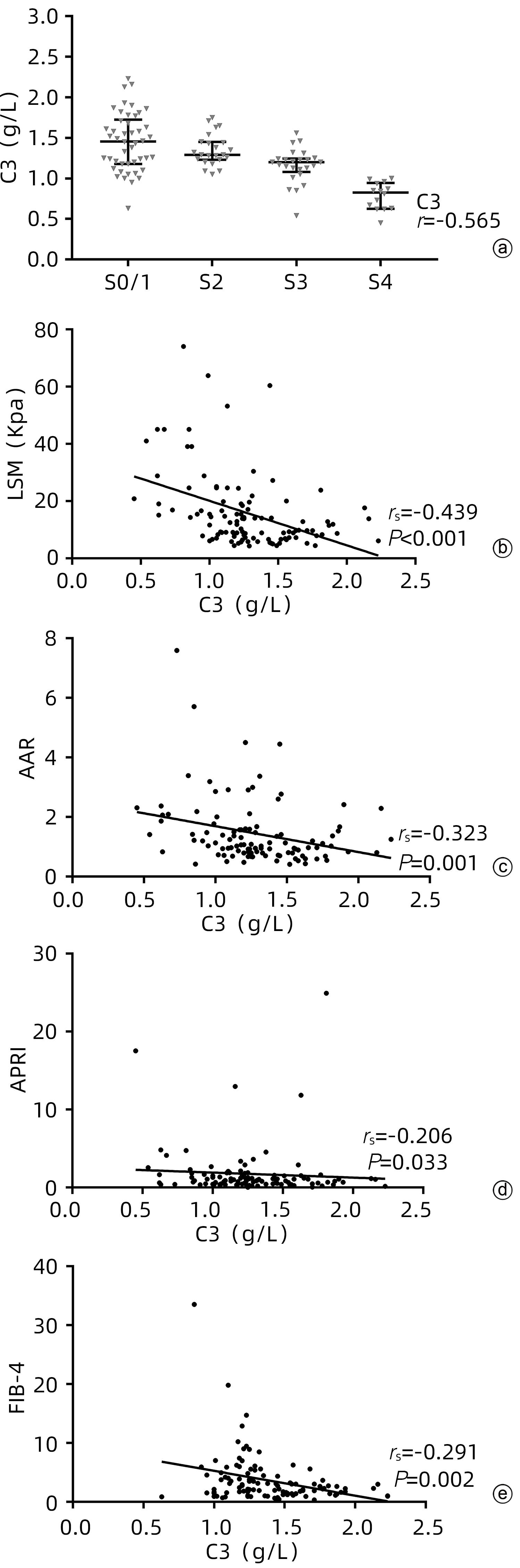
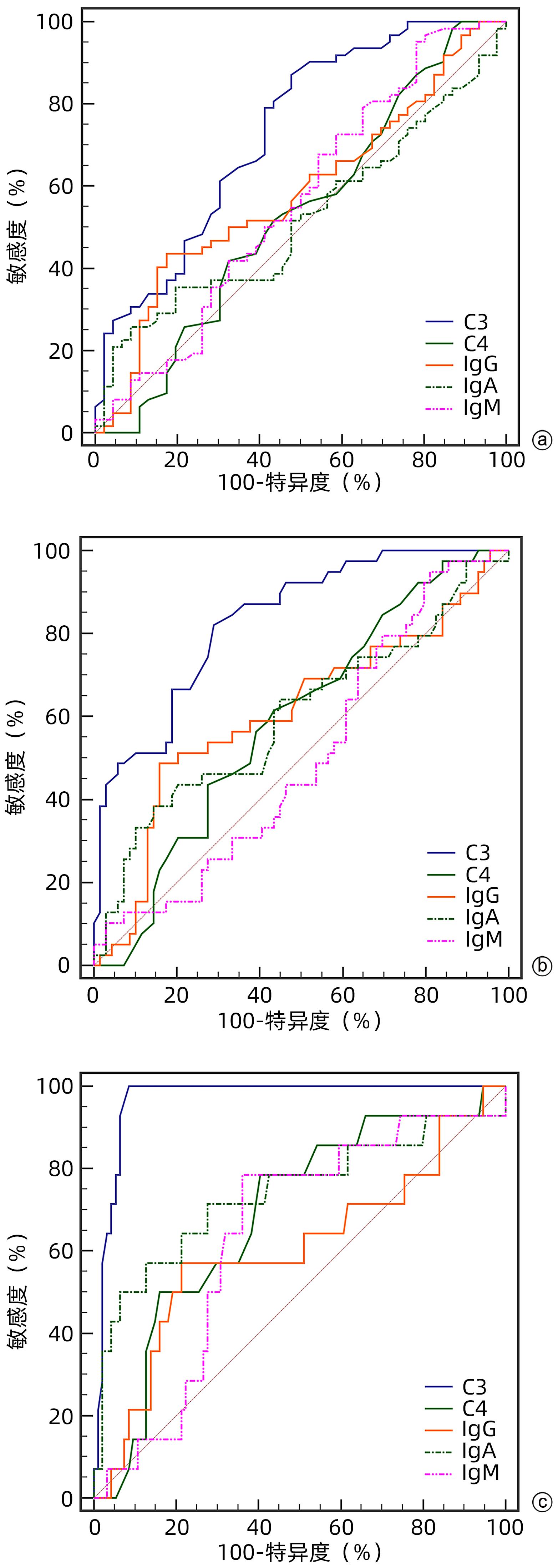
  •   Objective  To investigate the value of serum complement C3 level in determining the stage of liver fibrosis in primary biliary cholangitis (PBC).  Methods  Clinical data were collected from 108 patients with PBC who attended Tianjin Second People’s Hospital and underwent liver biopsy from January 2012 to October 2022. The degree of liver fibrosis (S0-4) was assessed according to the Scheuer scoring system, with ≥S2 defined as significant liver fibrosis, ≥S3 defined as progressive liver fibrosis, and S4 defined as liver cirrhosis. The independent samples t-test was used for comparison of normally distributed continuous data between two groups, and a one-way analysis of variance was used for comparison between multiple groups; the Mann-Whitney U test was used for comparison of non-normally distributed continuous data between two groups, and the Kruskal-Wallis H test was used for comparison between multiple groups; the chi-square test or the Fisher’s exact test was used for comparison of categorical data between groups. The area under the ROC curve (AUC) was used to evaluate the efficacy of complement C3 in the diagnosis of liver fibrosis in patients with PBC. The Spearman correlation analysis was used to investigate the correlation between complement C3 and liver fibrosis stage.  Results  Among the 108 patients with PBC, there were 87 (80.6%) female patients and 102 patients (94.4%) with positive autoantibody. As for the stage of liver fibrosis, there were 5 patients (4.6%) in S0 stage, 41 (38.0%) in S1 stage, 23 (21.3%) in S2 stage, 25 (23.1%) in S3 stage, and 14 (13.0%) in S4 stage. There was a significant difference in the level of complement C3 between the patients with different liver fibrosis stages (H=42.891, P<0.001). The level of complement C3 gradually decreased with the aggravation of liver fibrosis, with a negative correlation between them (r=-0.565, P<0.001). Liver stiffness measurement (LSM), aspartate aminotransferase/alanine aminotransferase ratio, aspartate aminotransferase-to-platelet ratio index, and fibrosis-4 were negatively correlated with complement C3, with a correlation coefficient of -0.439 (P<0.001), -0.323 (P=0.001), -0.206 (P=0.033), and -0.291 (P=0.002), respectively. The multivariate logistic regression analysis showed that complement C3 level was an independent predictive factor for significant liver fibrosis, progressive liver fibrosis, and liver cirrhosis, while LSM was an independent predictive factor for significant liver fibrosis and progressive liver fibrosis. The ROC curve analysis showed that complement C3 had an AUC of 0.731, 0.832, and 0.968, respectively, in the diagnosis of significant liver fibrosis, progressive liver fibrosis, and liver cirrhosis, with a corresponding cut-off value of 1.445, 1.235, and 1.005, respectively, and complement C3 combined with LSM had an AUC of 0.811, 0.941, and 0.976, respectively, in the diagnosis of significant liver fibrosis, progressive liver fibrosis, and liver cirrhosis. There was a significant difference in AUC between complement C3 combined with LSM and complement C3 alone in the diagnosis of significant liver fibrosis (Z=2.604, P=0.009), and there was also a significant difference in AUC between complement C3 combined with LSM and complement C3 alone in the diagnosis of progressive liver fibrosis (Z=3.033, P=0.002); there was no significant difference in AUC between complement C3 combined with LSM and complement C3 alone in the diagnosis of liver cirrhosis (Z=1.050, P=0.294), while There was a significant difference in AUC between complement C3 combined with LSM and LSM alone in the diagnosis of liver cirrhosis (Z=2.326, P=0.020).  Conclusion  Serum complement C3 level has a certain clinical value in assessing the degree of liver fibrosis in patients with PBC, and complement C3 combined with LSM can further improve the efficacy of complement C3 or LSM in the diagnosis of liver fibrosis in PBC.


  • 目前我国慢性HBV感染者约0.7亿人,其中慢性乙型肝炎(CHB)患者为0.2亿~0.3亿例[1]。自1998年第一个核苷(酸)类似物(NAs)抗HBV药物拉米夫定(LAM)在我国临床应用至今,先后有阿德福韦酯(ADV)、替比夫定(LdT)、恩替卡韦(ETV)、替诺福韦酯(TDF)和丙酚替诺福韦(TAF)陆续应用于临床。作为逆转录酶抑制剂,NAs可强效抑制HBV复制,目前临床接受抗病毒治疗的患者中约80%应用NAs治疗[2]。但由于单纯的NAs治疗难以获得持久的免疫学应答,HBsAg阴转率仅0~3%,停药后容易复发,需要长期甚至终身服药。因此如何进一步提高NAs经治患者的疗效并尽可能实现临床治愈是临床亟待解决的热点和难点问题[2-4]。本研究选取NAs经治HBeAg阴性CHB低水平HBsAg患者进行临床研究,探讨如何选取优势人群和应用更短疗程获得CHB临床治愈的方法。

    选取2017年10月—2019年10月在鹤壁市第三人民医院接受治疗的CHB患者。入组标准:诊断均符合《慢性乙型肝炎防治指南(2015年更新版)》[5]中的诊断标准。NAs治疗均48周以上,入组时HBeAg阴性,HBsAg精准定量检测<1500 IU/ml,HBV DNA<200 IU/ml。排除标准:重叠或混合甲、丙、丁、戊等其他类型病毒感染者;有肝硬化或其他严重器质性病变的患者;妊娠或哺乳期妇女;精神异常者等。

    根据患者意愿,分为联合治疗组(A组)和NAs单药治疗组(B组)。A组,在NAs治疗基础上联合聚乙二醇干扰素(PEG-IFN)α-2b(厦门特宝生物工程股份有限公司,国药准字S20160001) 进行治疗,用药方式为皮下注射,180 μg/次,1次/周,联合治疗48周。联合治疗期间获得HBsAg阴转、抗-HBs>100 IU/ml,满足联合治疗24周,可考虑停用联合治疗方案,并观察至48周,否则继续联合治疗至48周。B组,NAs单药治疗,观察48周。观察记录所有入组患者12、24、36、48周乙型肝炎五项精准定量结果、生化学指标和用药的不良反应。乙型肝炎五项精准定量检测采用美国雅培全自动化学发光分析仪微粒子酶免疫化学发光法;高灵敏度HBV DNA的检测使用COBASAmpliPrep/COBASTaqMan分析仪,检测下限:0~20 IU/ml。

    观察两组乙型肝炎五项血清学指标动态变化。主要指标:HBsAg阴转率和抗-HBs阳转率。判定标准:HBsAg<0.05 IU/ml视为阴性,抗-HBs>10 IU/ml视为HBsAg血清学转换。抗-HBs>100 IU/ml,视为稳定阈值[6]

    本研究获得鹤壁市第三人民医院伦理委员会批准,批号:论研批第2017-RS001。纳入患者均签署知情同意书。

    采用SPSS 17.0统计学软件进行数据分析。计量资料以x±s表示,两组间比较采用t检验;计数资料组间比较采用Fisher精确检验。影响因素相关分析采用logistic回归法。P<0.05为差异有统计学意义。

    选取接受治疗的CHB患者44例,其中男36例,女8例,年龄(47.51±7.21)岁。排除1例患者在应用NAs联合PEG-IFN治疗4周后癫痫发作而停药;另1例患者联合治疗6周后因白细胞及血小板过低而停药,共42例患者完成规定疗程进入本研究,其中A组22例,B组20例。A组中接受ETV治疗18例、LAM 1例、ADV 3例,疗程1~14(5.8±3.99)年。B组中接受LAM治疗1例、LAM+ADV 1例、TDF 1例、ETV 17例,疗程2~9(6.1±1.97)年。两组在年龄、性别、BMI、NAs治疗时间及HBsAg水平方面差异均无统计学意义(P值均>0.05)(表 1)。

    表  1  基线时两组患者的临床资料对比
    组别 例数 年龄
    (岁)
    男性
    [例(%)]
    BMI
    (kg/m2)
    NAs治疗时间
    (年)
    HBsAg
    (log10 IU/ml)
    总体 42 47.43±7.10 35(83.33) 25.20±3.01 5.91±3.17 2.12±0.93
    A组 22 44.82±6.20 19(86.36) 24.69±3.33 5.72±4.00 2.02±1.06
    B组 20 47.70±5.11 16(80.00) 25.39±2.66 6.10±1.97 2.23±0.78
    统计值 t=-1.634 t=-0.746 t=-0.388 t=-0.744
    P 0.110 0.691 0.460 0.701 0.462
    下载: 导出CSV 
    | 显示表格

    24周时A组的HBsAg清除率和HBsAg血清学转换率高于B组(P值均<0.05);48周时A组的HBsAg清除率达45.5%(10/22),HBsAg血清学转换率达到31.8%(7/22),均明显高于B组(P值均<0.01)(表 2)。

    表  2  两组不同时间节点临床治愈结果比较
    组别 例数 24周 48周
    HBsAg清除[例(%)] 抗-HBs阳性[例(%)] HBsAg清除[例(%)] 抗-HBs阳性[例(%)]
    A组 22 6(27.3) 5(22.7) 10(45.5) 7(31.8)
    B组 20 0 0 0 0
    P 0.022 0.049 0.001 0.009
    下载: 导出CSV 
    | 显示表格

    对可能影响HBsAg清除的因素进行多因素logsitic回归分析,结果显示:性别、BMI、ALT或AST升高以及NAs治疗时长均对HBsAg清除均无明显影响(P值均>0.05);既往应用不同抗病毒药物LAM/ADV或ETV/TDF均对HBsAg清除无明显影响(P值均>0.05)。开始联合治疗时的年龄对HBsAg清除存在影响[OR(95%CI)=0.877(0.781~0.985),P=0.026],HBsAg阴转患者的年龄集中在36~49(44.20±4.49)岁;基线HBsAg水平也对HBsAg清除存在影响[OR(95%CI)=0.996(0.992~1.000),P=0.050)。

    本研究以不同节点及不同区间的HBsAg水平来划分联合治疗患者以更好的观察HBsAg清除率。48周“节点水平”的HBsAg检测值虽可对HBsAg清除有效预测,但该方法包括了重叠人群。再以“区间水平”对人群进行分段观察,HBsAg水平500~1000 IU/ml、100~500 IU/ml、10~100 IU/ml、<10 IU/ml人群治疗48周时,HBsAg清除率分别为33.3%、50%、40%和100%(表 34)。

    表  3  联合治疗基线值不同HBsAg定量节点的患者在12、24和48周获得临床治愈率比较
    HBsAg节点 例数 12周治愈率 24周治愈率 48周治愈率
    <1000 IU/ml 19 26.3%(5/19) 31.6%(6/19) 52.6%(10/19)
    <500 IU/ml 13 38.5%(5/13) 46.2%(6/13) 61.5%(8/13)
    <100 IU/ml 9 55.6%(5/9) 55.6%(5/9) 66.7%(6/9)
    <10 IU/ml 4 100%(4/4) 100%(4/4) 100%(4/4)
    下载: 导出CSV 
    | 显示表格
    表  4  联合治疗基线值不同HBsAg定量区间的患者在12、24和48周获得临床治愈率比较
    HBsAg区间 例数 12周治愈率 24周治愈率 48周治愈率
    500~1000 IU/ml 6 0(0/6) 0(0/6) 33.3%(2/6)
    100~500 IU/ml 4 0(0/4) 25.0%(1/4) 50.0%(2/4)
    10~100 IU/ml 5 20.0%(1/5) 20.0%(1/5) 40.0%(2/5)
    <10 IU/ml 4 100%(4/4) 100%(4/4) 100%(4/4)
    下载: 导出CSV 
    | 显示表格

    基线HBsAg<10 IU/ml患者共4例,均于联合治疗12周取得临床治愈,巩固至24周,停药后抗-HBs持续升高,观察至48周均达到稳定阈值(抗-HBs>100 IU/ml),其中2例超过1000 IU/ml。

    在NAs联合PEG-IFN治疗期间,15例(15/24, 62.5%)于治疗早期出现发热,治疗过程中有10例(10/24, 41.7%)出现乏力症状,7例(7/24, 29.2%)出现食欲下降,4例(4/24, 16.7%)出现脱发现象。治疗过程中大多数患者都有WBC、PLT下降,给予对症治疗均能很快恢复正常。未见明显骨髓抑制、精神异常、自身免疫性疾病等严重不良反应。

    目前国内外有关乙型肝炎防治指南均较关注CHB的临床治愈问题,并强调在CHB的治疗和管理过程中,对于部分适合的患者,应尽可能追求临床治愈。针对HBeAg阴性CHB的抗病毒治疗, 采用NAs单药或IFN单药固定疗程(48周)的方法获得的HBsAg清除率极低[7-8];IFN能够增强HBV特异性细胞毒性T淋巴细胞的功能,又可以通过其受体经信号转导等生物化学过程启动多种抗病毒蛋白的合成,对HBV的复制和转录产生抑制,达到抗病毒及免疫调节的双重作用[9-10]。NAs主要是通过竞争性抑制HBVDNA多聚酶及反转录酶直接抑制病毒复制,并可直接增强PEG-IFN诱导的固有免疫激活效应[11]。联合两类不同作用机制的抗病毒药物治疗CHB可能存在协同作用,为快速实现临床治愈奠定理论基础[12]

    本研究选取NAs经治48周以上HBeAg阴性、HBsAg<1500 IU/ml的CHB患者,在NAs治疗基础上联合PEG-IFN治疗48周,与NAs单药组作对比,联合治疗组HBsAg清除率达到45.5%,HBsAg血清学转换率达到31.8%,NAs单药组无1例HBsAg清除,差异具有显著性。说明选取NAs经治的HBeAg阴性的CHB患者,给予积极联合PEG-IFN治疗,可以获得更高的临床治愈率。本研究治愈率较高与更多的患者HBsAg<1000 IU/ml有关(19/22),结果与宁琴主持OSST研究[13]和任红主持New Switch研究[14]一致。HBsAg清除的多因素logsitic回归结果显示,性别、BMI、ALT或AST升高、NAs治疗时长、既往应用不同抗病毒药物LAM/ADV或ETV/TDF均对HBsAg清除无明显影响(P值均>0.05)。开始联合治疗时的年龄<45岁,基线HBsAg水平<500 lU/ml临床治愈率高。提示开始联合治疗时的年龄越低、HBsAg水平越低,更容易获得HBsAg清除。原因可能是中青年人群,新陈代谢机能旺盛,免疫功能良好,对PEG-IFN应答率高;在长期使用NAs治疗,对HBV DNA控制良好情况下,T淋巴细胞和NK细胞的特异性免疫应答功能得以恢复,也能够提高患者对PEG-IFN的应答率[15-16]。很多研究已表明,基线HBsAg水平与HBsAg清除密切相关,基线水平越低,HBsAg清除率越高[10]

    本研究HBsAg<500 IU/ml水平的患者获得约61.5%的临床治愈率,HBsAg在500~1000 IU/ml、100~500 IU/ml、10~100 IU/ml及<10 IU/ml人群的HBsAg清除率分别为33.3%、50.0%、40.0%和100%。陈新月研究团队[17]对HBsAg低水平的HBeAg阴性CHB获得临床治愈优势人群的研究,采用“区间水平”划分人群,联合治疗48周100 IU/ml<HBsAg<1000 IU/ml、10 IU/ml<HBsAg<100 IU/ml以及HBsAg<10 IU/ml人群的HBsAg清除率分别为6.7%、31.8%和67.7%。本研究结果与其相似,但本研究中HBsAg越低越容易获得临床治愈,更高的HBsAg清除率是在HBsAg<10 IU/ml人群,且获得HBsAg血清学转换。说明基线HBsAg水平越低,获得临床治愈的联合治疗时间越短,与既往NAs用药时间长短无关。

    本研究观察到联合治疗组在应用IFN早期会出现发热现象,部分患者治疗过程中出现乏力、食欲下降、脱发等临床表现,大多数患者有WBC、PLT水平下降情况,给予对症治疗均能很快恢复正常。未见明显骨髓抑制、精神异常、自身免疫性疾病等严重不良反应。联合用药的患者对于IFN的相关不良反应大多能够承受,为赢得临床治愈创造了机会。尽管本研究样本量较少,但优化出的无论哪种核苷类似物,基线HBsAg低水平(<500 IU/ml)时加用PEG-IFN联合治疗24~48周,能够获得约61.5%的临床治愈率对基层临床医生具有重要的指导意义。

  • [1]
    COLAPIETRO F, BERTAZZONI A, LLEO A. Contemporary epidemiology of primary biliary cholangitis[J]. Clin Liver Dis, 2022, 26( 4): 555- 570. DOI: 10.1016/j.cld.2022.06.001.
    [2]
    Chinese Society of Hepatology, Chinese Society of Gastroenterology, Chinese Society of Infectious Diseases. Consensus on the diagnosis and treatment of primary biliary cirrhosis(also known as primary biliary cholangitis)(2015)[J]. J Clin Hepatol, 2015, 31( 12): 1980- 1988. DOI: 10.3969/j.issn.1001-5256.2015.12.004.

    中华医学会肝病学分会, 中华医学会消化病学分会, 中华医学会感染病学分会. 原发性胆汁性肝硬化(又名原发性胆汁性胆管炎)诊断和治疗共识(2015)[J]. 临床肝胆病杂志, 2015, 31( 12): 1980- 1988. DOI: 10.3969/j.issn.1001-5256.2015.12.004.
    [3]
    Chinese Medical Association, Branch Hepatology. Guidelines for the diagnosis and treatment of primary biliary cholangitis(2021)[J]. J Clin Hepatol, 2022, 38( 1): 35- 41. DOI: 10.3969/j.issn.1001-5256.2022.01.007.

    中华医学会肝病学分会. 原发性胆汁性胆管炎的诊断和治疗指南(2021)[J]. 临床肝胆病杂志, 2022, 38( 1): 35- 41. DOI: 10.3969/j.issn.1001-5256.2022.01.007.
    [4]
    Chinese Society of Hepatology, Chinese Society of Gastroenterology, Chinese Society of Infectious Diseases. Consensus on the diagnosis and treatment of liver fibrosis(2019)[J]. J Clin Hepatol, 2019, 35( 10): 2163- 2172. DOI: 10.3969/j.issn.1001-5256.2019.10.007.

    中华医学会肝病学分会, 中华医学会消化病学分会, 中华医学会感染病学分会. 肝纤维化诊断及治疗共识(2019年)[J]. 临床肝胆病杂志, 2019, 35( 10): 2163- 2172. DOI: 10.3969/j.issn.1001-5256.2019.10.007.
    [5]
    TRIVEDI PJ, HIRSCHFIELD GM. Recent advances in clinical practice: Epidemiology of autoimmune liver diseases[J]. Gut, 2021, 70( 10): 1989- 2003. DOI: 10.1136/gutjnl-2020-322362.
    [6]
    HOHENESTER S, OUDE-ELFERINK RPJ, BEUERS U. Primary biliary cirrhosis[J]. Semin Immunopathol, 2009, 31( 3): 283- 307. DOI: 10.1007/s00281-009-0164-5.
    [7]
    YANG YL, ZHAO RC, ZHANG FC. Potential mesenchymal stem cell therapeutics for treating primary biliary cholangitis: Advances, challenges, and perspectives[J]. Front Cell Dev Biol, 2022, 10: 933565. DOI: 10.3389/fcell.2022.933565.
    [8]
    BUGDACI MS, ALKIM C, KARACA C, et al. Could complement C4 be an alternative to biopsy for chronic hepatitis B histopathologic findings?[J]. J Clin Gastroenterol, 2011, 45( 5): 449- 455. DOI: 10.1097/MCG.0b013e31820f7ee5.
    [9]
    LI Q, LU Q, ZHU MQ, et al. Lower level of complement component C3 and C3a in the plasma means poor outcome in the patients with hepatitis B virus related acute-on-chronic liver failure[J]. BMC Gastroenterol, 2020, 20( 1): 106. DOI: 10.1186/s12876-020-01258-3.
    [10]
    CHEN C, YUAN Z, LI WX, et al. Complement C3 facilitates stratification of stages of chronic hepatitis B and signifies development of acute-on-chronic liver failure in acute decompensated cirrhosis[J]. Adv Ther, 2023, 40( 3): 1171- 1186. DOI: 10.1007/s12325-022-02416-7.
    [11]
    RICKLIN D, HAJISHENGALLIS G, YANG K, et al. Complement: A key system for immune surveillance and homeostasis[J]. Nat Immunol, 2010, 11( 9): 785- 797. DOI: 10.1038/ni.1923.
    [12]
    TROUW LA, DAHA MR. Role of complement in innate immunity and host defense[J]. Immunol Lett, 2011, 138( 1): 35- 37. DOI: 10.1016/j.imlet.2011.02.014.
    [13]
    SAHU A, LAMBRIS JD. Structure and biology of complement protein C3, a connecting link between innate and acquired immunity[J]. Immunol Rev, 2001, 180: 35- 48. DOI: 10.1034/j.1600-065x.2001.1800103.x.
    [14]
    LUBBERS R, van ESSEN MF, van KOOTEN C, et al. Production of complement components by cells of the immune system[J]. Clin Exp Immunol, 2017, 188( 2): 183- 194. DOI: 10.1111/cei.12952.
    [15]
    SELMI C, BOWLUS CL, GERSHWIN ME, et al. Primary biliary cirrhosis[J]. Lancet, 2011, 377( 9777): 1600- 1609. DOI: 10.1016/S0140-6736(10)61965-4.
    [16]
    SCHLESINGER M, BENBASSAT C, SHOENFELD Y. Complement profile in primary biliary cirrhosis[J]. Immunol Res, 1992, 11( 2): 98- 103. DOI: 10.1007/BF02918614.
    [17]
    BARAK V, SELMI C, SCHLESINGER M, et al. Serum inflammatory cytokines, complement components, and soluble interleukin 2 receptor in primary biliary cirrhosis[J]. J Autoimmun, 2009, 33( 3-4): 178- 182. DOI: 10.1016/j.jaut.2009.09.010.
    [18]
    BIEWENGA M, FARINA SARASQUETA A, TUSHUIZEN ME, et al. The role of complement activation in autoimmune liver disease[J]. Autoimmun Rev, 2020, 19( 6): 102534. DOI: 10.1016/j.autrev.2020.102534.
    [19]
    HOMANN C, VARMING K, HØGÅSEN K, et al. Acquired C3 deficiency in patients with alcoholic cirrhosis predisposes to infection and increased mortality[J]. Gut, 1997, 40( 4): 544- 549. DOI: 10.1136/gut.40.4.544.
    [20]
    GARRED P, LYON H, CHRISTOFFERSEN P, et al. Deposition of C3, the terminal complement complex and vitronectin in primary biliary cirrhosis and primary sclerosing cholangitis[J]. Liver, 1993, 13( 6): 305- 310. DOI: 10.1111/j.1600-0676.1993.tb00650.x.
    [21]
    YUAN H, LI YW, LI J, et al. Clinical feature of patients with primary biliary cholangitis and primary biliary cirrhosis[J]. J Pract Hepatol, 2022, 25( 3): 403- 406. DOI: 10.3969/j.issn.1672-5069.2022.03.025.

    袁慧, 李毓雯, 李军, 等. 原发性胆汁性胆管炎与肝硬化患者临床指标差异分析[J]. 实用肝脏病杂志, 2022, 25( 3): 403- 406. DOI: 10.3969/j.issn.1672-5069.2022.03.025.
  • Relative Articles

    [1]Pinsheng HAN, Long YANG, Tao CUI, Yamin ZHANG. Role of tumor-associated macrophages in cholangiocarcinoma[J]. Journal of Clinical Hepatology, 2023, 39(2): 469-473. doi: 10.3969/j.issn.1001-5256.2023.02.035
    [2]Chaoran YANG, Sirou LI, Yuan LIU, Zhiyuan HOU, Yuan WANG, Jihong YANG. Role of non-coding RNA on immune cells in tumor microenvironment of hepatocellular carcinoma[J]. Journal of Clinical Hepatology, 2023, 39(4): 961-967. doi: 10.3969/j.issn.1001-5256.2023.04.033
    [3]Yuhan LI, Beichen GUO, Feng ZHOU, Tao HAN. Research advances in long non-coding small nucleolar RNA host gene in hepatocellular carcinoma[J]. Journal of Clinical Hepatology, 2023, 39(8): 1977-1982. doi: 10.3969/j.issn.1001-5256.2023.08.032
    [4]Weiyu CHEN, Xiaobin QIN, Yingyu LE, Han WANG, Xiaorong LONG, Dewen MAO. Mechanism of action of macrophage polarization in non-neoplastic liver diseases and related targeted therapies[J]. Journal of Clinical Hepatology, 2022, 38(11): 2649-2653. doi: 10.3969/j.issn.1001-5256.2022.11.042
    [5]Baolin XU, Yu CHENG, Yong WEI. The role of competitive endogenous RNA in the development of cholangiocarcinoma[J]. Journal of Clinical Hepatology, 2022, 38(11): 2659-2662. doi: 10.3969/j.issn.1001-5256.2022.11.044
    [6]Yu LIANG, Yuanhong ZHAO, Zheng LI. Recent advancement in research on intrahepatic cholangiocarcinoma: Risk factors and pathogenesis[J]. Journal of Clinical Hepatology, 2022, 38(10): 2391-2395. doi: 10.3969/j.issn.1001-5256.2022.10.037
    [7]Mingyu YANG, Zhen YANG, Wanhua REN. Mechanism of action of ferroptosis in cholangiocarcinoma[J]. Journal of Clinical Hepatology, 2022, 38(4): 951-955. doi: 10.3969/j.issn.1001-5256.2022.04.043
    [8]Ziyue WANG, Daqun LIU, Jiaao YU, Jingxuan ZHANG, Guoyue LYU. A case of huge intrahepatic biliary papillomatosis[J]. Journal of Clinical Hepatology, 2021, 37(11): 2651-2652. doi: 10.3969/j.issn.1001-5256.2021.11.034
    [9]Sheng CHEN, Guoxiang CHEN, Zhongming HE, Shujie CHENG, Jisen ZHAO. Role of miRNA in the development and progression of cholangiocarcinoma[J]. Journal of Clinical Hepatology, 2021, 37(9): 2241-2245. doi: 10.3969/j.issn.1001-5256.2021.09.048
    [10]Wang ShiDong, Wang XiaoWei, Liu GuoHong, Tian ZiBin, Zhao QingXi. Research advances in intraductal papillary neoplasm of the bile duct[J]. Journal of Clinical Hepatology, 2020, 36(2): 476-480. doi: 10.3969/j.issn.1001-5256.2020.02.054
    [11]NIU XingJie, LIU ZhiHui, CUI FengMei, LIU YaoMin, WANG YanFei, ZHANG GuoMin, LIU JinXia. Value of peripheral blood long non-coding RNA-LET in the diagnosis of chronic hepatitis B cirrhosis[J]. Journal of Clinical Hepatology, 2020, 36(12): 2709-2713. doi: 10.3969/j.issn.1001-5256.2020.12.014
    [12]Zhang LiYun, Deng JiaQi, Zhang Shuang, Deng HuaJiang, Li XiaoHong. Expression and significance of the long non-coding RNA EXOC7 in nonalcoholic fatty liver disease[J]. Journal of Clinical Hepatology, 2020, 36(2): 391-394. doi: 10.3969/j.issn.1001-5256.2020.02.032
    [13]LU NenYuan, ZHONG FangFei, DENG QingMei, YANG YanQi, ZHENG Yang, ZHAO TieJian, LIANG TianJian. Regulatory effect of long non-coding RNAs on liver fibrosis[J]. Journal of Clinical Hepatology, 2020, 36(12): 2811-2814. doi: 10.3969/j.issn.1001-5256.2020.12.035
    [14]Tan ZhengRong, Feng DaLi, Zhang Qing. Expression of intergenic long non-coding RNA RP11-488L18.10 in cholangiocarcinoma and its association with prognosis[J]. Journal of Clinical Hepatology, 2020, 36(5): 1087-1090. doi: 10.3969/j.issn.1001-5256.2020.05.027
    [15]Wang Jing, Zhang BiQing, Pan YunZhi, Zhu ChuanWu. Role of microRNA in the pathogenesis, diagnosis, and outcome evaluation of primary biliary cholangitis[J]. Journal of Clinical Hepatology, 2019, 35(7): 1636-1639. doi: 10.3969/j.issn.1001-5256.2019.07.047
    [16]Duan YiFei, Zhao QingXi, Jing Xue. Effect of long non-coding RNAs on glycolytic pathway in primary liver cancer and related mechanisms[J]. Journal of Clinical Hepatology, 2019, 35(6): 1374-1376. doi: 10.3969/j.issn.1001-5256.2019.06.043
    [17]Cha YuFeng, He XiaoHu, Tian Mian, Gao Jun, Wang Gang. Value of preoperative and postoperative carbohydrate antigen 19-9 levels in predicting prognosis and early recurrence in patients with hilar cholangiocarcinoma[J]. Journal of Clinical Hepatology, 2018, 34(5): 1046-1051. doi: 10.3969/j.issn.1001-5256.2018.05.024
    [18]Chen Jie. Pathological and differential diagnoses of exocrine pancreatic tumors[J]. Journal of Clinical Hepatology, 2013, 29(1): 45-49.
    [19]Zhu Zhen, Cong WenMing. Clinicopathological study of intrahepatic bile duct cancers from 1982 to 2011[J]. Journal of Clinical Hepatology, 2013, 29(1): 42-44.
    [20]Zhang Wei, Han XinWei, Wu Gang, Li Zhen, Ding PengXu. Analysis of the clinical symptom and its relationship with the clinical classification of hilar cholangiocarcinoma[J]. Journal of Clinical Hepatology, 2011, 27(4): 405-407.
  • Cited by

    Periodical cited type(2)

    1. 刘华梅,孙家敏. 核苷(酸)类似物联合Peg-IFNα-2b治疗HBsAg低表达型慢性乙型肝炎临床效果. 中国现代医药杂志. 2024(05): 57-59 .
    2. 李瑾,董京涛,董振博,张云,李志媛,王洁莹. 聚乙二醇干扰素α-2a联合恩替卡韦对慢性乙型肝炎患者相关指标的影响. 实用临床医药杂志. 2023(02): 92-96 .

    Other cited types(2)

  • 加载中

Catalog

    通讯作者: 陈斌, bchen63@163.com
    • 1. 

      沈阳化工大学材料科学与工程学院 沈阳 110142

    1. 本站搜索
    2. 百度学术搜索
    3. 万方数据库搜索
    4. CNKI搜索

    Figures(2)  / Tables(6)

    Article Metrics

    Article views (434) PDF downloads(29) Cited by(4)
    Proportional views
    Related

    /

    DownLoad:  Full-Size Img  PowerPoint
    Return
    Return