| [1] |
QUEK J, CHAN KE, WONG ZY, et al. Global prevalence of non-alcoholic fatty liver disease and non-alcoholic steatohepatitis in the overweight and obese population: A systematic review and meta-analysis[J]. Lancet Gastroenterol Hepatol, 2023, 8( 1): 20- 30. DOI: 10.1016/S2468-1253(22)00317-X. |
| [2] |
WONG VWS, EKSTEDT M, WONG GLH, et al. Changing epidemiology, global trends and implications for outcomes of NAFLD[J]. J Hepatol, 2023, 79( 3): 842- 852. DOI: 10.1016/j.jhep.2023.04.036. |
| [3] |
CHEN JH, ZHOU ZH. Research progress of abnormal lipid metabolism and mitochondrial dysfunction in the pathogenesis and disease process of NAFLD[J]. Chin Hepatol, 2023, 28( 11): 1372- 1375. DOI: 10.3969/j.issn.1008-1704.2023.11.027. |
| [4] |
REN QN, SUN QM, FU JF. Dysfunction of autophagy in high-fat diet-induced non-alcoholic fatty liver disease[J]. Autophagy, 2024, 20( 2): 221- 241. DOI: 10.1080/15548627.2023.2254191. |
| [5] |
XIN YJ, CHEN YY, YANG HL, et al. Effect of Xuanfuhua Decoction on a mouse model of nonalcoholic steatohepatitis induced by high-fat, high-fructose, and high-cholesterol diet[J]. J Clin Hepatol, 2023, 39( 6): 1340- 1350. DOI: 10.3969/j.issn.1001-5256.2023.06.014. |
| [6] |
ZHANG QY, ZHOU ZH. Mechanism of non-alcoholic steatohepatitis improved by Qizhu prescription based on AMPK signaling pathway[J]. Chin J Exp Tradit Med Formulae, 2024, 30( 8): 49- 56. DOI: 10.13422/j.cnki.syfjx.20232237. |
| [7] |
FAKHOURY-SAYEGH N, TRAK-SMAYRA V, SAYEGH R, et al. Fructose threshold for inducing organ damage in a rat model of nonalcoholic fatty liver disease[J]. Nutr Res, 2019, 62: 101- 112. DOI: 10.1016/j.nutres.2018.11.003. |
| [8] |
CHEN Z, TIAN RF, SHE ZG, et al. Role of oxidative stress in the pathogenesis of nonalcoholic fatty liver disease[J]. Free Radic Biol Med, 2020, 152: 116- 141. DOI: 10.1016/j.freeradbiomed.2020.02.025. |
| [9] |
SCHUSTER S, CABRERA D, ARRESE M, et al. Triggering and resolution of inflammation in NASH[J]. Nat Rev Gastroenterol Hepatol, 2018, 15( 6): 349- 364. DOI: 10.1038/s41575-018-0009-6. |
| [10] |
ZHANG DW, ZHAO HY, LI QS, et al. Effect of AstragalosideIV and ginsenoside Rg1 on autophagy of myocardial tissue injury induced by ischemia-reperfusion injury in hyperlipidemic mice[J]. Chin Arch Tradit Chin Med, 2020, 38( 3): 60- 64. DOI: 10.13193/j.issn.1673-7717.2020.03.016. |
| [11] |
SU J, GAO CT, XIE L, et al. Astragaloside II ameliorated podocyte injury and mitochondrial dysfunction in streptozotocin-induced diabetic rats[J]. Front Pharmacol, 2021, 12: 638422. DOI: 10.3389/fphar.2021.638422. |
| [12] |
LI SD, CHEN XJ, LIU JK, et al. Signaling pathways involved in the active components of Polygonum cuspidatum in treatment of nonalcoholic fatty liver disease and their interaction[J]. J Clin Hepatol, 2022, 38( 4): 902- 907. DOI: 10.3969/j.issn.1001-5256.2022.04.033. |
| [13] |
WANG Y, YANG ZY, LI SQ, et al. Regulatory mechanism of Polygonidine on endoplasmic reticulum stress in non⁃alcoholic fatty hepatocytes[J]. Anatomy Research, 2023, 45( 1): 57- 62. DOI: 10.20021/j.cnki.1671-0770.2023.01.10. |
| [14] |
ZHANG PF, CHENG XY, SUN HM, et al. Atractyloside protect mice against liver steatosis by activation of autophagy via ANT-AMPK-mTORC1 signaling pathway[J]. Front Pharmacol, 2021, 12: 736655. DOI: 10.3389/fphar.2021.736655. |
| [15] |
UENO T, KOMATSU M. Autophagy in the liver: Functions in health and disease[J]. Nat Rev Gastroenterol Hepatol, 2017, 14( 3): 170- 184. DOI: 10.1038/nrgastro.2016.185. |
| [16] |
MA XW, MCKEEN T, ZHANG JH, et al. Role and mechanisms of mitophagy in liver diseases[J]. Cells, 2020, 9( 4): 837. DOI: 10.3390/cells9040837. |
| [17] |
LU YY, LI ZJ, ZHANG SQ, et al. Cellular mitophagy: Mechanism, roles in diseases and small molecule pharmacological regulation[J]. Theranostics, 2023, 13( 2): 736- 766. DOI: 10.7150/thno.79876. |
| [18] |
LI L, JIA QL, WANG XX, et al. Chaihu Shugan San promotes gastric motility in rats with functional dyspepsia by regulating Drp-1-mediated ICC mitophagy[J]. Pharm Biol, 2023, 61( 1): 249- 258. DOI: 10.1080/13880209.2023.2166966. |
| [19] |
LIU T, YU JN, LIU Y, et al. Effects of serum-free starvation on proliferative capacity of muscle satellite cells and expression of autophagy-related proteins LC3 and Beclin1[J]. Chin J Tissue Eng Res, 2019, 23( 11): 1657- 1661. DOI: 10.3969/j.issn.2095-4344.1062. |
| [20] |
SUI XY, XU P, DUAN CZ, et al. Advances in molecular function of p62 protein and its role in diseases[J]. Chin J Biotechnol, 2023, 39( 4): 1374- 1389. DOI: 10.13345/j.cjb.220681. |
| [21] |
YAO RQ, REN C, XIA ZF, et al. Organelle-specific autophagy in inflammatory diseases: A potential therapeutic target underlying the quality control of multiple organelles[J]. Autophagy, 2021, 17( 2): 385- 401. DOI: 10.1080/15548627.2020.1725377. |


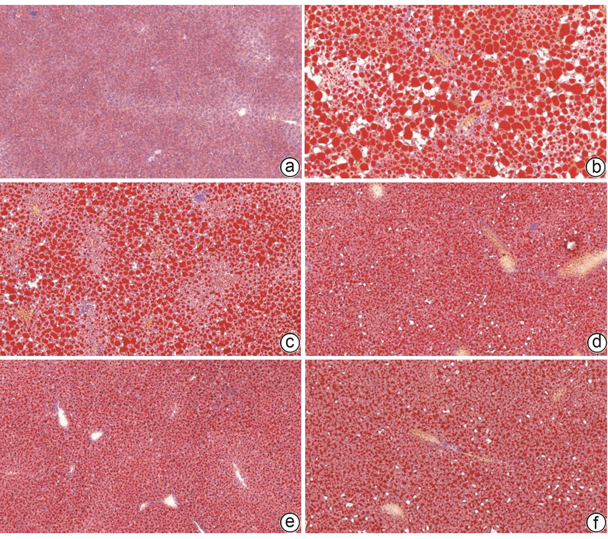
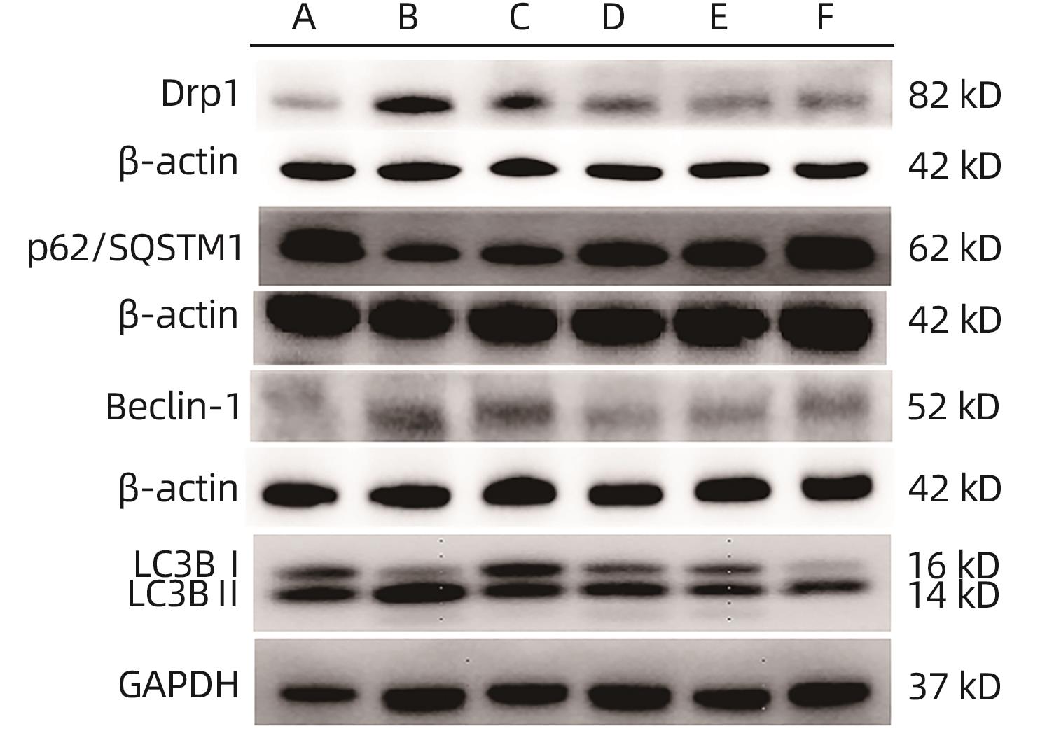
 DownLoad:
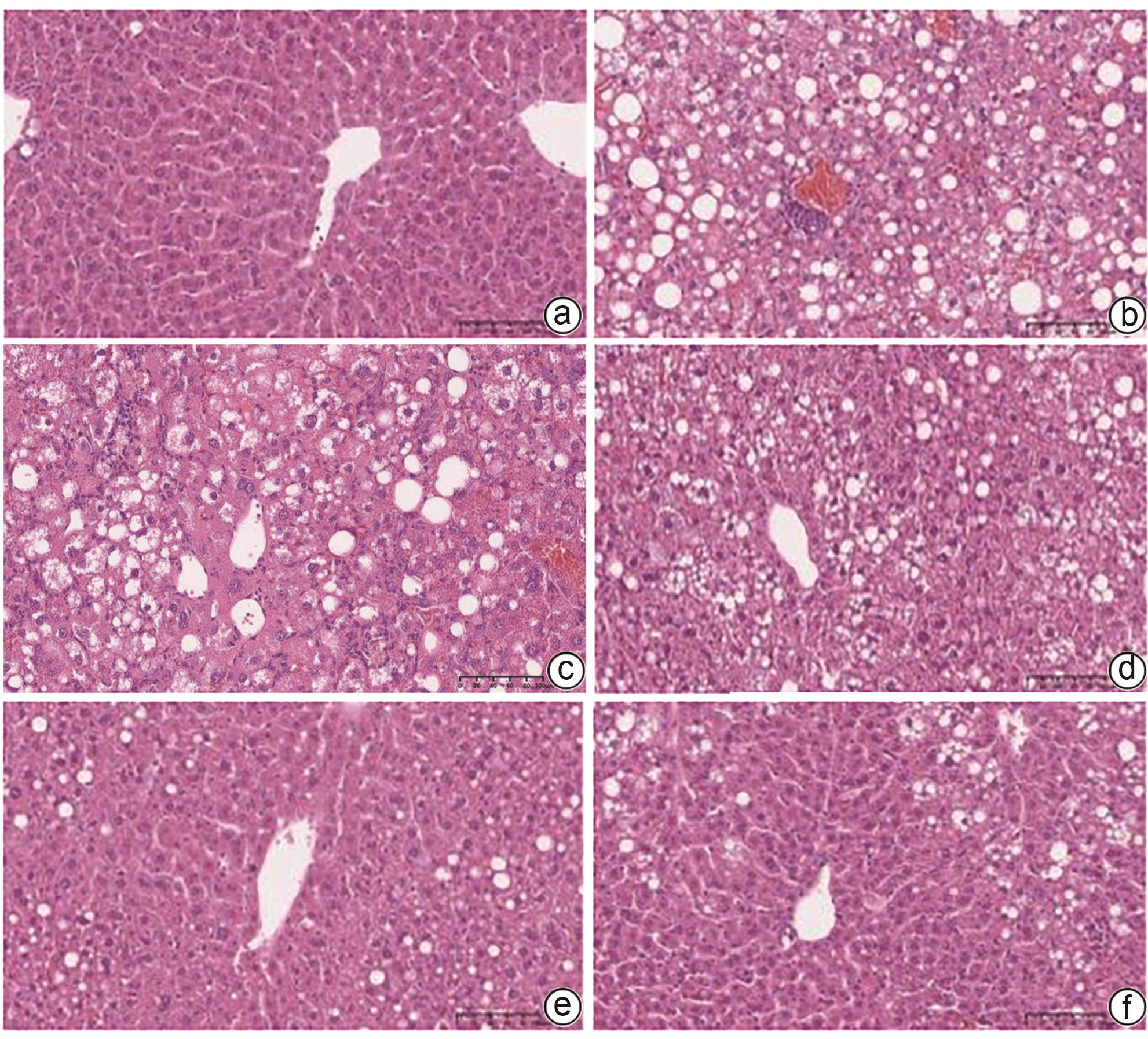