中文English
ISSN 1001-5256 (Print)
ISSN 2097-3497 (Online)
CN 22-1108/R

Prognosis and adverse reactions of patients with acute-on-chronic liver failure receiving artificial liver support therapy stratified by international normalized ratio

DOI: 10.3969/j.issn.1001-5256.2022.10.020
Research funding:

Science and Technology Project of Health Commission of Jiangxi Province in 2021 (202140131)

More Information
  • Corresponding author: XIONG Molong, xml86005@163.com(ORCID: 0000-0003-4701-6002)
  • Received Date: 2022-02-15
  • Accepted Date: 2022-03-20
  • Published Date: 2022-10-20
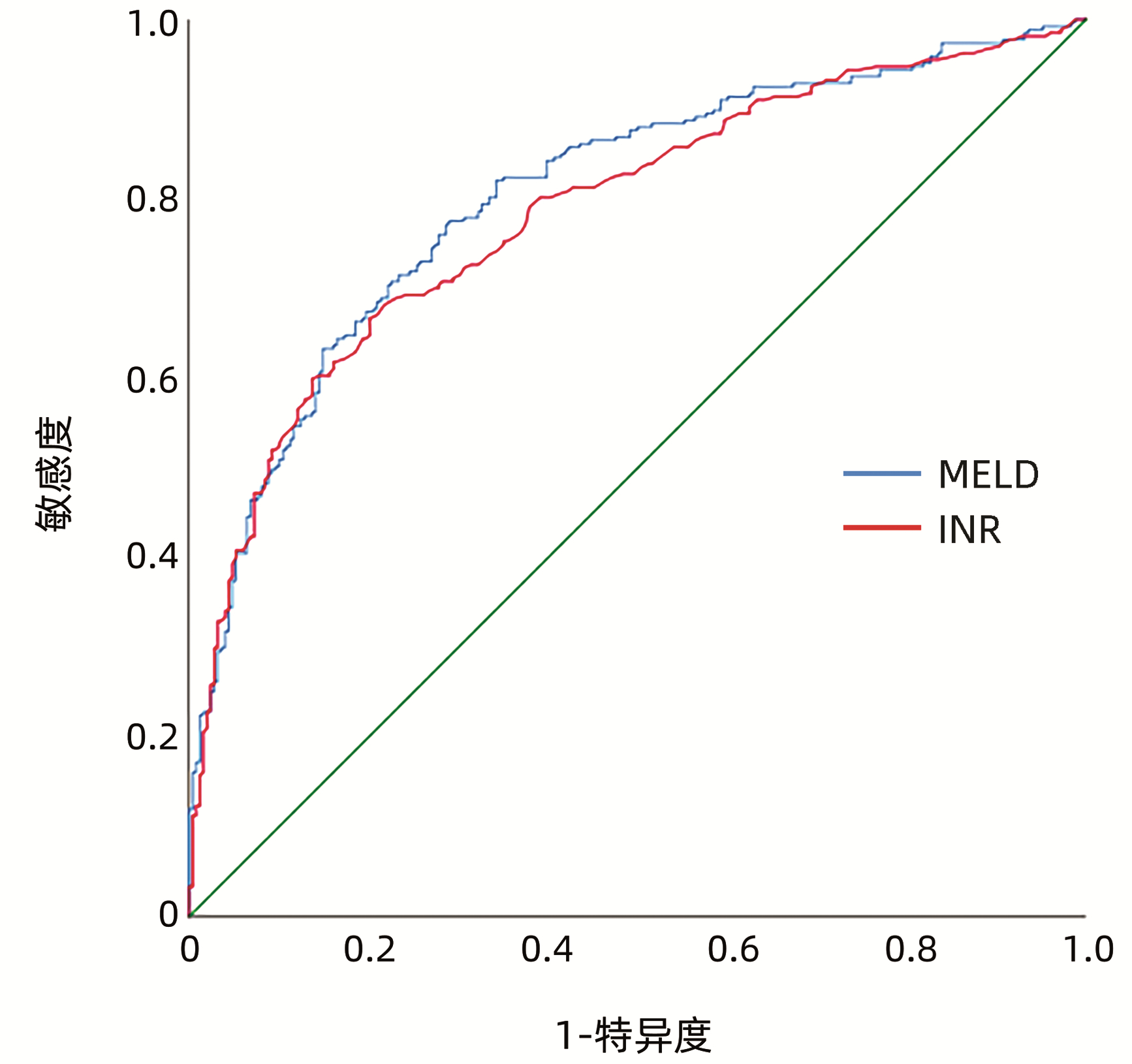
  •   Objective  To investigate the prognosis and adverse reactions of patients with acute-on-chronic liver failure (ACLF) receiving artificial liver support therapy stratified by international normalized ratio (INR).  Methods  A total of 515 ACLF patients who received artificial liver support therapy in Department of Severe liver Disease, The Ninth Hospital of Nanchang, from January 2010 to May 2020 were enrolled, and according to the level of INR, they were divided into group A with 20 patients (INR < 1.5), group B with 115 patients (1.5≤INR < 1.9), group C with 179 patients (1.9≤INR < 2.6), group D with 61 patients (2.6≤INR < 3.2), group E with 75 patients (3.2≤INR < 4.2), and group F with 65 patients (INR≥4.2). All patients received multimodality medical treatment combined with artificial liver support therapy. The one-way analysis of variance was used for comparison of normally distributed continuous data between multiple groups; the Kruskal-Wallis H test was used for comparison of non-normally distributed continuous data between multiple groups。The chi-square test was used for comparison of categorical data between groups. Bonferroni correction was used for further comparison between two groups. and the receiver operating characteristic (ROC) curve was used to evaluate the value of INR and MELD scoring system in predicting the prognosis of ACLF patients.  Results  As for 90-day mortality rate, there was a significant difference between the six groups stratified by INR (χ2=124.84, P < 0.001); there was no significant difference between groups A(25%), B(25.2%), and C(39.7%) (P > 0.05), and there was a significant difference between groups D/E/F(65.6%, 82.7%, and 92.3%, respectively) and groups A/B/C (all P < 0.05); there was no significant difference between groups D and E and between groups E and F (P > 0.05), and there was a significant difference between groups D and F (P < 0.05). There was no significant difference in the incidence rate of intraoperative adverse reactions between the six groups (χ2=8.956, P=0.111). INR had an area under the ROC curve of 0.786 (95% confidence interval: 0.746-0.825, P < 0.001) in predicting the prognosis of patients with ACLF receiving artificial liver support therapy, with a sensitivity of 66.7% and a specificity of 79.8%.  Conclusion  INR has a good value in predicting the prognosis of ACLF patients receiving artificial liver support therapy, and the artificial liver has good safety.


  • 慢性乙型肝炎(CHB)相关的慢加急性肝衰竭(acute-on-chronic hepatitis B liver failure,ACHBLF)是在慢性HBV感染引起的CHB基础上出现的急性严重肝功能障碍临床综合征,病死率极高。因我国慢性HBV的高感染率,ACHBLF已成为影响患者生存质量的重要因素[1]。在CHB向ACHBLF进展过程中,存在着患者肝功能急剧恶化,但尚未达到肝衰竭的“肝衰竭前期(pre-ACHBLF)”阶段[2],如能在此阶段进行预警及干预,则有可能预防进一步发展为肝衰竭。

    目前普遍认为细胞免疫功能紊乱是ACHBLF发生的病理机制之一,许多免疫细胞如髓系抑制性细胞(myeloid-derived suppressor cells, MDSC)、调节性T淋巴细胞(Treg)、分泌IL-17的CD4 T淋巴细胞(IL-17-producing CD4 T cells,Th17)和细胞毒性T淋巴细胞等在肝衰竭的发病中发挥重要作用[3-5]。尽管既往许多研究已证实肝衰竭发病与免疫密切相关,但pre-ACHBLF阶段的免疫状态及其与疾病进展的关系尚不清楚,因此本研究探讨了MDSC、Th17、Treg和分泌IL-17的CD8 T淋巴细胞(IL-17-producing CD8 T cells, Tc17)在pre-ACHBLF和ACHBLF患者中的表达,以期为ACHBLF的早期治疗提供思路。

    选取2018年8月—2019年5月于石家庄市第五医院住院患者45例,其中ACHBLF患者、pre-ACHBLF患者和CHB患者各15例,同时选取于本院健康体检者15例作为健康对照组(HC组)。ACHBLF和pre-ACHBLF的临床诊断均符合《肝衰竭诊治指南(2018年版)》[6];CHB的临床诊断符合《慢性乙型肝炎防治指南(2015年版)》[7]。排除标准为合并其他病毒性肝炎或由酒精、药物等原因导致的肝脏疾病;肝癌或其他恶性肿瘤伴有肝脏转移;患有自身免疫性疾病、HIV感染、妊娠或有严重的心、肺、肾脏、神经系统等疾病。

    采用EDTA-K2抗凝的真空采血管于清晨空腹采集外周静脉血约3 mL,取100 μL用于流式细胞仪检测外周血MDSC频数,剩余全血分离外周血单个核细胞(peripheral blood mononuclear cells, PBMC)。所有标本均为新鲜抗凝血,且在采集后4 h内处理。

    MDSC细胞检测:取100 μL全血加至流式管中,然后分别加入CD11b-APC、CD33-PE和HLA-DR-PE/Cy7流式抗体各2 μL,避光孵育15 min后,每管加入溶血素500 μL,室温避光静置15 min,离心洗涤,加入流式鞘液300 μL重悬细胞,流式细胞仪检测MDSC细胞比例。

    Treg细胞检测:收集PBMC,加入CD4-FITC和CD25-APC抗体各2 μL,4 ℃避光孵育30 min。经固定破膜处理后,离心洗涤2次,重悬细胞,然后加入10 μL Foxp3-PE抗体,4 ℃避光孵育45 min,离心洗涤,加入流式鞘液200 μL重悬细胞,流式细胞仪检测。

    Th17、Tc17细胞检测:收集PBMC,用佛波醇酯和离子霉素刺激,同时加入GolgiStop,4 h后收集细胞至流式管,加入CD4-FITC和CD8a-PerCP/Cy5.5抗体各2 μL,4 ℃避光孵育30 min。经固定破膜处理后,离心洗涤2次,然后加入10 μL IL-17-APC抗体,避光孵育30 min,离心洗涤,加入流式鞘液200 μL重悬细胞,流式细胞仪检测。

    肝功能检测采用美国雅培公司C8000型生化分析仪,HBeAg检测采用瑞士罗氏公司全自动Cobase601型电化学发光免疫分析仪,血常规检测采用日本希森美康公司XN1000血液分析仪,并计算炎症指标,包括中性粒细胞/淋巴细胞比值(neutrophil-to-lymphocyte ratio, NLR)、单核细胞/淋巴细胞比值(monocyte-to-lymphocyte ratio, MLR)、血小板/淋巴细胞比值(platelet-to-lymphocyte ratio, PLR)与全身免疫炎症指数(systemic immune inflammation index,SIRS),SIRS=中性粒细胞×血小板/淋巴细胞。

    采用SPSS 21.0统计软件进行数据分析。符合正态分布的计量资料以x±s表示,多组间比较采用独立样本方差分析,组间进一步两两比较采用LSD-t检验;不服从正态分布的计量资料以M(P25~P75)表示,多组间比较采用Kruskal-Wallis H检验,进一步两两比较采用Nemenyi检验。变量间的相关性采用Pearson线性相关或Spearman秩相关分析,P<0.05为差异有统计学意义。

    四组间年龄与性别构成具有可比性,差异均无统计学意义(P值均>0.05)。ACHBLF组、pre-ACHBLF组和CHB组患者ALT与AST水平、HBeAg阳性比均明显高于HC组(P值均<0.05)。ACHBLF组和pre-ACHBLF组患者TBil水平均明显高于HC组(P值均<0.05)。ACHBLF组和pre-ACHBLF组患者PTA水平均明显低于CHB组(P值均<0.05)(表 1)。

    表  1  四组研究对象一般特征比较
    Table  1.  Comparison of general characteristics of four groups
    组别 例数 年龄(岁) 性别(男/女,例) ALT(U/L) AST(U/L) HBeAg (+/-) TBil(μmol/L) PTA(%)
    ACHBLF组 15 45.00±10.86 11/4 67.00(33.00~132.00)1) 88.00(57.70~117.00)1) 10/51) 279.00(241.00~337.00)1) 38.00(25.00~65.50)2)
    pre-ACHBLF组 15 48.50±7.47 11/4 57.40(31.20~114.00)1) 54.90(34.00~112.00)1) 10/51) 76.40(54.70~129.00)1) 43.90(37.50~51.00)2)
    CHB组 15 43.10±11.72 12/3 57.00(44.00~261.00)1) 46.00(29.00~135.00)1) 12/31) 20.00(12.00~33.00)1) 84.00(70.50~91.20)
    HC组 15 42.10±6.37 12/3 21.10(11.00~32.30) 21.70(17.20~27.90) 0/15 11.60(9.90~14.40) -
    统计值 F=1.326 χ2=0.373 F=18.594 F=27.525 χ2=23.571 F=48.781 F=26.217
    P 0.275 0.946 <0.001 <0.001 <0.001 <0.001 <0.001
    注:与HC组比较,1)P<0.05;与CHB组比较,2)P<0.05。
    下载: 导出CSV 
    | 显示表格

    MDSC、Th17、Treg和Tc17细胞水平在四组间差异均有统计学意义(P值均<0.01)。与CHB组比较,ACHBLF和pre- ACHBLF组的Th17、Treg和Tc17细胞水平均明显升高(P值均<0.05),同时pre-ACHBLF患者的MDSC细胞水平明显升高,差异有统计学意义(P<0.05)(图 1)。

    图  1  四组研究对象MDSC、Th17、Treg和Tc17表达水平
    Figure  1.  The expression levels of MDSC, Th17, Treg and TC17 in four groups

    相关性分析显示,在pre-ACHBLF患者中,MDSC与白细胞数、中性粒细胞数及NLR、MLR、SIRS呈正相关(P值均<0.05),而Treg细胞仅与白细胞数呈正相关(P=0.043)。相反,Th17/Treg值和Tc17细胞水平与淋巴细胞数呈负相关(P值均<0.05)(表 2)。

    表  2  pre-ACHBLF患者免疫细胞水平与炎症指标的关系
    Table  2.  The relationship between the expression of immune cells and the degree of inflammation in patients with pre-ACHBLF
    炎症指标 MDSC Th17 Treg Th17/Treg Tc17
    r P r P r P r P r P
    白细胞 0.775 0.001 0.167 0.668 0.618 0.043 -0.286 0.493 -0.533 0.139
    中性粒细胞 0.727 0.002 0.250 0.516 0.491 0.125 -0.238 0.570 -0.417 0.265
    淋巴细胞 0.079 0.781 -0.611 0.081 0.333 0.318 -0.790 0.020 -0.795 0.010
    NLR 0.571 0.026 0.483 0.187 0.255 0.450 0.048 0.911 -0.150 0.700
    MLR 0.786 0.001 0.233 0.546 0.236 0.484 0.024 0.955 -0.433 0.244
    PLR 0.161 0.567 0.600 0.088 0.018 0.958 0.429 0.289 0.106 0.787
    SIRS 0.846 <0.001 0.233 0.546 0.336 0.312 0.024 0.955 -0.433 0.244
    下载: 导出CSV 
    | 显示表格

    ACHBLF的发生发展涉及固有免疫和获得性免疫的多个环节,已有许多研究[8]证实免疫细胞如树突状细胞、巨噬细胞、MDSC、T淋巴细胞等与ACHBLF的进展及预后密切相关,但尚未有研究评价这些细胞在pre-ACHBLF中的变化。本研究发现pre-ACHBLF患者中MDSC、Th17、Treg和Tc17均比CHB患者明显升高,提示ACHBLF前期已存在严重的细胞免疫功能紊乱。因此,关注患者的病期变化,对pre-ACHBLF患者及早进行干预,维持细胞免疫平衡,可能对防止疾病进展起到重要作用。

    MDSC是机体重要的免疫抑制细胞,具有抑制固有免疫和适应性免疫应答的作用,参与多种炎症和免疫性疾病的发生发展。近年来,一些研究[9]相继分析了MDSC在ACHBLF中的作用,发现ACHBLF患者MDSC频数明显升高,且随患者病期程度加重,本研究也证实了以上结果。为了探讨MDSC在ACHBLF疾病进程中的变化,本研究还观察了pre-ACHBLF患者中MDSC的频数,结果发现pre-ACHBLF患者中MDSC频数明显高于CHB患者,且高于ACHBLF患者。推测随着各种诱因导致CHB患者病情加重,肝细胞坏死及炎症反应产生的多种因子诱导机体MDSC快速升高,以抑制过强的免疫反应;而在ACHBLF发生时,机体以免疫炎症反应占主导,使MDSC频数相应降低。目前国内外关于MDSC在CHB-pre-ACHBLF-ACHBLF进程中如何发挥作用的研究还很少,尚需要进一步深入探讨。

    值得注意的是,笔者团队观察到MDSC和Treg细胞的变化过程存在差异,MDSC水平在pre-ACHBLF患者中最高,而Treg细胞在ACHBLF患者中更高,此结果提示MDSC的增殖具有相对前置性,这两类重要的抑制性细胞在ACHBLF进展的不同时期分别发挥作用。此外还发现pre-ACHBLF患者中MDSC水平与炎症指标之间存在正相关性,而Treg水平仅与白细胞数呈正相关。目前炎症和免疫反应是ACHBLF发生发展过程中的重要病理生理机制,随着系统性炎症反应综合征的发生,炎症介质的持续释放导致ACHBLF患者出现血管内皮障碍,毛细血管渗漏和组织灌注不足而引起微循环紊乱, 继而引发器官衰竭。既往研究表明循环炎症介质及肝细胞坏死释放的损伤相关模式分子等可促进MDSC的增殖和分化[10],而MDSC具有诱导Treg细胞扩增和分化的作用[11-12]。因此,这些研究提示在肝衰竭前期,过强的炎症反应激活MDSC使其表达升高,形成以MDSC为主的免疫抑制状态;随着疾病进展,MDSC诱导Treg细胞表达,转变为以Treg细胞为主的状态。

    Th17和Tc17是以分泌IL-17为主要特征的T淋巴细胞亚群,他们有着共同的分化刺激因子及分化调节通路,共同在炎症性疾病、病毒感染、自身免疫性疾病、肿瘤等疾病中发挥作用[13]。但是,这两类细胞在许多方面如分子和代谢调节网络、可塑性、表达变化也存在差异[14]。一项肝癌相关的研究[15]显示,肝癌组织中分泌IL-17的细胞主要是Tc17细胞,而外周血中则主要是Th17细胞。在ACHBLF病理损伤过程中,已有大量研究证实ACHBLF患者的Th17细胞明显升高,且与预后密切相关[16],但仅有极少数研究报道了Tc17细胞在ACHBLF发生发展中的作用[17-18], 特别是罕有研究观察Tc17细胞在pre-ACHBLF患者的表达。本研究发现pre-ACHBLF和ACHBLF患者外周血Th17与Tc17细胞频数显著高于CHB患者,且Tc17的变化更明显,推测Th17与Tc17细胞可能协同参与ACHBLF的疾病进程。

    综上所述,本研究发现pre-ACHBLF患者外周血MDSC、Treg、Th17细胞和Tc17细胞水平均明显升高,特别是MDSC表达与炎症指标呈密切相关性。尽管本研究病例数偏少,但研究结果对理解ACHBLF的疾病进展具有重要意义。将来进一步扩大样本量进行验证,并深入探讨这些免疫细胞的作用机制,将为ACHBLF早期抗免疫治疗提供更多的理论依据。

  • [1]
    Chinese Society of Infectious Diseases, Chinese Medical Association; Chinese Society of Hepatology, Chinese Medical Association. Guidelines for the prevention and treatment of chronic hepatitis B (version 2019)[J]. J Clin Hepatol, 2019, 35(12): 2648-2669. DOI: 10.3969/j.issn.1001-5256.2019.12.007.

    中华医学会感染病学分会, 中华医学会肝病学分会. 慢性乙型肝炎防治指南(2019年版)[J]. 临床肝胆病杂志, 2019, 35(12): 2648-2669. DOI: 10.3969/j.issn.1001-5256.2019.12.007.
    [2]
    XIAO LL, XU XW, HUANG KZ, et al. Artificial liver support system improves short-term outcomes of patients with HBV-associated acute-on-chronic liver failure: A propensity score analysis[J]. Biomed Res Int, 2019, 2019: 3757149. DOI: 10.1155/2019/3757149.
    [3]
    FAN Q, LI Z. Liver transplantation for acute-on-chronic liver failure[J]. Ogran Transplant, 2022, 13(3): 333-337. DOI: 10.3969/j.issn.1674-7445.2022.03.008.

    范祺, 李照. 慢加急性肝衰竭的肝移植治疗[J]. 器官移植, 2022, 13(3): 333-337. DOI: 10.3969/j.issn.1674-7445.2022.03.008.
    [4]
    ALSHAMSI F, ALSHAMMARI K, BELLEY-COTE E, et al. Extracorporeal liver support in patients with liver failure: a systematic review and meta-analysis of randomized trials[J]. Intensive Care Med, 2020, 46(1): 1-16. DOI: 10.1007/s00134-019-05783-y.
    [5]
    Liver Failure and Artificial Liver Group, Chinese Society of Infectious Diseases, Chinese Medical Association; Severe Liver Disease and Artificial Liver Group, Chinese Society of Hepatology, Chinese Medical Association. Guideline for diagnosis and treatment of liver failure(2018)[J]. J Clin Hepatol, 2019, 35(1): 38-44. DOI: 10.3969/j.issn.1001-5256.2019.01.007.

    中华医学会感染病学分会肝衰竭与人工肝学组, 中华医学会肝病学分会重型肝病与人工肝学组. 肝衰竭诊治指南(2018年版)[J]. 临床肝胆病杂志, 2019, 35(1): 38-44. DOI: 10.3969/j.issn.1001-5256.2019.01.007.
    [6]
    Chinese Society of Hepatology, Chinese Medical Association. Chinese guidelines on the management of liver cirrhosis[J]. J Clin Hepatol, 2019, 35(11): 2408-2425. DOI: 10.3969/j.issn.1001-5256.2019.11.006.

    中华医学会肝病学分会. 肝硬化诊治指南[J]. 临床肝胆病杂志, 2019, 35(11): 2408-2425. DOI: 10.3969/j.issn.1001-5256.2019.11.006.
    [7]
    PORTE RJ, LISMAN T, TRIPODI A, et al. The International Normalized Ratio (INR) in the MELD score: problems and solutions[J]. Am J Transplant, 2010, 10(6): 1349-1353. DOI: 10.1111/j.1600-6143.2010.03064.x.
    [8]
    MOREAU R, JALAN R, GINES P, et al. Acute-on-chronic liver failure is a distinct syndrome that develops in patients with acute decompensation of cirrhosis[J]. Gastroenterology, 2013, 144(7): 1426-1437. DOI: 10.1053/j.gastro.2013.02.042.
    [9]
    CLÀRIA J, STAUBER RE, COENRAAD MJ, et al. Systemic inflammation in decompensated cirrhosis: Characterization and role in acute-on-chronic liver failure[J]. Hepatology, 2016, 64(4): 1249-1264. DOI: 10.1002/hep.28740.
    [10]
    RUESCHENBAUM S, CIESEK S, QUECK A, et al. Dysregulated adaptive immunity is an early event in liver cirrhosis preceding acute-on-chronic liver failure[J]. Front Immunol, 2021, 11: 534731. DOI: 10.3389/fimmu.2020.534731.
    [11]
    ZHANG CX, GENG JW, XIE Q. Global disease burden and regional differences of acute-on-chronic liver failure: a review[J]. Chin Hepatol, 2021, 26(4): 355-358. DOI: 10.3969/j.issn.1008-1704.2021.04.003.

    张宸溪, 耿嘉蔚, 谢青. 慢加急性肝衰竭全球疾病负担及地域性差异研究进展[J]. 肝脏, 2021, 26(4): 355-358. DOI: 10.3969/j.issn.1008-1704.2021.04.003.
    [12]
    YADAV SK, SARAF N, CHOUDHARY NS, et al. Living donor liver transplantation for acute-on-chronic liver failure[J]. Liver Transpl, 2019, 25(3): 459-468. DOI: 10.1002/lt.25395.
    [13]
    DU FJ, SHAO C, ZHENG GZ. Therapeutic effect and nursing observation of artificial liver plasma replacement in severe hepatitis[J]. J Changchun Univ Chin Med, 2020, 36(2): 381-384. DOI: 10.13463/j.cnki.cczyy.2020.02.051.

    杜粉静, 邵灿, 郑鸽之. 人工肝血浆置换术在重症肝炎中的治疗效果及护理[J]. 长春中医药大学学报, 2020, 36(2): 381-384. DOI: 10.13463/j.cnki.cczyy.2020.02.051.
    [14]
    NOVELLI G, ANNESINI MC, MORABITO V, et al. Cytokine level modifications: molecular adsorbent recirculating system versus standard medical therapy[J]. Transplant Proc, 2009, 41(4): 1243-1248. DOI: 10.1016/j.transproceed.2009.03.035.
    [15]
    XU KL, LEI M, YUAN WF, et al. Effect of dual plasma molecular adsorption system in the treatment of hyperbilirubinemia in patients with liver failure[J]. Traum Crit Med, 2020, 8(2): 91-93, 96. DOI: 10.16048/j.issn.2095-5561.2020.02.08.

    许开亮, 雷鸣, 袁维方, 等. 双重血浆分子吸附系统治疗肝衰竭高胆红素患者疗效研究[J]. 创伤与急危重病医学, 2020, 8(2): 91-93, 96. DOI: 10.16048/j.issn.2095-5561.2020.02.08.
    [16]
    WIESNER R, EDWARDS E, FREEMAN R, et al. Model for end-stage liver disease (MELD) and allocation of donor livers[J]. Gastroenterology, 2003, 124(1): 91-96. DOI: 10.1053/gast.2003.50016.
    [17]
    HERNAEZ R, SOLÀ E, MOREAU R, et al. Acute-on-chronic liver failure: an update[J]. Gut, 2017, 66(3): 541-553. DOI: 10.1136/gutjnl-2016-312670.
    [18]
    JALAN R, SALIBA F, PAVESI M, et al. Development and validation of a prognostic score to predict mortality in patients with acute-on-chronic liver failure[J]. J Hepatol, 2014, 61(5): 1038-1047. DOI: 10.1016/j.jhep.2014.06.012.
    [19]
    DU L, MA Y, ZHOU S, et al. A prognostic score for patients with acute-on-chronic liver failure treated with plasma exchange-centered artificial liver support system[J]. Sci Rep, 2021, 11(1): 1469. DOI: 10.1038/s41598-021-81019-8.
    [20]
    MA S, XIE Z, ZHANG H, et al. Characterization of an artificial liver support system-related vasovagal reaction[J]. Biomed Res Int, 2020, 2020: 6313480. DOI: 10.1155/2020/6313480.
  • Relative Articles

    [1]Jun LIU, Ling WANG, Yuhuan JIANG, Jingzhi WANG, Huiming LI. Establishment of a risk model based on immunogenic cell death-related genes and its value in predicting the prognosis and tumor microenvironment characteristics of hepatocellular carcinoma[J]. Journal of Clinical Hepatology, 2024, 40(12): 2473-2483. doi: 10.12449/JCH241218
    [2]Xiaohua ZHANG, Ying FENG, Xianbo WANG. Role of the Hedgehog signaling pathway in hepatocellular carcinoma and its tumor microenvironment[J]. Journal of Clinical Hepatology, 2024, 40(4): 822-827. doi: 10.12449/JCH240429
    [3]Pinsheng HAN, Long YANG, Tao CUI, Yamin ZHANG. Role of tumor-associated macrophages in cholangiocarcinoma[J]. Journal of Clinical Hepatology, 2023, 39(2): 469-473. doi: 10.3969/j.issn.1001-5256.2023.02.035
    [4]Guodong SHI, Zipeng LU, Kuirong JIANG. Diagnosis and treatment of dyspepsia associated with pancreatic tumors[J]. Journal of Clinical Hepatology, 2023, 39(12): 2770-2774. doi: 10.3969/j.issn.1001-5256.2023.12.004
    [5]Peng WANG, Jiannan QIU, Zhongxia WANG, Junhua WU, Chunping JIANG. Research advances in tumor-associated macrophages in hepatocellular carcinoma microenvironment[J]. Journal of Clinical Hepatology, 2023, 39(5): 1212-1218. doi: 10.3969/j.issn.1001-5256.2023.05.033
    [6]Yanxin TIAN, Na LI, Lei GAO, Jia WU, Ying ZHU. Influence of the interaction between tumor microenvironment and liver cancer stem cells on the development and progression of hepatocellular carcinoma[J]. Journal of Clinical Hepatology, 2023, 39(3): 684-692. doi: 10.3969/j.issn.1001-5256.2023.03.032
    [7]Chaoran YANG, Sirou LI, Yuan LIU, Zhiyuan HOU, Yuan WANG, Jihong YANG. Role of non-coding RNA on immune cells in tumor microenvironment of hepatocellular carcinoma[J]. Journal of Clinical Hepatology, 2023, 39(4): 961-967. doi: 10.3969/j.issn.1001-5256.2023.04.033
    [8]Jiani YANG, Hairong ZHANG. Research advances in the mechanism of tumor microenvironment and targeted therapy for pancreatic neuroendocrine tumor[J]. Journal of Clinical Hepatology, 2023, 39(8): 2005-2011. doi: 10.3969/j.issn.1001-5256.2023.08.036
    [9]Jing WANG, Bin TAN, Zhijie ZHAO, Haochen ZHONG, Linlin QU. Genomic profile of pancreatic tumor in the coastal regions of Eastern China: A multicenter analysis of 40 cases[J]. Journal of Clinical Hepatology, 2022, 38(2): 409-414. doi: 10.3969/j.issn.1001-5256.2022.02.028
    [10]Shun CHEN, Jun WANG. Advances in tumor microenvironment and immunotherapy of Cholangiocarcinoma[J]. Journal of Clinical Hepatology, 2022, 38(10): 2428-2432. doi: 10.3969/j.issn.1001-5256.2022.10.044
    [11]Yiyi ZHANG, Min XU. Research advances in the mechanism of tumor microenvironment in pancreatic cancer and related targeted therapy[J]. Journal of Clinical Hepatology, 2022, 38(4): 965-968. doi: 10.3969/j.issn.1001-5256.2022.04.046
    [12]Xiao HU, Dewen MAO, Chengyu YA. Association between cancer-associated fibroblasts and liver cancer[J]. Journal of Clinical Hepatology, 2021, 37(2): 444-447. doi: 10.3969/j.issn.1001-5256.2021.02.041
    [13]Feiyu ZHANG, Yakepu ADILA, Jinming ZHAO, Yanhang GAO. New advances in the treatment of pancreatic cancer by targeting tumor microenvironment[J]. Journal of Clinical Hepatology, 2021, 37(9): 2246-2248. doi: 10.3969/j.issn.1001-5256.2021.09.049
    [14]Hu Lei, Gao Li, Chen AiDong. Regulatory effect of tumor-associated neutrophils in the development and progression of liver cancer[J]. Journal of Clinical Hepatology, 2020, 36(9): 2100-2102. doi: 10.3969/j.issn.1001-5256.2020.09.043
    [15]Zheng SuLi, Zhang TaiPing. An excerpt of laparoscopic surgery for pancreatic neoplasms: the European Association for Endoscopic Surgery clinical consensus conference(2017)[J]. Journal of Clinical Hepatology, 2017, 33(5): 816-819. doi: 10.3969/j.issn.1001-5256.2017.05.004
    [17]Hou XiaoJia, Jin ZhenDong. Progress in additional new techniques of endoscopic ultrasound in treatment of pancreatic tumors[J]. Journal of Clinical Hepatology, 2015, 31(5): 671-673. doi: 10.3969/j.issn.1001-5256.2015.05.008
    [18]Ma DeNing, Zhu XiaoDong, Tang ZhaoYou. Role of cancer- associated fibroblasts in growth and metastasis of hepatocellular carcinoma[J]. Journal of Clinical Hepatology, 2014, 30(9): 865-868. doi: 10.3969/j.issn.1001-5256.2014.09.007
    [19]Chen Jie. Pathological and differential diagnoses of exocrine pancreatic tumors[J]. Journal of Clinical Hepatology, 2013, 29(1): 45-49.
    [20]Zhang WenYing, Jin ZhenDong. Endoscopic ultrasound -guided treatment for pancreatic tumor[J]. Journal of Clinical Hepatology, 2012, 28(8): 573-575+578.
  • Cited by

    Periodical cited type(2)

    1. 张雄乐,陈芬兰,林文. 甲泼尼龙联合抗病毒药物治疗对乙型肝炎早期肝衰竭患者肝功能及炎性因子的影响. 中国医药指南. 2023(34): 98-101 .
    2. 吴刚,叶晓玲,张宇,石磬. 单免疫球蛋白-白介素1受体蛋白在慢性乙肝患者中的表达水平变化及临床意义研究. 罕少疾病杂志. 2023(12): 72-74 .

    Other cited types(3)

  • 加载中

Catalog

    通讯作者: 陈斌, bchen63@163.com
    • 1. 

      沈阳化工大学材料科学与工程学院 沈阳 110142

    1. 本站搜索
    2. 百度学术搜索
    3. 万方数据库搜索
    4. CNKI搜索

    Figures(1)  / Tables(3)

    Article Metrics

    Article views (475) PDF downloads(44) Cited by(5)
    Proportional views
    Related

    /

    DownLoad:  Full-Size Img  PowerPoint
    Return
    Return