中文English
ISSN 1001-5256 (Print)
ISSN 2097-3497 (Online)
CN 22-1108/R

Effect of lysophosphatidic acid on hepatoma cells and related mechanism

DOI: 10.3969/j.issn.1001-5256.2023.11.016
Research funding:

Project of Jilin Provincial Health and Health Commission (2021LC093)

More Information
  • Corresponding author: CHENG Haitao, yychenghaitao@foxmail.com (ORCID: 0009-0004-2693-0449)
  • Received Date: 2023-02-09
  • Accepted Date: 2023-04-13
  • Published Date: 2023-11-28
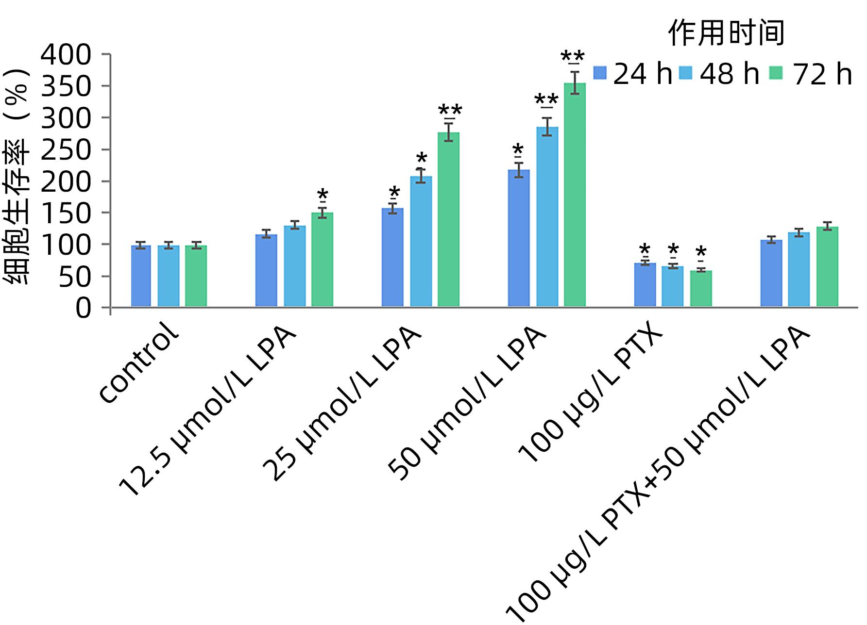
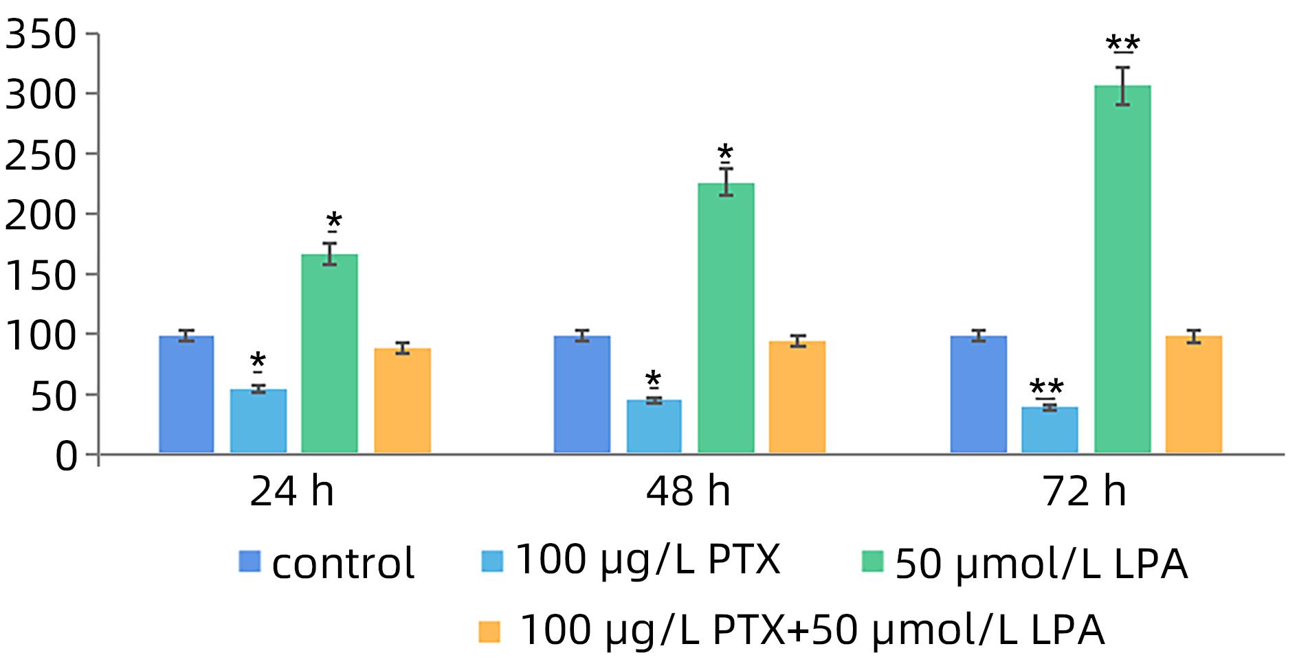
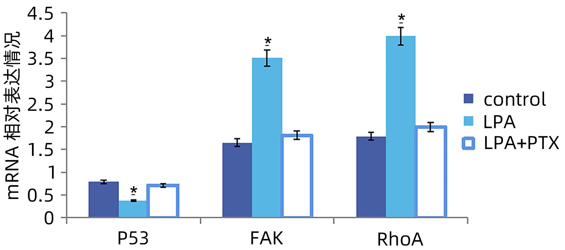
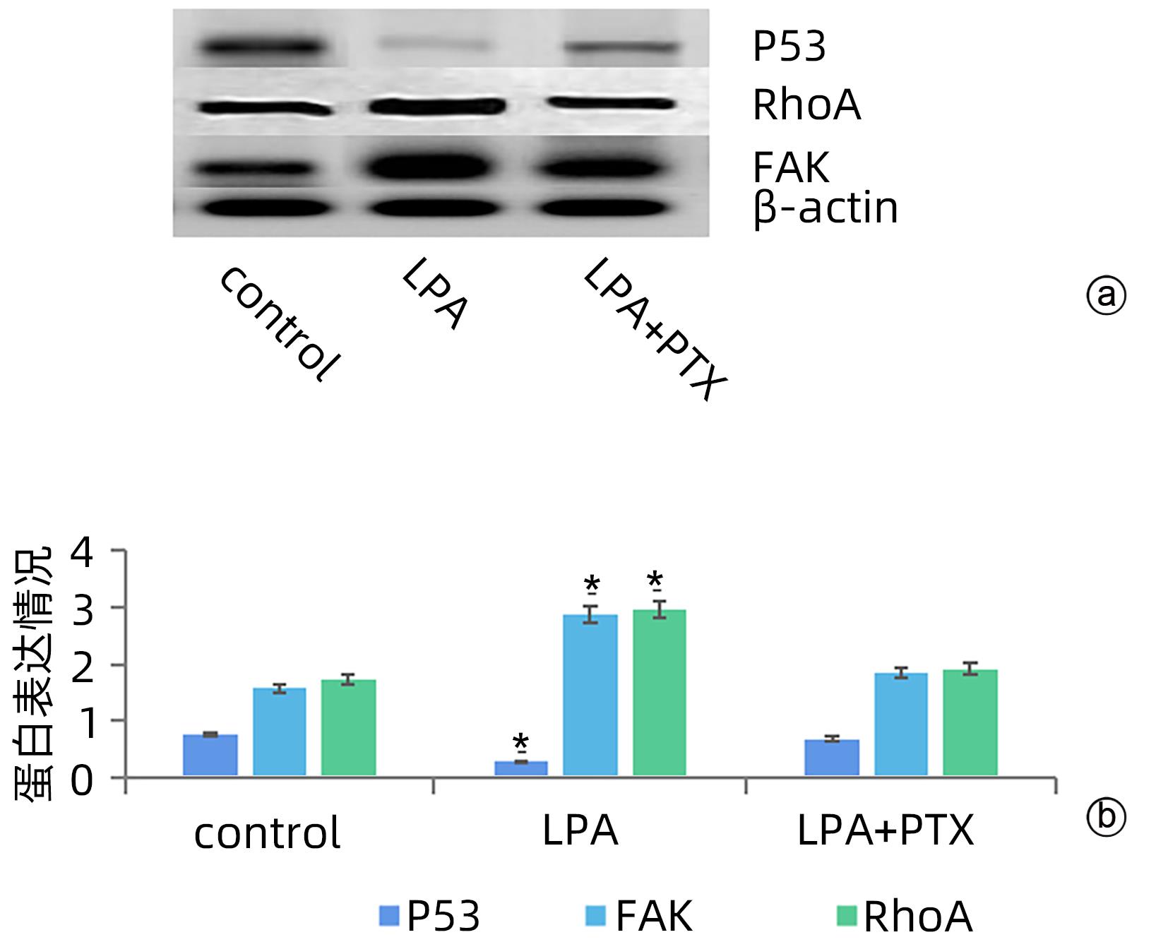
  •   Objective  To investigate the expression of lysophosphatidic acid (LPA) in patients with liver cancer, as well as its influence on malignant biological behavior of liver cancer and related regulatory mechanism.  Methods  From January 2016 to December 2022, 26 patients with liver cancer, 28 patients with liver cirrhosis, and 28 individuals undergoing physical examination were enrolled. ELISIA was used to measure the content of LPA in plasma and peritoneal effusion of the patients with liver cancer or liver cirrhosis accompanied by peritoneal effusion, and the content of LPA was measured in plasma of the normal population at the same time, so as to clarify the difference in the expression of LPA in different populations, such as the patients with liver cancer and those with liver cirrhosis. MTT cell proliferation assay and cell migration assay were used to observe the influence of LPA and its inhibitor pertussis toxin (PTX) on the proliferation, migration, and invasion of SMMC7721 cells. In order to investigate the effect of LPA on the expression of RhoA and its upstream and downstream molecules FAK and P53 after binding to its receptor, qPCR and Western blot were used to observe the effect of LPA on the mRNA and protein expression levels of P53, FAK, and RhoA in SMMC7721 cells. A one-way analysis of variance was used for comparison of the means of continuous data between multiple groups, and the SNK-q test was used for comparison between two groups.  Results  Compared with the patients with liver cirrhosis, the patients with liver cancer had a significantly higher concentration of LPA in plasma (4.99±0.55 μmol/L vs 2.63±0.43 μmol/L, P<0.05) and peritoneal effusion (5.19±0.63 μmol/L vs 2.91±0.46 μmol/L, P<0.05), and the patients with liver cancer also had a significantly higher level of plasma LPA than the normal population (4.99±0.55 μmol/L vs 1.61±0.39 μmol/L, P<0.05). The cell proliferation assay showed that LPA significantly promoted the proliferation of SMMC7721 cells, and cell proliferation rate increased with the increase in dose and time; in particular, the middle-and high-dose groups had a significantly higher proliferation rate than the control group (P<0.05). PTX inhibited the proliferative capacity of SMMC7721 cells in a time-dependent manner, and there was a significant difference between the groups (P<0.05). The proliferation rate of the 72-hour high-dose LPA group was 3.6 times that of the control group, while the proliferation rate of the PTX group was 0.6 times that of the control group; the proliferation rate of the 72-hour high-dose LPA+PTX group was 1.2 times that of the control group. In addition, LPA increased the migration ability of hepatoma cells, while PTX inhibited their migration, in a time-dependent manner, and there was a significant difference between the groups (P<0.05). The migration rate of the 72-hour high-dose LPA group was 3.09 times that of the control group, while the migration rate of the PTX group was 0.4 times that of the control group; the migration rate of the 72-hour high-dose LPA+PTX group was 0.99 times that of the control group. qPCR and Western blot showed that there were significant reductions in the mRNA and protein expression levels of P53 in SMMC7721 cells after LPA treatment, while there were significant increases in the mRNA and protein expression levels of FAK and RhoA; there was a significant difference between the LPA group and the control group (P<0.05).  Conclusion  There is an abnormal increase in the expression of LPA in patients with liver cancer, and LPA can promote the proliferation of liver cancer cells and increase their migration ability. At the same time, LPA changes the expression levels of P53, FAK, and RhoA, which may be associated with the promotion of tumor development and progression by LPA.


  • [1]
    LI X, RAMADORI P, PFISTER D, et al. The immunological and metabolic landscape in primary and metastatic liver cancer[J]. Nat Rev Cancer, 2021, 21( 9): 541- 557. DOI: 10.1038/s41568-021-00383-9.
    [2]
    XU Z, LIU Y, LIANG F. Clinical efficacy of Danshen Chuanxiongqin Injection and its effect on serum levels of LPA,Hcy and MCP-1 in patients with ischemic stroke[J]. J Changchun Univ Chin Med, 2021, 37( 1): 84- 87. DOI: 10.13463/j.cnki.cczyy.2021.01.023.

    许卓, 刘洋, 梁赋. 丹参川芎嗪注射液治疗缺血性脑卒中患者临床疗效及对血清LPA、Hcy、MCP-1水平的影响[J]. 长春中医药大学学报, 2021, 37( 1): 84- 87. DOI: 10.13463/j.cnki.cczyy.2021.01.023.
    [3]
    AIELLO S, CASIRAGHI F. Lysophosphatidic acid: Promoter of cancer progression and of tumor microenvironment development. A promising target for anticancer therapies?[J]. Cells, 2021, 10( 6): 1390. DOI: 10.3390/cells10061390.
    [4]
    RAY R, JANGDE N, SINGH SK, et al. Lysophosphatidic acid-RAGE axis promotes lung and mammary oncogenesis via protein kinase B and regulating tumor microenvironment[J]. Cell Commun Signal, 2020, 18( 1): 170. DOI: 10.1186/s12964-020-00666-y.
    [5]
    AMARAL RF, GERALDO LHM, EINICKER-LAMAS M, et al. Microglial lysophosphatidic acid promotes glioblastoma proliferation and migration via LPA1 receptor[J]. J Neurochem, 2021, 156( 4): 499- 512. DOI: 10.1111/jnc.15097.
    [6]
    KLYMENKO Y, BOS B, CAMPBELL L, et al. Lysophosphatidic acid modulates ovarian cancer multicellular aggregate assembly and metastatic dissemination[J]. Sci Rep, 2020, 10( 1): 10877. DOI: 10.1038/s41598-020-67565-7.
    [7]
    GNOCCHI D, CAVALLUZZI MM, MANGIATORDI GF, et al. Xanthenylacetic acid derivatives effectively target lysophosphatidic acid receptor 6 to inhibit hepatocellular carcinoma cell growth[J]. ChemMedChem, 2021, 16( 13): 2121- 2129. DOI: 10.1002/cmdc.202100032.
    [8]
    DEHGHAN M, SHAHBAZI S, SALEHNIA M. Effect of lysophosphatidic acid on the vascular endothelial growth factor expression in autotransplanted mouse ovaries encapsulated in sodium alginate[J]. J Family Reprod Health, 2021, 15( 2): 91- 98. DOI: 10.18502/jfrh.v15i2.6449.
    [9]
    XIE Y, WANG XC, WU XW, et al. Lysophosphatidic acid receptor 4 regulates osteogenic and adipogenic differentiation of progenitor cells via inactivation of RhoA/ROCK1/β-catenin signaling[J]. Stem Cells, 2020, 38( 3): 451- 463. DOI: 10.1002/stem.3128.
    [10]
    MINAMI K, UEDA N, ISHIMOTO K, et al. Cooperation of G12/13 and Gi proteins via lysophosphatidic acid receptor-2(LPA2) signaling enhances cancer cell survival to cisplatin[J]. Biochem Biophys Res Commun, 2020, 532( 3): 427- 432. DOI: 10.1016/j.bbrc.2020.08.087.
    [11]
    LEE SC, LIN KH, BALOGH A, et al. Dysregulation of lysophospholipid signaling by p53 in malignant cells and the tumor microenvironment[J]. Cell Signal, 2021, 78: 109850. DOI: 10.1016/j.cellsig.2020.109850.
    [12]
    ZHANG YZ, SHI HB, CHEN Y. Biological functions of apoptosis stimulating protein of p53 2 and its role in liver diseases[J]. J Clin Hepatol, 2018, 34( 11): 2443- 2447. DOI: 10.3969/j.issn.1001-5256.2018.11.039.

    张译之, 时红波, 陈煜. p53凋亡刺激蛋白2的生物学功能及其在肝病中的作用[J]. 临床肝胆病杂志, 2018, 34( 11): 2443- 2447. DOI: 10.3969/j.issn.1001-5256.2018.11.039.
    [13]
    HUANG CC, TSENG TT, LIU SC, et al. S1P increases VEGF production in osteoblasts and facilitates endothelial progenitor cell angiogenesis by inhibiting miR-16-5p expression via the c-src/FAK signaling pathway in rheumatoid arthritis[J]. Cells, 2021, 10( 8): 2168. DOI: 10.3390/cells10082168.
    [14]
    WEI YH, WANG YF, LIU NB, et al. A FAK inhibitor boosts anti-PD1 immunotherapy in a hepatocellular carcinoma mouse model[J]. Front Pharmacol, 2022, 12: 820446. DOI: 10.3389/fphar.2021.820446.
    [15]
    LIAO Y, LIU L, YANG JY, et al. ATX/LPA axis regulates FAK activation, cell proliferation, apoptosis, and motility in human pancreatic cancer cells[J]. Vitro Cell Dev Biol Anim, 2022, 58( 4): 307- 315. DOI: 10.1007/s11626-022-00660-3.
    [16]
    SUMITOMO A, SIRIWACH R, THUMKEO D, et al. LPA induces keratinocyte differentiation and promotes skin barrier function through the LPAR1/LPAR5-RHO-ROCK-SRF axis[J]. J Invest Dermatol, 2019, 139( 5): 1010- 1022. DOI: 10.1016/j.jid.2018.10.034.
    [17]
    KIM D, KIM HJ, BAEK JO, et al. Lysophosphatidic acid mediates imiquimod-induced psoriasis-like symptoms by promoting keratinocyte proliferation through LPAR1/ROCK2/PI3K/AKT signaling pathway[J]. Int J Mol Sci, 2021, 22( 19): 10777. DOI: 10.3390/ijms221910777.
    [18]
    NAKAJIMA K, OKA S, TANIKAWA T, et al. Lysophosphatidylinositol induced morphological changes and stress fiber formation through the GPR55-RhoA-ROCK pathway[J]. Int J Mol Sci, 2022, 23( 18): 10932. DOI: 10.3390/ijms231810932.
    [19]
    INABA A, HARADA H, IKEZAKI S, et al. LPA6-RhoA signals regulate junctional complexes for polarity and morphology establishment of maturation stage ameloblasts[J]. J Oral Biosci, 2022, 64( 1): 85- 92. DOI: 10.1016/j.job.2022.01.004.
    [20]
    BUTERA A, ROY M, ZAMPIERI C, et al. p53-driven lipidome influences non-cell-autonomous lysophospholipids in pancreatic cancer[J]. Biol Direct, 2022, 17( 1): 6. DOI: 10.1186/s13062-022-00319-9.
  • Relative Articles

    [1]Ke SHI, Qun ZHANG, Xianbo WANG, Ying FENG. Effect of intestinal flora and metabolites on the development and progression of acute-on-chronic liver failure[J]. Journal of Clinical Hepatology, 2025, 41(3): 568-573. doi: 10.12449/JCH250327
    [2]Viral Hepatitis Group of Professional Committee of Liver Disease (combined traditional Chinese and western medicine) of Chinese Research Hospital Association, Zhejiang Provincial Key Laboratory for Accurate Diagnosis and Treatment of Chronic Liver Diseases. Guidelines for fecal microbiota transplantation therapy in patients with liver cirrhosis[J]. Journal of Clinical Hepatology, 2025, 41(3): 424-431. doi: 10.12449/JCH250306
    [3]Xiaoming WU, Qiang HE, Linyi HOU, Yan HU, Xiaofang ZHEN, Jing HAO, Yan SHENG. Effect of Yudantong decoction on intestinal flora and intestinal barrier function in mice with cholestasis induced by α-naphthyl isothiocyanate[J]. Journal of Clinical Hepatology, 2023, 39(4): 864-875. doi: 10.3969/j.issn.1001-5256.2023.04.018
    [4]Hongkai XU, Chunfu WANG, Ye ZHANG, Jianqi LIAN. Role of fecal microbiota transplantation in chronic liver diseases[J]. Journal of Clinical Hepatology, 2023, 39(9): 2237-2243. doi: 10.3969/j.issn.1001-5256.2023.09.031
    [5]Guirong CHEN, Minggang WANG, Huaming LIN, Huiping YAN, Xiufeng WANG. Changes and pathogenic mechanism of intestinal flora in acute-on-chronic liver failure[J]. Journal of Clinical Hepatology, 2023, 39(8): 1992-1998. doi: 10.3969/j.issn.1001-5256.2023.08.034
    [6]Quan ZHOU, Chunlin CAI, Jinqiang LI. Gut-liver axis: Intestinal microbial homeostasis and hepatocellular carcinoma[J]. Journal of Clinical Hepatology, 2023, 39(11): 2710-2717. doi: 10.3969/j.issn.1001-5256.2023.11.029
    [7]Yixin HOU, Qun ZHANG, Yuyong JIANG, Hao YU, Yuying YANG, Xianbo WANG. Effect of Liangxue Jiedu decoction on intestinal flora in patients with hepatitis B virus-related acute-on-chronic liver failure: An analysis based on high-throughput sequencing[J]. Journal of Clinical Hepatology, 2022, 38(6): 1280-1287. doi: 10.3969/j.issn.1001-5256.2022.06.013
    [8]Fuchun WANG, Ziyi LI, Wanjie ZHANG, Xiaorong MAO, Junfeng LI. The significance of gut microbiota in acute-on-chronic liver failure[J]. Journal of Clinical Hepatology, 2022, 38(7): 1667-1670. doi: 10.3969/j.issn.1001-5256.2022.07.040
    [9]Transplantation Immunology Committee of Branch of Organ Transplantation Physician of Chinese Medical Doctor Association, Organ Transplant Rehabilitation Committee of China Association Rehabilitation Medicine, Branch of Organ Transplantation Physician of Guangdong Medical Doctor Association. Expert consensus on liver transplantation perioperative evaluation and rehabilitation for acute-on-chronic liver failure[J]. Journal of Clinical Hepatology, 2022, 38(12): 2701-2708. doi: 10.3969/j.issn.1001-5256.2022.12.005
    [10]Hui DENG, Bin ZHANG, Bin ZHU, Zhayier DILIHUMAER, Weixian WANG, Chunxia GUO, Dongliang YANG, Xin ZHENG, Junzhong WANG, Baoju WANG. Research advances in the role of gut microbiota in chronic hepatitis B, chronic hepatitis C, and related liver diseases[J]. Journal of Clinical Hepatology, 2022, 38(5): 1143-1147. doi: 10.3969/j.issn.1001-5256.2022.05.035
    [11]Mingyin MAN, Nana LI, Yue BU, Kaijiang YU. Influence of mitochondria-targeted antioxidant SS-31 on acute liver injury in a mouse model of sepsis[J]. Journal of Clinical Hepatology, 2022, 38(2): 392-396. doi: 10.3969/j.issn.1001-5256.2022.02.025
    [12]Xujuan LUO, Xue BAI, Zenghui LI, Fan LIU, Hao TANG, Ruoxin LI, Guodong YANG. Clinical effect of fecal microbiota transplantation versus the traditional Chinese medicine Rheum officinale in a rat model of hyperlipidemic acute pancreatitis[J]. Journal of Clinical Hepatology, 2022, 38(12): 2767-2773. doi: 10.3969/j.issn.1001-5256.2022.12.016
    [13]Ziwei GUO, Jiaxin ZHANG, Shuo LI, Xiaobin LI, Shun ZHU, Qian JIN, Xiaoke LI, Yongan YE. Research advances in intestinal flora and the development and prognosis of chronic hepatitis B[J]. Journal of Clinical Hepatology, 2022, 38(5): 1137-1142. doi: 10.3969/j.issn.1001-5256.2022.05.034
    [14]Lingyan XIAO, Awen XING, Shanzhong TAN. Research advances in the association between liver failure and intestinal barrier injury[J]. Journal of Clinical Hepatology, 2021, 37(11): 2710-2714. doi: 10.3969/j.issn.1001-5256.2021.11.049
    [15]Yanyan CHEN, Yanmei LAN, Minggang WANG, Dewen. MAO. Mechanism of action of bile acid-farnesoid X receptor-intestinal microecological axis in the development of liver failure and liver regeneration[J]. Journal of Clinical Hepatology, 2021, 37(2): 480-484. doi: 10.3969/j.issn.1001-5256.2021.02.049
    [16]Menghao LI, Kai LI, Shihao TANG, Zhengyu WANG, Wengang GUO, Zhanxin YIN, Guohong HAN. Changes in gut microbiota after transjugular intrahepatic portosystemic shunt in cirrhotic patients with mild hepatic encephalopathy in different prognosis groups[J]. Journal of Clinical Hepatology, 2021, 37(2): 326-330. doi: 10.3969/j.issn.1001-5256.2021.02.016
    [17]Li HongShan, Hu YiYang. Gut microecology: An important target of traditional Chinese medicine in the treatment of nonalcoholic fatty liver disease[J]. Journal of Clinical Hepatology, 2020, 36(1): 14-18. doi: 10.3969/j.issn.1001-5256.2020.01.002
    [18]Huang YunYi, Liu Yao, Zhang Qun, Shi Ke, Wang XianBo. Association of intestinal microecology with hepatic encephalopathy[J]. Journal of Clinical Hepatology, 2020, 36(4): 912-914. doi: 10.3969/j.issn.1001-5256.2020.04.045
    [19]Huang Qian, Zhang HaiBo, Li JingTao, Wei HaiLiang, Yan ShuGuang, Hui Yi, Chang ZhanJie. Research advances in the mechanism of action of intestinal microecology in intrahepatic cholestasis[J]. Journal of Clinical Hepatology, 2019, 35(10): 2355-2359. doi: 10.3969/j.issn.1001-5256.2019.10.050
    [20]Zheng Wei, Zhang YongHong, Zhao Yan. Role of intestinal microflora in the pathogenesis of hepatocellular carcinoma[J]. Journal of Clinical Hepatology, 2019, 35(7): 1613-1615. doi: 10.3969/j.issn.1001-5256.2019.07.041
  • Cited by

    Periodical cited type(9)

    1. 刘韦,白浪. 慢加急性肝衰竭动物模型研究现状. 临床肝胆病杂志. 2024(01): 187-192 . 本站查看
    2. 陈然,王帅,高扬,杨志琴,徐严. HBV相关慢加急性肝衰竭合并脓毒症的诊疗进展. 肝脏. 2024(07): 867-870 .
    3. 刘扬,洪文,黄克林,杨博,刘亚坡,路明. 粪菌移植对顽固性便秘小鼠的肠道菌群和肠道动力以及TLR4/NF-κB通路蛋白的影响. 现代生物医学进展. 2024(16): 3020-3024 .
    4. 于丹丹,刘亚坡,洪文,杨博,张媛. 粪菌移植对顽固性便秘患者肠道功能、免疫功能及炎症反应的改善效果. 结直肠肛门外科. 2024(05): 578-583 .
    5. 阮浩龙,王宁,于鸿浩,岳鹏鹏. 基于基因编辑技术研究特定基因对小鼠肠道微生物的影响. 中国医学工程. 2024(11): 44-49 .
    6. 徐洪凯,汪春付,张野,连建奇. 粪菌移植在慢性肝病治疗中的应用. 临床肝胆病杂志. 2023(09): 2237-2243 . 本站查看
    7. 周荃,蔡春琳,李金强. 肠-肝轴:肠道微生物稳态与肝细胞癌. 临床肝胆病杂志. 2023(11): 2710-2717 . 本站查看
    8. 周宜,刘晓琴,吴学敏,张浩,王海琴,王红亮,孟祥龙. 乳香及其炮制品对葡聚糖硫酸钠诱导小鼠炎症性肠病的保护作用. 现代药物与临床. 2022(07): 1432-1438 .
    9. 刘蕾,高越颖,郭琳,邱立朋,李会,耿燕. 酒精性肝损伤保护的药理实验教学设计. 实验室研究与探索. 2022(05): 238-243 .

    Other cited types(2)

  • 加载中
    Created with Highcharts 5.0.7Chart context menuAccess Class DistributionFULLTEXT: 24.6 %FULLTEXT: 24.6 %META: 71.8 %META: 71.8 %PDF: 3.6 %PDF: 3.6 %FULLTEXTMETAPDF
    Created with Highcharts 5.0.7Chart context menuAccess Area Distribution其他: 5.4 %其他: 5.4 %其他: 0.9 %其他: 0.9 %Bolivia: 0.1 %Bolivia: 0.1 %Central District: 0.1 %Central District: 0.1 %China: 0.1 %China: 0.1 %Herndon: 0.1 %Herndon: 0.1 %India: 0.1 %India: 0.1 %Malvern: 0.0 %Malvern: 0.0 %Matawan: 0.0 %Matawan: 0.0 %United States: 0.2 %United States: 0.2 %[]: 0.5 %[]: 0.5 %上海: 6.5 %上海: 6.5 %上饶: 0.0 %上饶: 0.0 %东京: 0.1 %东京: 0.1 %东莞: 0.6 %东莞: 0.6 %丽水: 0.0 %丽水: 0.0 %乌鲁木齐: 0.1 %乌鲁木齐: 0.1 %佛山: 0.2 %佛山: 0.2 %佳木斯: 0.0 %佳木斯: 0.0 %保定: 0.1 %保定: 0.1 %六安: 0.0 %六安: 0.0 %兰州: 0.2 %兰州: 0.2 %北京: 4.3 %北京: 4.3 %南京: 1.2 %南京: 1.2 %南宁: 0.4 %南宁: 0.4 %南昌: 0.3 %南昌: 0.3 %南阳: 0.0 %南阳: 0.0 %厦门: 0.0 %厦门: 0.0 %台北: 0.1 %台北: 0.1 %台州: 0.0 %台州: 0.0 %台湾: 0.1 %台湾: 0.1 %台湾省: 0.0 %台湾省: 0.0 %合肥: 0.2 %合肥: 0.2 %吉林: 0.1 %吉林: 0.1 %周口: 0.0 %周口: 0.0 %呼和浩特: 0.1 %呼和浩特: 0.1 %咸阳: 0.0 %咸阳: 0.0 %哈尔滨: 0.1 %哈尔滨: 0.1 %哥伦布: 0.1 %哥伦布: 0.1 %嘉兴: 0.0 %嘉兴: 0.0 %图尔库: 0.0 %图尔库: 0.0 %圣彼得堡: 0.1 %圣彼得堡: 0.1 %大连: 0.1 %大连: 0.1 %大阪: 0.1 %大阪: 0.1 %天津: 0.5 %天津: 0.5 %太原: 0.1 %太原: 0.1 %孝感: 0.2 %孝感: 0.2 %孟买: 0.0 %孟买: 0.0 %宁波: 0.1 %宁波: 0.1 %安康: 0.1 %安康: 0.1 %安顺: 0.1 %安顺: 0.1 %宣城: 0.1 %宣城: 0.1 %密蘇里城: 0.1 %密蘇里城: 0.1 %常德: 0.1 %常德: 0.1 %广州: 1.3 %广州: 1.3 %廊坊: 0.1 %廊坊: 0.1 %弗吉尼亚州: 0.3 %弗吉尼亚州: 0.3 %张家口: 1.2 %张家口: 1.2 %成都: 0.3 %成都: 0.3 %扬州: 0.1 %扬州: 0.1 %新乡: 0.0 %新乡: 0.0 %新德里: 0.0 %新德里: 0.0 %无锡: 0.2 %无锡: 0.2 %昆明: 1.0 %昆明: 1.0 %昌吉: 0.1 %昌吉: 0.1 %晋中: 0.1 %晋中: 0.1 %晋城: 0.1 %晋城: 0.1 %普洱: 0.1 %普洱: 0.1 %曼谷: 0.1 %曼谷: 0.1 %朝阳: 0.2 %朝阳: 0.2 %杭州: 1.0 %杭州: 1.0 %松原: 0.0 %松原: 0.0 %柏林: 0.0 %柏林: 0.0 %柳州: 0.0 %柳州: 0.0 %梧州: 0.0 %梧州: 0.0 %武汉: 1.3 %武汉: 1.3 %汕头: 0.0 %汕头: 0.0 %江门: 0.0 %江门: 0.0 %沈阳: 0.1 %沈阳: 0.1 %法兰克福: 0.0 %法兰克福: 0.0 %法尔肯施泰因: 0.5 %法尔肯施泰因: 0.5 %泰安: 0.0 %泰安: 0.0 %泰州: 0.0 %泰州: 0.0 %洛杉矶: 0.2 %洛杉矶: 0.2 %洛阳: 0.1 %洛阳: 0.1 %济南: 0.1 %济南: 0.1 %海口: 0.1 %海口: 0.1 %海得拉巴: 0.0 %海得拉巴: 0.0 %深圳: 0.6 %深圳: 0.6 %温州: 0.1 %温州: 0.1 %湘潭: 0.0 %湘潭: 0.0 %漯河: 0.0 %漯河: 0.0 %珠海: 0.2 %珠海: 0.2 %石家庄: 0.4 %石家庄: 0.4 %福州: 0.8 %福州: 0.8 %秦皇岛: 0.1 %秦皇岛: 0.1 %米兰: 0.1 %米兰: 0.1 %绍兴: 0.5 %绍兴: 0.5 %绥化: 0.1 %绥化: 0.1 %美国: 0.0 %美国: 0.0 %美国伊利诺斯芝加哥: 0.0 %美国伊利诺斯芝加哥: 0.0 %聊城: 0.0 %聊城: 0.0 %芒廷维尤: 38.9 %芒廷维尤: 38.9 %芝加哥: 0.5 %芝加哥: 0.5 %苏州: 0.3 %苏州: 0.3 %荆州: 0.0 %荆州: 0.0 %莫斯科: 0.8 %莫斯科: 0.8 %衡水: 0.2 %衡水: 0.2 %衢州: 0.0 %衢州: 0.0 %襄阳: 0.0 %襄阳: 0.0 %西宁: 12.9 %西宁: 12.9 %西安: 0.3 %西安: 0.3 %贵阳: 0.1 %贵阳: 0.1 %赤峰: 0.1 %赤峰: 0.1 %辽源: 0.1 %辽源: 0.1 %运城: 0.5 %运城: 0.5 %遵义: 0.0 %遵义: 0.0 %郑州: 2.4 %郑州: 2.4 %重庆: 1.3 %重庆: 1.3 %钦州: 0.1 %钦州: 0.1 %银川: 0.0 %银川: 0.0 %长春: 0.7 %长春: 0.7 %长沙: 1.1 %长沙: 1.1 %长治: 0.1 %长治: 0.1 %防城港: 0.0 %防城港: 0.0 %阿什本: 0.0 %阿什本: 0.0 %青岛: 0.1 %青岛: 0.1 %香港: 0.2 %香港: 0.2 %香港特别行政区: 0.0 %香港特别行政区: 0.0 %驻马店: 0.0 %驻马店: 0.0 %高雄: 0.1 %高雄: 0.1 %黄冈: 0.1 %黄冈: 0.1 %其他其他BoliviaCentral DistrictChinaHerndonIndiaMalvernMatawanUnited States[]上海上饶东京东莞丽水乌鲁木齐佛山佳木斯保定六安兰州北京南京南宁南昌南阳厦门台北台州台湾台湾省合肥吉林周口呼和浩特咸阳哈尔滨哥伦布嘉兴图尔库圣彼得堡大连大阪天津太原孝感孟买宁波安康安顺宣城密蘇里城常德广州廊坊弗吉尼亚州张家口成都扬州新乡新德里无锡昆明昌吉晋中晋城普洱曼谷朝阳杭州松原柏林柳州梧州武汉汕头江门沈阳法兰克福法尔肯施泰因泰安泰州洛杉矶洛阳济南海口海得拉巴深圳温州湘潭漯河珠海石家庄福州秦皇岛米兰绍兴绥化美国美国伊利诺斯芝加哥聊城芒廷维尤芝加哥苏州荆州莫斯科衡水衢州襄阳西宁西安贵阳赤峰辽源运城遵义郑州重庆钦州银川长春长沙长治防城港阿什本青岛香港香港特别行政区驻马店高雄黄冈

Catalog

    通讯作者: 陈斌, bchen63@163.com
    • 1. 

      沈阳化工大学材料科学与工程学院 沈阳 110142

    1. 本站搜索
    2. 百度学术搜索
    3. 万方数据库搜索
    4. CNKI搜索

    Figures(4)  / Tables(1)

    Article Metrics

    Article views (342) PDF downloads(14) Cited by(11)
    Proportional views
    Related

    /

    DownLoad:  Full-Size Img  PowerPoint
    Return
    Return